At a Glance
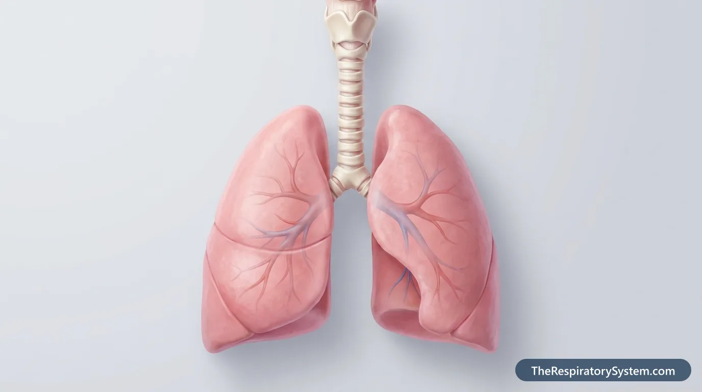
The lower respiratory tract consists of the trachea, the two main bronchi and their branches, the bronchioles and the alveoli, all housed in the lungs. It begins just below the larynx in the neck and ends in about 300 to 500 million alveoli, where gas exchange with the blood takes place.
- Location
- From the lower edge of the larynx in the neck down into both lungs in the chest
- Parts
- Trachea, bronchi, bronchioles, alveoli, lungs
- Function
- Conducts, cleans and warms air, then exchanges oxygen and carbon dioxide with the blood
- Zones
- Conducting zone (trachea to terminal bronchioles) and respiratory zone (respiratory bronchioles to alveoli)
- Driven by
- Diaphragm and intercostal muscles
- Part of
- The respiratory system, below the upper respiratory tract
On this page
- What Is the Lower Respiratory Tract?
- What Does the Lower Respiratory Tract Consist Of?
- Where Is the Lower Respiratory Tract?
- What Does the Lower Respiratory Tract Do?
- How Does Air Move Through the Lower Respiratory Tract?
- Upper Respiratory Tract vs Lower Respiratory Tract
- Explore Every Part of the Lower Respiratory Tract
- Key Facts
- FAQ
What Is the Lower Respiratory Tract?
The lower respiratory tract is the branching system of airways that starts at the trachea, just below the voice box, and ends in the microscopic air sacs of the lungs. It is one of the two main divisions of the respiratory system; the other is the upper respiratory tract, which includes the nose, sinuses, pharynx and larynx.
Because its airways divide again and again, the lower tract is often called the respiratory tree or bronchial tree. The trachea is the trunk, the bronchi are the large branches, the bronchioles are the twigs, and the alveoli are the leaves at the tips where the real work of breathing happens.
Conducting zone and respiratory zone
- Conducting zone: the trachea, bronchi and bronchioles down to the terminal bronchioles. These airways only carry air, and on the way they warm, moisten and clean it.
- Respiratory zone: the respiratory bronchioles, alveolar ducts, alveolar sacs and alveoli. Here the walls are thin enough for oxygen and carbon dioxide to pass between the air and the blood.
Explained simply
Picture an upside-down tree inside your chest. Air enters at the trunk in your neck, flows out along thinner and thinner branches, and reaches millions of tiny bubble-like leaves. Each leaf is wrapped in blood vessels, and that is where the blood picks up fresh oxygen and drops off carbon dioxide.
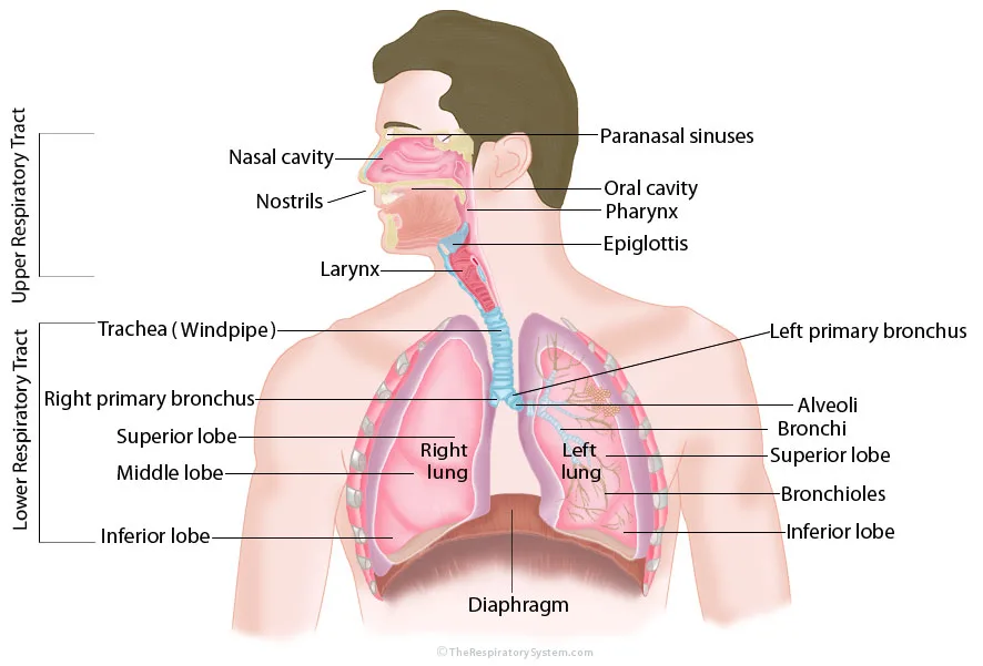
Explore Every Part of the Lower Respiratory Tract (24)


What Does the Lower Respiratory Tract Consist Of?
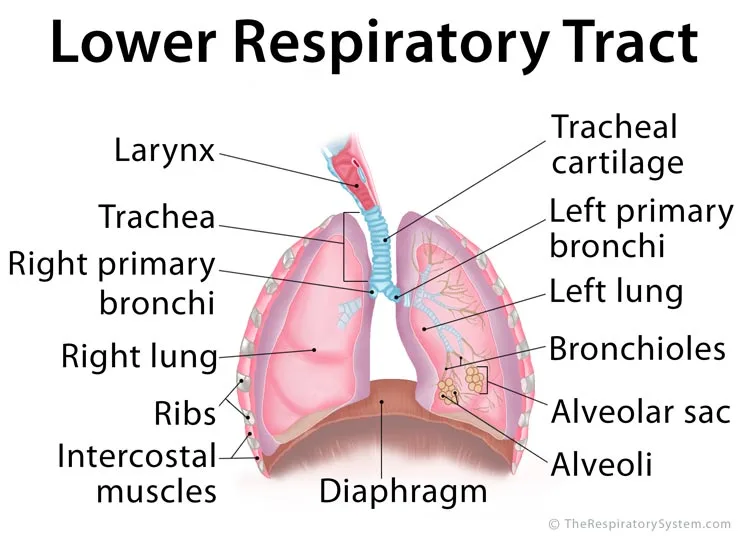
The lower respiratory tract consists of five main structures, from top to bottom: the trachea, the bronchi, the bronchioles, the alveoli and the lungs that contain them. Two breathing muscles, the diaphragm and the intercostal muscles, are not airways but power the airflow through it.
- Trachea (windpipe): the largest airway, a tube about 10 to 12 cm long held open by 16 to 20 C-shaped rings of hyaline cartilage. It carries air from the larynx into the chest.
- Bronchi: the trachea divides into the right and left main bronchi, thick tubes that enter the lungs and branch into lobar and segmental bronchi.
- Bronchioles: narrower tubes, about 1 mm wide or less, with no cartilage in their walls. Their smooth muscle widens or narrows them to adjust airflow.
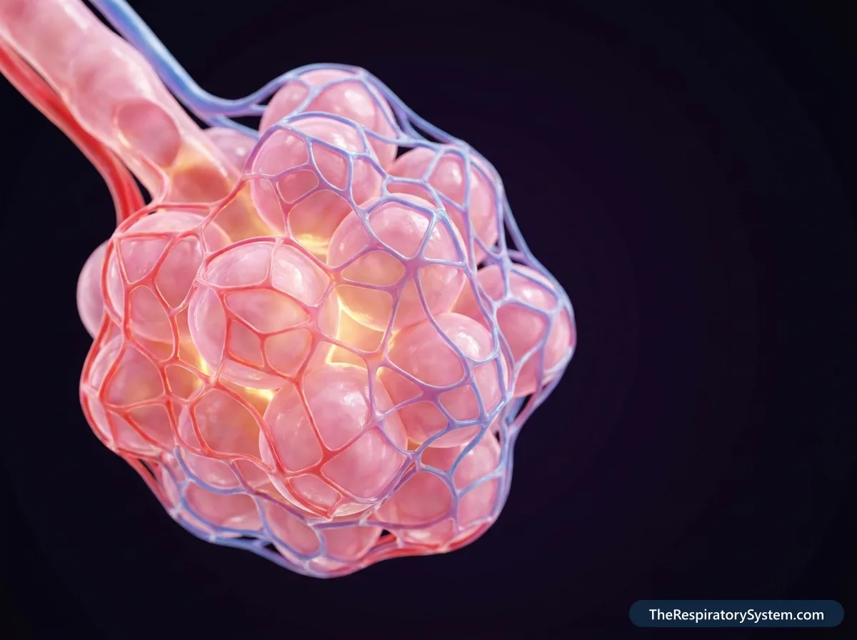
- Alveoli: tiny, thin-walled air sacs at the ends of the smallest airways, wrapped in capillaries. An adult has about 300 to 500 million of them.
- Lungs: the two spongy organs on either side of the heart that hold the bronchial tree and alveoli. The right lung has three lobes and the left lung has two.
The muscles that move air
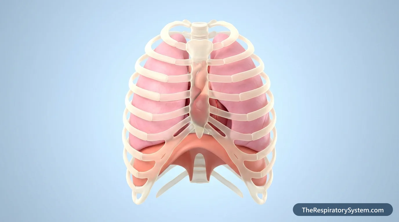
The airways cannot pump air on their own. The dome-shaped diaphragm under the lungs contracts and flattens to enlarge the chest, and the intercostal muscles between the ribs lift and spread the rib cage. Together they create the pressure changes that pull air into the lower tract and push it out again.
| Part | Location | Wall support | Main job |
|---|---|---|---|
| Trachea | From the larynx (C6) to the sternal angle | 16 to 20 C-shaped hyaline cartilage rings | Carries air into the chest; mucus and cilia clean it |
| Main bronchi | From the split of the trachea to each lung's hilum | Cartilage rings | Deliver air to the right and left lungs |
| Lobar and segmental bronchi | Inside each lung | Plates of cartilage | Supply each lobe and bronchopulmonary segment |
| Bronchioles | Deep in the lungs, about 1 mm wide or less | Smooth muscle, no cartilage | Adjust airflow by widening or narrowing |
| Terminal bronchioles | End of the conducting zone | Smooth muscle, club cells | Last airways that only carry air |
| Respiratory bronchioles | Start of the respiratory zone | Thin wall with a few alveoli | First site of some gas exchange |
| Alveolar ducts and sacs | At the tips of the respiratory bronchioles | Thin walls lined by alveoli | Lead air into clusters of alveoli |
| Alveoli | Tips of the bronchial tree, about 300 to 500 million | Type I and type II pneumocytes | Exchange oxygen and carbon dioxide; type II cells make surfactant |
| Lungs | Either side of the heart in the chest | Elastic tissue covered by pleura | House the airways and alveoli |
| Diaphragm (muscle) | Under the lungs, between chest and abdomen | Skeletal muscle and central tendon | Main muscle of inhalation |
| Intercostal muscles | Between the ribs | Three layers of skeletal muscle | Move the ribs to widen and narrow the chest |
| Source: Typical adult anatomy, our editors | |||
Where Is the Lower Respiratory Tract?
The lower respiratory tract lies mostly in the chest (thoracic cavity), starting in the lower front of the neck and branching into both lungs inside the rib cage, above the diaphragm. Only the upper part of the trachea lies in the neck.
Where does the lower respiratory tract begin?
It begins where the larynx ends, at the lower border of the cricoid cartilage, the ring-shaped cartilage at the base of the voice box, at about the level of the 6th cervical vertebra (C6). From there the trachea runs down in front of the esophagus into the chest. Some textbooks count the larynx as part of the lower tract because it lies below the pharynx, but most, including our site, place it in the upper tract and draw the dividing line just below it.
Where do the airways divide?
The trachea ends in the chest at about the level of the sternal angle, the ridge where the handle and body of the breastbone meet, where it splits into the two main bronchi. The bronchi enter each lung at the hilum, on the inner surface facing the heart, and then branch throughout the lung tissue all the way to its outer edges near the ribs.
What Does the Lower Respiratory Tract Do?
The main function of the lower respiratory tract is gas exchange: it delivers inhaled air to the alveoli, where oxygen passes into the blood and carbon dioxide passes out to be breathed away. An adult at rest breathes about 12 to 20 times a minute, moving roughly 11,000 liters of air through these airways every day.
- Conducting air: the branching airways guide air to every part of both lungs and back out again.
- Gas exchange: in the alveoli, oxygen diffuses into the capillaries and carbon dioxide diffuses into the air spaces.
- Cleaning the air: mucus traps dust and microbes, and the beating cilia of the lining push it up toward the throat, a system called the mucociliary escalator. Alveolar macrophages clear particles that reach the air sacs.
- Warming and moistening: the airways finish the warming and humidifying begun in the nose, so the air reaching the alveoli is at body temperature and fully moist.
- Protecting the airway: the cough reflex, triggered by irritation of the trachea and bronchi, forcefully clears mucus and foreign material.
- Acid-base balance: by adjusting how much carbon dioxide is breathed out, the lungs help keep the pH of the blood stable.
- Speech: air pushed up from the lungs through the trachea vibrates the vocal folds in the larynx above.
Our page on respiratory system functions explains how these jobs work together with the upper tract.
How Does Air Move Through the Lower Respiratory Tract?
Air moves through the lower respiratory tract in a fixed order, from the trachea through about 23 generations of ever smaller branches to the alveoli, and then back out the same way. The pressure changes made by the breathing muscles keep it flowing.
- Larynx to trachea: air that has passed through the nose or mouth, the pharynx and the larynx enters the trachea in the neck.
- Trachea to main bronchi: at the sternal angle the trachea splits into the right and left main bronchi. The right one is shorter, wider and more vertical.
- Lobar and segmental bronchi: each main bronchus divides into lobar bronchi, three on the right and two on the left, then into segmental bronchi for each bronchopulmonary segment.
- Bronchioles: the bronchi keep dividing until the cartilage disappears and the tubes become bronchioles, ending in terminal bronchioles, the last purely conducting airways.
- Respiratory bronchioles and alveolar ducts: these thin airways have a few alveoli budding from their walls and lead into alveolar ducts and sacs.
- Alveoli: oxygen crosses the thin alveolar and capillary walls into the blood, and carbon dioxide crosses the other way.
- Back out: when the diaphragm relaxes and the elastic lungs recoil, air rich in carbon dioxide flows back up the same path and out through the upper tract.
Upper Respiratory Tract vs Lower Respiratory Tract
The upper respiratory tract (nose, nasal cavity, sinuses, pharynx and larynx) prepares the air and directs it into the airway, while the lower respiratory tract (trachea, bronchi, bronchioles, alveoli and lungs) carries it into the chest and exchanges gases with the blood. The dividing line is just below the larynx.
Anatomy and function
The upper tract lies in the head and neck and shares space with the digestive system in the pharynx; it filters, warms and moistens air, gives us the sense of smell and makes the voice. The lower tract lies mostly in the chest, carries only air, and contains the only part of the airway where gas exchange happens. Its walls also change on the way down: thick tubes held open by cartilage give way to air sacs whose walls are a single thin layer of cells.
Upper vs lower respiratory tract infections
Infections are grouped the same way. Upper respiratory tract infections, such as the common cold, sinusitis and sore throat, affect the nose, sinuses or throat and are usually mild. Lower respiratory tract infections, such as acute bronchitis, bronchiolitis and pneumonia, affect the airways or air sacs in the chest; they more often cause a deep cough, shortness of breath or chest discomfort and can be more serious, especially in young children and older adults. Our respiratory diseases section describes these conditions, and anyone with breathing difficulty should see a doctor.
Our upper vs lower respiratory tract page compares the two halves side by side.
Key Facts
- The lower respiratory tract includes the trachea, bronchi, bronchioles, alveoli and lungs.
- It begins just below the larynx, at the lower border of the cricoid cartilage, at about the level of the C6 vertebra.
- The airways branch about 23 times between the trachea and the alveoli.
- The conducting zone ends at the terminal bronchioles; gas exchange happens only in the respiratory zone beyond them.
- An adult has about 300 to 500 million alveoli, the only place in the airway where oxygen enters the blood.
- The diaphragm and intercostal muscles power the airflow; the airways themselves do not pump air.
Frequently Asked Questions
Is the lower respiratory tract sterile?
It was long believed to be sterile, but modern genetic methods show that the healthy lower airways carry a small, low-density community of microbes, mostly arriving from the mouth and throat with tiny amounts of inhaled or swallowed material. The mucociliary escalator, coughing and immune cells constantly clear them, so the numbers stay far lower than in the upper respiratory tract.
Is the larynx part of the upper or lower respiratory tract?
Most anatomy texts, and this site, place the larynx in the upper respiratory tract and start the lower tract just below it, at the trachea. Some sources, especially older ones and some medical classifications, count the larynx as the first part of the lower tract because it lies below the pharynx. Both systems agree that the trachea, bronchi, bronchioles and alveoli belong to the lower tract.
What is a lower respiratory tract infection?
A lower respiratory tract infection is any infection of the airways or lung tissue below the larynx. The main types are acute bronchitis (infection of the bronchi), bronchiolitis (infection of the small airways, common in babies) and pneumonia (infection of the alveoli). They are caused mainly by viruses and bacteria and typically bring a cough, sometimes with fever and shortness of breath.
Why does the lower respiratory tract branch so many times?
Branching packs an enormous surface into a small space. Each division multiplies the number of airways, so after about 23 generations a single trachea leads to hundreds of millions of alveoli. Their combined surface is roughly 50 to 75 square meters, far more than the chest could hold if the airways were simple tubes, and it lets enough oxygen reach the blood for the whole body.
What lines the lower respiratory tract?
The trachea and bronchi are lined by pseudostratified ciliated columnar epithelium with mucus-producing goblet cells. Lower down, the lining thins to simple cuboidal cells in the small bronchioles, with dome-shaped club cells that help protect the airways. The alveoli are lined mostly by very flat type I pneumocytes, with type II pneumocytes that produce surfactant to keep the air sacs from collapsing.
Which is bigger, the right or left lung?
The right lung is slightly larger and heavier. It has three lobes and is wider, although it is a little shorter because the liver pushes the right side of the diaphragm upward. The left lung has two lobes and a hollow, the cardiac notch, that makes room for the heart, which sits slightly to the left in the chest.
Keep Exploring


