Intercostal Muscles
Published on May 28th 2018 by admin under
What Are the Intercostal Muscles
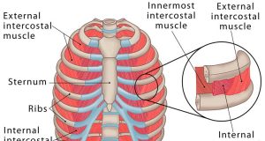
The small muscles located in the intercostal spaces between each of the 12 ribs in the human body are known as the intercostal muscles [1, 2].
Anatomy and Structure of Intercostal Muscles
These muscles are divided into three layers [3]:
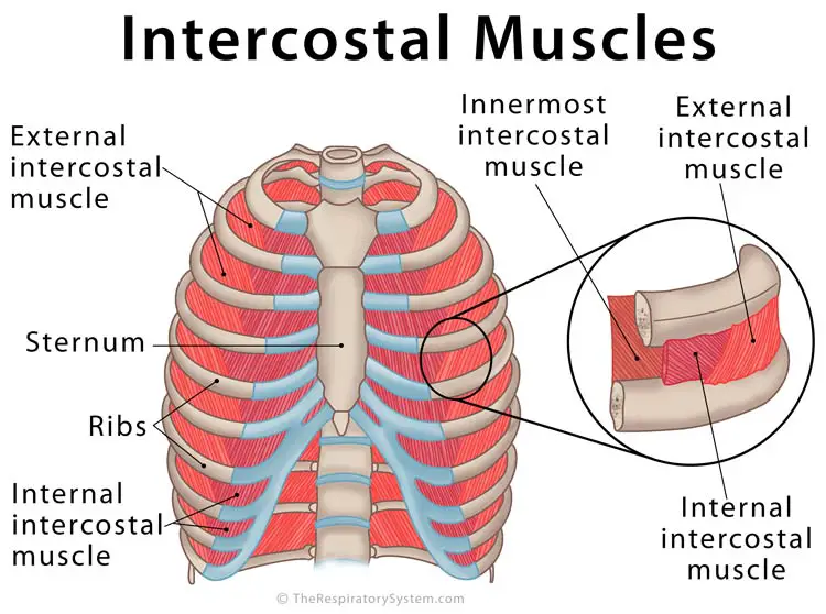
Intercostal Muscles
1. External Intercostal Muscles
The most superficial of all the intercostal muscles, there are 11 pairs of external intercostal muscles in the human body [4].
Origin: The inferior or lower border of each rib [5]
Insertion: The superior or upper border of the rib below [5]
The muscle fibers run slantingly, in an inward (toward the center of the body) and downward direction [2], connecting the 12 ribs to each other. The external intercostal muscles form the external intercostal membrane that extends to attach to the sternum [6].
2. Internal Intercostal Muscles
There are 11 internal intercostal muscles located on each side of the rib cage [7], forming the second or middle layer of the intercostal muscles [8].
Origin: The superior or upper border of each rib, at the costal groove [9]
Insertion: The inferior or lower border of the rib above [9]
The fibers of the internal intercostals lie perpendicular to those of the external intercostal muscles, running down in a sideways and backward direction (opposite to the external intercostals) [7, 10].
3. Innermost Intercostal Muscles
As the name suggests, these form the innermost layer of the intercostal muscles. They are poorly developed in the upper parts of the rib cage, becoming more pronounced toward the lower levels [4], with the muscle fibers at the lateral thoracic wall being most prominent.
Origin: The inner border of the ribs, at the costal groove [11]
Insertion: The upper border of the next rib below [12]
The innermost intercostal muscle fibers have the similar alignment as the internal intercostal muscles, also running in the same direction [11, 13].
Innervation and Blood Supply
All the three layers of the intercostal muscles are innervated by the intercostal nerves arising from thoracic nerves T1 to T11 [14]. The blood supply in this area is carried out by the intercostal arteries and veins [15].
Function: What Do the Intercostal Muscles Do in the Respiratory System
Along with the diaphragm, these three muscles play a vital role in respiration, with the three layers serving their own purpose during breathing in and out [16].
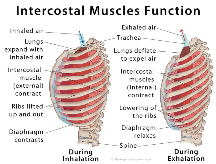
Intercostal Muscles Function
During Inhalation: The external intercostals connect all the ribs in such a way that when these muscles contract, the ribs are lifted up (they have no other way to move in as they are fixed to the vertebral column at the back) [17]. The diaphragm also contracts at the same time, flattening towards the abdominal cavity [13]. This expands the thoracic cavity, causing air to rush into the lungs [17]. Being the strongest of all the three intercostal muscles [18], these are the primary intercostal muscles used in regular breathing [16]. The internal muscles remain in a relaxed position during inhalation [19].
During Exhalation: As the internal intercostals are perpendicular to the external intercostals, the ribcage comes down when the former contracts and the latter relaxes. This reduces the volume of the chest cavity, putting pressure on the lungs so the air can be expelled (following gas exchange) [7]. The diaphragm also relaxes during this time, applying more pressure on the rib cage.
The innermost intercostals are the weakest of the three [11], and are used only during deep breathing. As they run in a similar direction as the innermost intercostals, they work similarly as well [20].
They also assist in protecting the lungs and solidifying the thoracic region [2].
Associated Conditions
An intercostal muscle strain is the most common associated condition. Apart from that, intercostal pain or spasms may occur due to certain underlying problems like multiple sclerosis, or some chest trauma.
References
- https://www.medicinenet.com/script/main/art.asp?articlekey=3991
- http://www.innerbody.com/image_chest1/chest01.html
- https://www.healthline.com/health/intercostal-muscle-strain
- https://www.sciencedirect.com/topics/neuroscience/innermost-intercostal-muscle
- https://www.getbodysmart.com/muscles-that-act-on-the-chest/external-intercostal-muscles
- https://www.imaios.com/en/e-Anatomy/Anatomical-Parts/External-intercostal-membrane
- https://radiopaedia.org/articles/internal-intercostal-muscle
- http://anatomyzone.com/anatomy-feed/internal-intercostal-muscle/
- http://gmch.gov.in/e-study/e%20lectures/Anatomy/Intercostal%20Spaces%20-%20Avinash.pdf
- https://www.sciencedirect.com/topics/neuroscience/external-intercostal-muscles
- https://radiopaedia.org/articles/innermost-intercostal-muscles
- https://www.earthslab.com/anatomy/innermost-intercostal-muscles/
- http://anatomy.uams.edu/muscles_thorax.html
- http://teachmeanatomy.info/thorax/muscles/thoracic-cage/
- https://www.kenhub.com/en/library/anatomy/intercostal-arteries-and-veins
- https://study.com/academy/lesson/intercostal-muscles-definition-function-location.html
- http://www.yorku.ca/earmstro/journey/external.html
- https://radiopaedia.org/articles/external-intercostal-muscle
- https://www.bbc.com/education/guides/z6h4jxs/revision
- https://www.kenhub.com/en/library/anatomy/intercostal-muscles


