At a Glance
The inferior nasal concha (inferior turbinate) is a paired, scroll-shaped facial bone on the lower lateral wall of each nasal cavity. It is the largest concha and joins four bones: the maxilla, palatine, lacrimal and ethmoid. The inferior meatus beneath it receives the nasolacrimal (tear) duct, and its swelling mucosa does most of the warming and humidifying of inhaled air.
- Location
- Lower part of the lateral wall of each nasal cavity, just above the nasal floor
- Function
- Warms, humidifies and filters most inhaled air; adjusts nasal airflow by swelling and shrinking
- Bone type
- Independent, paired facial bone; thin, scroll-shaped and porous
- Articulates with
- Maxilla, palatine, lacrimal and ethmoid bones
- Latin name
- Concha nasalis inferior (concha nasi inferior)
- Part of
- Nasal cavity, upper respiratory tract
On this page
- What Is the Inferior Nasal Concha?
- Where Is the Inferior Nasal Concha Located?
- Inferior Nasal Concha Anatomy: Surfaces, Borders and Lining
- What Bones Does the Inferior Nasal Concha Articulate With?
- Inferior Nasal Concha and Meatus: What Drains There?
- What Does the Inferior Nasal Concha Do?
- How Does the Inferior Nasal Concha Develop?
- Common Conditions That Affect the Inferior Nasal Concha
- Key Facts
- Quiz
- FAQ
What Is the Inferior Nasal Concha?
The inferior nasal concha is a thin, curled, spongy-looking bone attached to the lower part of the lateral wall of each nasal cavity, and it is the largest of the three nasal conchae. There are two of them, one in each half of the nose, and they are mirror images of each other. Together with its thick covering of mucous membrane, the bone is called the inferior turbinate.
Is the inferior nasal concha its own bone?
Yes. The superior nasal concha and the middle nasal concha are only parts of the ethmoid bone, but the inferior concha is an independent bone. It is one of the 14 facial bones of the skull, a paired bone like the maxillae, nasal bones and palatine bones. It is not a cranial bone, and it does not touch the nasal septum: it hangs from the side wall and curls toward the septum, with an air channel between them.
Explained simply
Imagine a rolled-up leaf fixed along one edge to the outer wall of a narrow tunnel, near the floor. That is the inferior concha. Its wet, warm, spongy covering acts like a radiator and a humidifier at the same time, so cold, dry air passing over it comes out warmer and moister.
| Feature | Inferior concha | Middle concha |
|---|---|---|
| Bone | Independent, paired facial bone | Part of the ethmoid, a cranial bone |
| Size | Largest concha | Medium |
| Position | Lowest, just above the nasal floor | Between the superior and inferior conchae |
| Ossification | Own center, about 5th fetal month | With the ethmoid labyrinth, about 4th to 5th fetal month |
| Meatus below | Inferior meatus | Middle meatus |
| Drains below it | Nasolacrimal duct | Frontal, maxillary and anterior ethmoidal sinuses |
| Erectile tissue | Thickest in the nose | Thinner |
| Main job | Warming, humidifying, controlling airflow | Shielding sinus openings, guiding airflow |
| Typical problem | Turbinate hypertrophy | Concha bullosa |
| Source: Typical adult anatomy, our editors | ||
Where Is the Inferior Nasal Concha Located?
The inferior nasal concha is located along the lower part of the lateral wall of each nasal cavity, below the middle concha and just above the floor of the nose, which is formed by the hard palate. It runs front to back along most of the length of the cavity.
Borders and neighbors
- Above: the middle meatus and the middle concha.
- Below and lateral: the inferior meatus, the air passage between the concha and the nasal floor.
- Medially: the nasal septum, formed lower down by the vomer, with the common meatus between them.
- In front: the nasal vestibule just inside the nostrils.
- Behind: the choanae; its back end lies close to the opening of the auditory tube in the nasopharynx.
- Laterally, beyond the wall: the maxillary sinus.
Inferior nasal concha on the skull
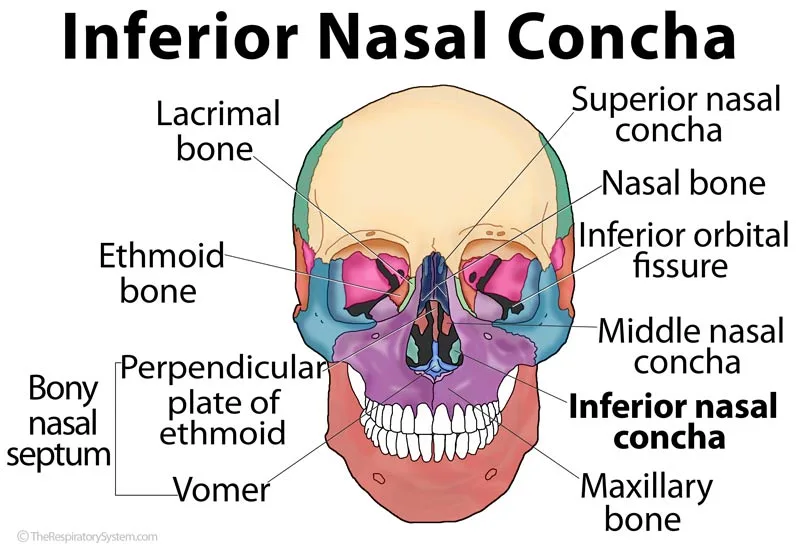
In an anterior view of the skull, the inferior conchae are the two curled bones easily seen in the lower part of the pear-shaped nasal opening, one on each side of the vomer. In a living person, the front end of the inferior turbinate is the pink, rounded swelling seen on the outer wall when a light is shone into the nostril.
Inferior Nasal Concha Anatomy: Surfaces, Borders and Lining
The inferior nasal concha is a thin, scroll-shaped plate with two surfaces, two borders and two ends, and it is covered by a thick mucous membrane rich in blood vessels. The bone itself is light and porous, and its texture under the covering looks spongy.
Surfaces
- Medial surface: convex and facing the septum. It is pitted with many small openings and marked by long grooves that run front to back and carry blood vessels to the mucous layer covering the bone.
- Lateral surface: concave, facing the side wall, and forming part of the boundary of the inferior meatus.
Borders and ends
The upper border is thin and irregular and is joined to several bones along the lateral nasal wall. The lower border is free, thick and porous, especially in its middle part. The front end is broader and blunter, while the back end tapers to a point.
Lining and blood supply
The respiratory mucosa over the inferior concha is the thickest in the nose. Under its ciliated pseudostratified columnar epithelium and goblet cells lies a lamina propria filled with glands and wide venous sinusoids, often called swell bodies or erectile tissue, which fill with blood and empty again under the control of autonomic nerves. Blood arrives mainly through branches of the sphenopalatine artery that enter at the back end of the concha. Sensation is carried by small nasal branches of the maxillary nerve, including branches from the greater palatine nerve, and by the anterior ethmoidal nerve at the front.
What Bones Does the Inferior Nasal Concha Articulate With?
The inferior nasal concha articulates with four bones: the maxilla, the palatine bone, the lacrimal bone and the ethmoid bone. All of these joints lie along its upper border, which can be divided into three portions.
- Front portion: joins the conchal crest of the maxilla.
- Back portion: joins the conchal crest of the palatine bone.
- Middle portion: carries three processes of different size and shape, described below.
The three processes
The lacrimal process is a small, pointed projection toward the front. Its tip meets the lacrimal bone and its edges meet the maxilla, and together these bones complete the nasolacrimal canal that carries the tear duct. The ethmoidal process, a thin, broad plate behind it, rises to join the uncinate process of the ethmoid bone. The maxillary process is a thin sheet that curves downward and outward to meet the maxilla, partly closing the large opening in the maxilla and forming part of the medial wall of the maxillary sinus.
| Part of the concha | Joins | Result |
|---|---|---|
| Front portion of the upper border | Conchal crest of the maxilla | Anchors the front of the concha |
| Back portion of the upper border | Conchal crest of the palatine bone | Anchors the back of the concha |
| Lacrimal process, tip | Lacrimal bone | Helps form the nasolacrimal canal |
| Lacrimal process, edges | Frontal process of the maxilla | Completes the walls of the nasolacrimal canal |
| Ethmoidal process | Uncinate process of the ethmoid bone | Links the concha to the ethmoid on the lateral wall |
| Maxillary process | Body of the maxilla | Forms part of the medial wall of the maxillary sinus |
| Source: Typical adult anatomy, our editors | ||
Inferior Nasal Concha and Meatus: What Drains There?
The inferior meatus, the passage under the inferior nasal concha, receives the opening of the nasolacrimal duct, which carries tears from the eye into the nose. It is the largest of the meatuses and runs along almost the whole length of the lateral wall.
The duct opens under the front part of the concha, where a small fold of mucous membrane, the lacrimal fold (also called the valve of Hasner), helps stop air and mucus from being pushed back up toward the eye. No paranasal sinus drains here; the sinuses open into the meatuses and recess higher up. This is why heavy crying makes the nose run, and why some eye drops can be tasted at the back of the throat. Our nasal meatus page compares all of the meatuses in detail.
What Does the Inferior Nasal Concha Do?
The inferior nasal concha warms, humidifies and filters most of the air that enters the nose, and by swelling and shrinking it adjusts how much air can flow through each side. Of the three conchae, it does most of this air-conditioning work because it is the largest and has the thickest, most blood-rich lining.
Function of the inferior nasal concha
- Warming: blood flowing through its sinusoids heats cold inhaled air toward body temperature.
- Humidifying: water from its glands and moist surface saturates the air, protecting the trachea and lower airways from drying.
- Filtering: dust, pollen and microbes stick to its mucus, and cilia sweep the mucus back to the pharynx.
- Controlling airflow: its front end lies close to the narrowest part of the nasal airway, so small changes in its size change nasal resistance a great deal.
- Recovering heat and moisture: on the way out, warm, moist air from the lungs gives some of its heat and water back to the cooler lining.
Swelling and the nasal cycle
The erectile tissue of the inferior turbinates is never still. In most people one side fills with blood while the other shrinks, and the pattern switches every few hours, a rhythm called the nasal cycle. Lying down, cold air, allergens and infections make the tissue swell, while exercise makes it shrink, which is why a stuffy nose often clears during a brisk walk. Together these changes are part of the respiratory system functions of the upper airway.
How Does the Inferior Nasal Concha Develop?
The inferior nasal concha develops from the maxilloturbinal, the lowest ridge on the side wall of the cartilaginous nasal capsule, and it ossifies from a single center that appears in about the fifth month of fetal life in that cartilaginous wall. This separate origin is why it becomes an independent bone, while the ethmoturbinals above it stay part of the ethmoid.
After birth the concha grows with the face and the maxillary sinus, which enlarges greatly during childhood and the eruption of the permanent teeth. In adults its size depends mostly on its soft-tissue covering rather than the bone. In older age the mucosa often becomes thinner, drier and less able to humidify air, which can leave the nose feeling dry and crusted.
Common Conditions That Affect the Inferior Nasal Concha
The inferior nasal concha is the turbinate most often involved in nasal blockage, because its thick, swelling lining sits right in the main airstream. Our respiratory diseases section covers related conditions, and a nose that stays blocked for weeks is a reason to see a doctor.
- Inferior turbinate hypertrophy: lasting enlargement or swelling of the inferior turbinates that narrows the airway, causing a blocked nose, breathing difficulty, mouth breathing, snoring and sometimes nosebleeds. Possible causes include colds and flu, allergies, nasal polyps and hormonal changes such as those of pregnancy.
- Boggy turbinates in allergic rhinitis: pale, soft, swollen inferior turbinates typical of an allergic reaction.
- Compensatory hypertrophy: enlargement of the inferior turbinate on the wider side of a deviated septum.
- Rebound congestion (rhinitis medicamentosa): swelling that returns more strongly after decongestant nasal sprays are overused.
- Empty nose syndrome: a rare condition in which the nose feels blocked or dry even though it is wide open, linked with removal of too much turbinate tissue.
Key Facts
- The inferior nasal concha is the largest of the three nasal conchae and the only one that is a separate bone.
- It is a paired facial bone that articulates with four bones: the maxilla, palatine, lacrimal and ethmoid.
- It attaches to the lateral wall of the nasal cavity, not to the septum.
- The nasolacrimal duct opens into the inferior meatus beneath it, so tears drain into the nose.
- Its maxillary process forms part of the medial wall of the maxillary sinus.
- It ossifies from a single center that appears in about the fifth month of fetal life.
- Its thick erectile lining swells and shrinks during the nasal cycle and is the usual site of turbinate hypertrophy.
Quick Quiz: Test What You Know About the Inferior Nasal Concha
6 questions. Tap an answer to check it.
How many bones does the inferior nasal concha articulate with?
It joins the maxilla, palatine, lacrimal and ethmoid bones.
What opens into the inferior meatus?
The nasolacrimal duct carries tears into the inferior meatus under the front part of the inferior concha.
Which statement about the inferior nasal concha is true?
The inferior concha is an independent, paired facial bone attached to the lateral nasal wall.
Which process of the inferior concha helps form the nasolacrimal canal?
The lacrimal process meets the lacrimal bone and the maxilla to complete the nasolacrimal canal.
Which turbinate is most often enlarged in turbinate hypertrophy?
The inferior turbinates have the thickest erectile lining and sit in the main airstream, so they are most often involved.
When does the ossification center of the inferior concha appear?
A single center appears in about the fifth month of fetal life in the lateral wall of the cartilaginous nasal capsule.
Frequently Asked Questions
Is the inferior nasal concha a paired bone?
Yes. There are two inferior nasal conchae, one in each nasal cavity, and they are mirror images of each other. Each attaches to the lateral wall of its own side, and together they count as two of the 14 facial bones. The other paired facial bones are the maxillae, zygomatic bones, nasal bones, lacrimal bones and palatine bones, while the vomer and mandible are single.
Is the inferior nasal concha a cranial bone?
No. The inferior nasal concha is a facial bone. The cranial bones are the eight bones that enclose the brain: the frontal, two parietal, two temporal, occipital, sphenoid and ethmoid bones. The superior and middle conchae belong to the ethmoid, so they are part of the cranium, but the inferior concha is a separate bone of the face that only touches the ethmoid at one small process.
Does the inferior nasal concha touch the vomer?
No. The vomer forms the lower back part of the nasal septum in the midline, while the inferior concha hangs from the lateral wall of each nasal cavity. Between them is an open air channel, the common meatus. Both are thin facial bones that sit low in the nasal cavity, which is why they appear close together in front views of the skull.
Is the inferior turbinate the same as the inferior nasal concha?
Yes, the two names describe the same structure. Inferior nasal concha is the anatomical name used in textbooks, while inferior turbinate is the name doctors usually use, often meaning the bone together with its thick, swelling mucous membrane. That is why medical terms such as turbinate hypertrophy and turbinate reduction refer to the soft tissue as well as the bone.
What does the inferior nasal concha look like on a radiograph?
On a plain X-ray of the face, the inferior conchae show as faint curved shadows along the lower lateral walls of the nasal cavity, partly hidden by overlapping bones. A CT scan shows them far better, as thin curved bony lines surrounded by a thick band of soft tissue, and it makes clear how much of a turbinate's size comes from bone and how much from its covering.
How do you pronounce inferior nasal concha?
Concha is pronounced KONG-kuh, and the plural, conchae, is pronounced KONG-kee. The whole term sounds like in-FEER-ee-er NAY-zul KONG-kuh. The name comes from the Latin word for a shell, because the curled bone resembles a seashell. The word turbinate, from a Latin word for a spinning top, is often used for the same structure.
Why does exercise clear a stuffy nose?
During exercise, the sympathetic nervous system tightens the blood vessels that feed the erectile tissue of the inferior turbinates. The venous spaces empty, the turbinates shrink, and the nasal airway widens so more air can flow through. The effect usually fades soon after exercise stops, when the normal swelling pattern of the nasal cycle returns.
What does it mean when the inferior turbinates are boggy?
Boggy is a term doctors use for inferior turbinates that look pale or bluish, soft and swollen, like a waterlogged sponge, rather than firm and pink. The appearance is typical of allergic rhinitis, in which fluid builds up in the lining. It describes what the turbinates look like during an examination rather than naming a separate disease.
Keep Exploring


