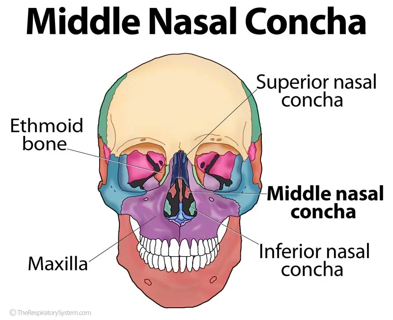
At a Glance
The middle nasal concha (middle turbinate) is a curved plate of the ethmoid bone on the lateral wall of each nasal cavity, lying between the superior and inferior conchae. It is not a separate bone. Beneath it lies the middle meatus, which drains the frontal, maxillary and anterior ethmoidal sinuses, and the concha protects these openings while guiding inhaled air.
- Location
- Lateral wall of each nasal cavity, between the superior and inferior conchae
- Function
- Shields the sinus openings in the middle meatus, directs airflow, warms and moistens air
- Bone
- Part of the ethmoid bone (a cranial bone), not a separate bone
- Tissue
- Thin curved bone covered by ciliated respiratory mucosa
- Latin name
- Concha nasalis media (concha media)
- Part of
- Nasal cavity, upper respiratory tract
On this page
- What Is the Middle Nasal Concha?
- Where Is the Middle Nasal Concha Located?
- Middle Nasal Concha Anatomy: Attachments and Lining
- Middle Nasal Concha and Meatus: What Drains There?
- What Does the Middle Nasal Concha Do?
- Middle Nasal Concha vs Superior and Inferior Nasal Concha
- How Does the Middle Nasal Concha Develop?
- Common Conditions That Affect the Middle Nasal Concha
- Key Facts
- Quiz
- FAQ
What Is the Middle Nasal Concha?
The middle nasal concha is the curved shelf of bone that lies, as its name suggests, between the superior nasal concha above and the inferior nasal concha below on the lateral wall of each nasal cavity. Covered by mucous membrane, it is also called the middle turbinate, and it is one of the three pairs of nasal conchae.
Is the middle nasal concha a separate bone?
No. The middle nasal concha is part of the ethmoid bone, not an individual bone. It is a thin, curled plate that hangs from the medial surface of the ethmoidal labyrinth (lateral mass), forming the lower edge of that surface. Because the ethmoid is a cranial bone, the middle concha is not a facial bone; of the three conchae, only the inferior one is a separate facial bone. In Latin it is the concha nasalis media.
Explained simply
Picture a curved awning fixed to a wall above a doorway. The middle concha is that awning, and under it lie the small doorways through which three of the four pairs of sinuses empty into the nose. Air rushing past is deflected by the awning, so the doorways stay sheltered while mucus from the sinuses drains out freely.
Where Is the Middle Nasal Concha Located?
The middle nasal concha is located on the lateral wall of each nasal cavity, below and in front of the superior concha and above the inferior concha, with its free curved border hanging down toward the middle of the cavity. Its front end lies in the front part of the lateral wall, and its back end reaches toward the choanae, the openings into the nasopharynx.
Neighbors of the middle concha
- Above: the superior meatus and the superior concha.
- Below and lateral: the middle meatus, the curved air passage under its free border.
- Below: the inferior concha.
- Medially: the nasal septum, formed above by the perpendicular plate of the ethmoid bone.
- Laterally, beyond the wall: the ethmoidal air cells and, lower down, the maxillary sinus.
Anterior and posterior views
In an anterior view of the skull, the middle concha is the curved bony ridge visible in the upper half of the nasal opening on each side of the septum, with the larger inferior concha below it. From behind, through the choanae, its rounded back end can be seen above the inferior concha. Unlike the superior concha, the front end of the middle turbinate can sometimes be seen in a living person by looking into the nostril with a light.
Middle Nasal Concha Anatomy: Attachments and Lining
The middle nasal concha is a thin plate of the ethmoid bone with a rough, grooved surface, attached above to the ethmoidal labyrinth and the skull base, and free below where its curved border overhangs the middle meatus. Its attachment twists along its length, which makes it an important landmark for surgeons.
How the middle concha is attached
- Front part: attaches almost vertically to the skull base at the side of the cribriform plate, and its front end joins the ethmoidal crest on the frontal process of the maxilla.
- Middle part: turns sideways as a thin sheet, the basal (ground) lamella, which runs across to the lamina papyracea, the paper-thin inner wall of the eye socket. This sheet divides the ethmoidal air cells into an anterior group and a posterior group.
- Back part: runs nearly horizontally along the lateral nasal wall and joins the ethmoidal crest of the perpendicular plate of the palatine bone.
Surface, lining and supply
The upper part of the medial surface of the ethmoidal labyrinth is marked by grooves that hold branches of the olfactory nerves on their way to the mucous membrane over the superior concha. The middle concha itself is covered mainly by respiratory mucosa: ciliated pseudostratified columnar epithelium with goblet cells, over a lamina propria with glands and a venous plexus that can swell, though less than over the inferior concha. In some people, olfactory epithelium extends onto the upper part of its medial surface. Blood comes mostly from posterior lateral nasal branches of the sphenopalatine artery, with help from the anterior ethmoidal artery, and sensation is carried by branches of the maxillary nerve and the anterior ethmoidal nerve.
Middle Nasal Concha and Meatus: What Drains There?
The middle meatus, the passage directly under the middle nasal concha, receives the drainage of the frontal sinus, the maxillary sinus and the anterior ethmoidal cells, which makes it the busiest drainage area in the nose. Lifting the middle concha out of the way, as a surgeon does with an endoscope, reveals several structures on the lateral wall:
- Ethmoidal bulla: a rounded bulge formed by ethmoidal air cells; the middle ethmoidal cells usually open on or above it.
- Uncinate process: a thin, hook-shaped plate of the ethmoid bone curving below and in front of the bulla.
- Semilunar hiatus: the crescent-shaped gap between the bulla and the uncinate process.
- Ethmoidal infundibulum: a funnel-shaped channel behind the hiatus into which the maxillary sinus opening and many anterior ethmoidal cells drain.
The frontal sinus drains downward through the frontal recess at the front of the middle meatus. Together these openings form the ostiomeatal complex, and our paranasal sinuses and nasal meatus pages follow each drainage route in more detail.
| Structure | Where it is | What it does or drains |
|---|---|---|
| Middle meatus | Air passage under the middle concha | Receives the frontal, maxillary and anterior ethmoidal sinuses |
| Ethmoidal bulla | Rounded bulge on the lateral wall, behind the hiatus | Contains ethmoidal cells; middle ethmoidal cells open on or above it |
| Uncinate process | Hook-shaped plate in front of and below the bulla | Forms the front edge of the semilunar hiatus |
| Semilunar hiatus | Crescent gap between bulla and uncinate process | Leads into the ethmoidal infundibulum |
| Ethmoidal infundibulum | Funnel-shaped channel behind the hiatus | Receives the maxillary sinus opening and anterior ethmoidal cells |
| Frontal recess | Front and top of the middle meatus | Drains the frontal sinus |
| Maxillary ostium | Deep in the infundibulum, on the lateral wall | Drains the maxillary sinus |
| Ostiomeatal complex | The combined area of these openings | Common drainage pathway of the front sinuses |
| Source: Typical adult anatomy, our editors | ||
What Does the Middle Nasal Concha Do?
The middle nasal concha shields the sinus openings in the middle meatus from the direct, pressurized airflow of each breath, and it helps warm, moisten, filter and direct the air flowing through the nose.
Function of the middle nasal concha
- Protecting the sinuses: by overhanging the middle meatus, the thin bone keeps the fast airstream from striking the openings of the maxillary, frontal and ethmoid sinuses, so their mucus can drain steadily.
- Guiding airflow: much of the air inhaled during each breath passes through the region between the middle and inferior turbinates and through the middle meatus, and the shape of the middle concha helps split and slow that stream.
- Conditioning air: its moist, blood-rich lining adds heat and water vapor, working together with the larger inferior concha.
- Filtering: particles caught in its mucus are swept back by cilia toward the pharynx and swallowed.
- Helping smell: by directing part of the airflow upward and, in some people, carrying a little olfactory lining on its upper surface.
Function of the superior and middle nasal conchae together
The two ethmoidal conchae split the work. The superior concha is mainly a smell surface over the posterior ethmoidal drainage, while the middle concha guards the much larger drainage area of the frontal, maxillary and anterior ethmoid sinuses. Together they make the lateral wall a series of sheltered pockets rather than a flat surface. This conditioning of air is part of the wider respiratory system functions of the upper airway.
Middle Nasal Concha vs Superior and Inferior Nasal Concha
The middle nasal concha is larger than the superior concha and smaller than the inferior concha, shares the ethmoid bone with the superior concha, and differs from the inferior concha, which is a separate facial bone. The table below compares the three side by side.
Two other structures are often confused with it. The perpendicular plate of the ethmoid bone is a flat midline sheet that forms the upper part of the nasal septum, while the middle concha hangs from the side of the same bone and curls toward that septum. The palatine bone is a separate bone at the back of the nasal cavity, and its ethmoidal crest is simply where the back end of the middle concha attaches.
| Feature | Superior concha | Middle concha | Inferior concha |
|---|---|---|---|
| Bone | Part of the ethmoid | Part of the ethmoid | Separate facial bone |
| Size | Smallest | Medium | Largest |
| Position | Highest, furthest back | Between the other two | Lowest, near the floor |
| Lining | Mainly olfactory | Respiratory, sometimes a little olfactory | Respiratory with thick erectile tissue |
| Meatus below | Superior meatus | Middle meatus | Inferior meatus |
| Drains below it | Posterior ethmoidal cells | Frontal, maxillary and anterior ethmoidal sinuses | Nasolacrimal duct |
| Main job | Smell | Shields sinus openings, guides airflow | Warms and humidifies most of the air |
| Typical variant | Supreme concha above it | Concha bullosa, paradoxical curve | Enlargement (hypertrophy) |
| Source: Typical adult anatomy, our editors | |||
How Does the Middle Nasal Concha Develop?
The middle nasal concha develops before birth from one of the ethmoturbinals, ridges on the side wall of the cartilaginous nasal capsule, and it ossifies with the ethmoidal labyrinth, whose ossification center appears around the fourth to fifth month of fetal life. The ethmoidal air cells grow into the surrounding bone after birth and through childhood, which is how air cells can come to invade the middle concha itself.
Common variants of the middle concha
- Concha bullosa: an air-filled cell inside the middle concha, formed when an ethmoidal air cell grows into it. It is one of the most common variants seen on sinus CT scans and may affect one or both sides.
- Paradoxical middle turbinate: a middle concha that curves outward, toward the lateral wall, instead of inward toward the septum, which can narrow the middle meatus.
- Bifid or lobulated middle concha: a concha with a groove or split along its lower edge.
Most of these variants cause no symptoms and are found by chance on imaging. In later life, the nasal lining over the middle concha, like the rest of the nose, tends to become thinner and drier.
Common Conditions That Affect the Middle Nasal Concha
Most problems linked to the middle nasal concha involve the middle meatus beneath it, because swelling or a change in shape there can block the sinus openings. Our respiratory diseases section covers related conditions, and a long-lasting blocked nose or facial pressure is a reason to see a doctor.
- Symptomatic concha bullosa: a large air cell can crowd the middle meatus, press on the septum, or itself become inflamed, sometimes contributing to nasal blockage or sinus problems.
- Chronic rhinosinusitis: long-term inflammation of the sinuses, often linked with swelling around the ostiomeatal complex under the middle concha.
- Nasal polyps: soft, pale growths that usually arise from the ethmoid region and bulge into the middle meatus, sometimes reaching the nasal cavity below.
- Septal contact: a deviated septum can touch the middle concha and narrow the airway.
Because its front part attaches to the skull base beside the thin cribriform plate, the middle concha is a key landmark in endoscopic sinus surgery, and surgeons work carefully around this attachment.
Key Facts
- The middle nasal concha is part of the ethmoid bone, not a separate bone, and lies between the superior and inferior conchae.
- The middle meatus beneath it drains the frontal sinus, the maxillary sinus and the anterior ethmoidal cells.
- Its basal lamella divides the ethmoidal air cells into anterior and posterior groups.
- Its front part attaches to the skull base beside the cribriform plate, and its back end joins the palatine bone.
- Concha bullosa, an air cell inside the middle concha, is one of the most common variants of nasal anatomy.
- The thin bone shields the sinus openings from the direct force of inhaled air.
- Much of each breath flows past the middle concha on its way to the pharynx and trachea.
Quick Quiz: Test What You Know About the Middle Nasal Concha
6 questions. Tap an answer to check it.
Which bone is the middle nasal concha part of?
The middle concha is a plate of the ethmoidal labyrinth. Only the inferior concha is a separate bone.
Which sinus does NOT drain into the middle meatus?
The sphenoid sinus opens into the sphenoethmoidal recess, above and behind the superior concha.
What is a concha bullosa?
A concha bullosa is an air cell inside a concha, most often the middle one, formed when an ethmoidal cell grows into it.
What does the basal (ground) lamella of the middle concha separate?
The basal lamella runs to the lamina papyracea and divides the ethmoidal air cells into anterior and posterior groups.
What is the crescent-shaped gap in the middle meatus called?
The semilunar hiatus lies between the ethmoidal bulla and the uncinate process and leads into the ethmoidal infundibulum.
Is the middle nasal concha a facial bone?
The middle concha belongs to the ethmoid bone, which is counted among the cranial bones.
Frequently Asked Questions
What bone forms the superior and middle nasal conchae?
Both are formed by the ethmoid bone. They are thin, curled projections from the medial surface of its labyrinth, the box of air cells on each side of the bone, which is why they are sometimes called the ethmoidal conchae. The perpendicular plate of the same bone forms the upper septum between them, and the cribriform plate forms the roof above.
What is the difference between the middle nasal concha and the perpendicular plate?
Both belong to the ethmoid bone, but they lie in different places. The perpendicular plate is a flat, vertical sheet in the midline that forms the upper part of the nasal septum between the two nasal cavities. The middle concha hangs from the side of the bone on the lateral wall of each cavity and curls toward the septum, with a narrow air channel between them.
Does the middle nasal concha articulate with the palatine bone?
Yes, at its back end. The posterior part of the middle concha attaches to the ethmoidal crest, a ridge on the perpendicular plate of the palatine bone. At its front end it joins the ethmoidal crest of the maxilla. The lower conchal crests on these same two bones hold the inferior concha, so each crest marks the line of one concha.
How do you pronounce middle nasal concha?
Concha is said KONG-kuh, and the plural, conchae, is said KONG-kee. The whole term sounds like MID-ul NAY-zul KONG-kuh. The word comes from Latin and Greek words for a shell or mussel, a reference to the curled shape of the bone in a cross-section of the nose, where it looks a little like a seashell.
Is concha bullosa dangerous?
In most people, no. A concha bullosa is a normal anatomical variant that is often found by chance on a CT scan done for another reason, and it usually causes no symptoms. When the air cell is large, it can narrow the middle meatus or press on the septum, and the cell itself can become inflamed like a small sinus.
What does the middle nasal concha look like?
Seen in a cross-section of the nose, the middle concha looks like a curled scroll or seashell hanging from the side wall, with a thicker rounded lower edge. Its bony surface is rough and pitted. In a living nose it is covered by pink, moist mucous membrane, and through an endoscope it appears as a smooth, rounded ridge running back toward the throat.
What is the agger nasi?
The agger nasi is a small ridge on the lateral nasal wall just in front of and above the place where the middle concha attaches. Inside it there is usually an air cell, the agger nasi cell, which is the most forward of the ethmoidal cells. Because it sits next to the drainage path of the frontal sinus, surgeons pay close attention to it.
Can you breathe normally with a concha bullosa or paradoxical middle turbinate?
Most people with these variants breathe normally and never know they have them. Breathing mainly depends on the space around the inferior turbinates and the nasal valve at the front of the nose. Problems arise only when a variant is large enough to narrow the middle meatus or crowd the septum, which can interfere with sinus drainage more than with airflow itself.
Keep Exploring


