At a Glance
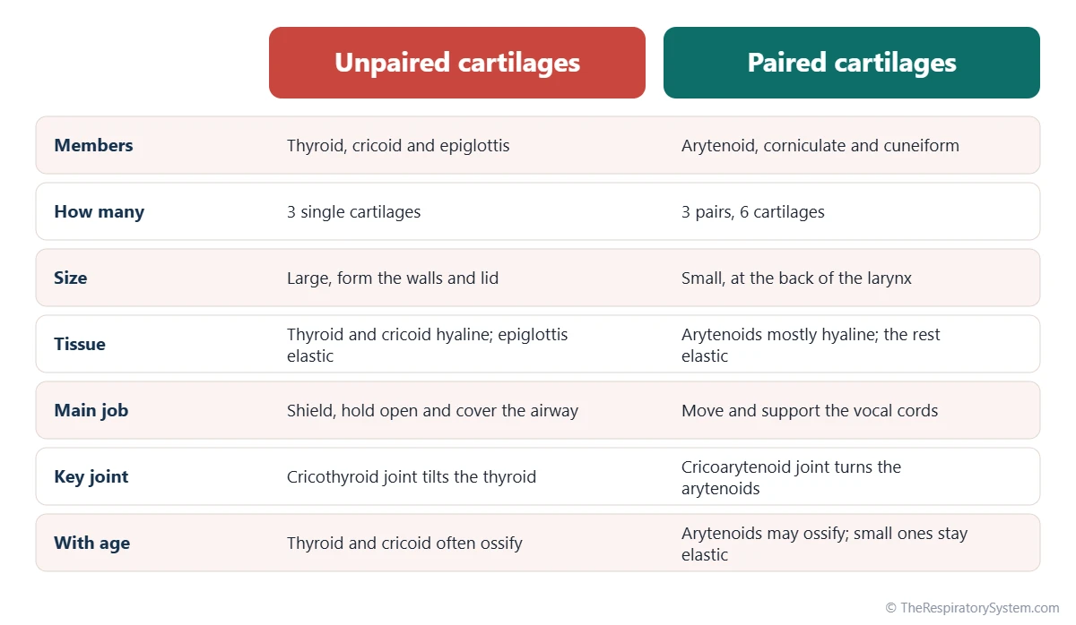
The larynx has 9 cartilages: 3 unpaired (the thyroid cartilage, the cricoid cartilage and the epiglottis) and 3 paired (the arytenoid, corniculate and cuneiform cartilages). The thyroid cartilage forms the Adam's apple, the cricoid cartilage is the only complete ring of cartilage in the airway, and the two arytenoid cartilages pivot and slide to open, close and tighten the vocal cords.
- Number
- 9 cartilages: 3 unpaired and 3 pairs
- Largest
- Thyroid cartilage, which forms the Adam's apple
- Tissue
- Hyaline cartilage (thyroid, cricoid, most of the arytenoids) and elastic cartilage (epiglottis, corniculate, cuneiform)
- Location
- Front of the neck, roughly from the third to the sixth cervical vertebra in adults
- Function
- Rigid frame for the airway, protection during swallowing, movement of the vocal cords
- Part of
- Larynx (voice box), upper respiratory tract
On this page
- What Are the 9 Cartilages of the Larynx?
- Thyroid Cartilage: What Is Its Function?
- What Is the Cricoid Cartilage and What Does It Do?
- What Is the Function of the Arytenoid Cartilage?
- Epiglottis, Corniculate and Cuneiform Cartilages
- How Are the Laryngeal Cartilages Connected and Moved?
- How Do the Laryngeal Cartilages Develop and Change With Age?
- Common Conditions That Affect the Laryngeal Cartilages
- Key Facts
- FAQ
What Are the 9 Cartilages of the Larynx?
The 9 cartilages of the larynx are the thyroid, cricoid and epiglottic cartilages, which occur once each, plus the arytenoid, corniculate and cuneiform cartilages, which occur in left and right pairs. Together they make a hollow, flexible box at the top of the trachea, the frame of the larynx.
- Unpaired (3): thyroid cartilage, cricoid cartilage, epiglottis.
- Paired (6): two arytenoid, two corniculate and two cuneiform cartilages.
The three unpaired cartilages are large and form the outer walls and lid of the larynx. The paired cartilages are small and sit on the back of the cricoid cartilage or inside the folds of the laryngeal inlet, where they move and support the vocal cords. Our larynx page shows how this frame fits with the muscles and lining of the whole voice box.
Explained simply
Picture the larynx as a small tent. The thyroid and cricoid cartilages are the stiff walls, the epiglottis is a flap over the door, and the tiny arytenoids are two tent pegs at the back. The vocal cords are strings tied from the front wall to those pegs, so when the pegs turn or slide, the strings open, close or tighten.
| Cartilage | Number | Tissue | Position | Key landmarks | Main function |
|---|---|---|---|---|---|
| Thyroid | 1 | Hyaline | Front and sides of the larynx, about C4 to C5 | Two laminae, laryngeal prominence, superior and inferior horns | Shields the larynx and anchors the front of the vocal cords |
| Cricoid | 1 | Hyaline | Base of the larynx, about C6 | Narrow arch in front, tall lamina behind, joint facets | Keeps the lower airway open; base for the joints |
| Epiglottis | 1 | Elastic | Behind the root of the tongue, stalk on the thyroid angle | Leaf-shaped blade, stalk, valleculae in front | Covers the laryngeal inlet during swallowing |
| Arytenoid | 2 | Mostly hyaline, elastic tips | On the upper back edge of the cricoid lamina | Apex, vocal process, muscular process | Open, close and tighten the vocal cords |
| Corniculate | 2 | Elastic | On the apex of each arytenoid | Small horn-shaped nodules | Support the back of the aryepiglottic folds |
| Cuneiform | 2 | Elastic | Inside the aryepiglottic folds | Small rods, seen as pale bumps | Stiffen the sides of the laryngeal inlet |
| Source: Typical adult anatomy, our editors | |||||
Thyroid Cartilage: What Is Its Function?
The function of the thyroid cartilage is to shield the front of the larynx and anchor the front ends of the vocal cords; it is the largest laryngeal cartilage and forms the Adam's apple. It is made of hyaline cartilage and lies at about the level of the fourth and fifth cervical vertebrae.
Shape and landmarks
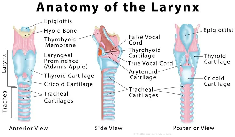
The thyroid cartilage is formed by two flat plates, the right and left laminae, which join in front at an angle and stay open at the back like a half-open book. Where they meet, the upper edge dips into the superior thyroid notch, and just below it the angle juts forward as the laryngeal prominence. A ridge called the oblique line runs across the outer surface of each lamina and gives attachment to neck and throat muscles. Its name comes from a Greek word for a shield, and the thyroid gland below it was later named after the cartilage.
Horns and attachments
The back edge of each lamina extends into two horns. The superior horns are tied to the hyoid bone by ligaments, so the larynx hangs from the hyoid. The inferior horns form small joints with the sides of the cricoid cartilage, the cricothyroid joints, which let the two cartilages tilt against each other. On the inside of the angle, the vocal ligaments, the vestibular ligaments and the stalk of the epiglottis all attach close together.
The Adam's apple
The Adam's apple is simply the laryngeal prominence seen through the skin. The angle between the two laminae is about 90 degrees in adult men and about 120 degrees in women, so the prominence sticks out more in men. It grows mainly at puberty, when the male larynx enlarges and the voice deepens.
What Is the Cricoid Cartilage and What Does It Do?
The cricoid cartilage is the ring-shaped cartilage at the base of the larynx, and its function is to keep the lower airway permanently open and to provide the platform on which the thyroid and arytenoid cartilages move. It lies at about the level of the sixth cervical vertebra, just above the first ring of the trachea.
The cricoid is shaped like a signet ring: a narrow arch in front and a tall, flat plate, the lamina, at the back. It is the only complete ring of cartilage anywhere in the airway, since the tracheal rings below are open at the back. It carries two pairs of smooth joint surfaces: one on each side, where the arch meets the lamina, for the inferior horns of the thyroid cartilage, and one on the upper edge of the lamina for the arytenoid cartilages. Its lower edge is joined to the trachea by the cricotracheal ligament.
In front, the gap between the cricoid arch and the thyroid cartilage above it is closed by the cricothyroid ligament, a firm membrane that can be felt as a soft dip just below the Adam's apple. Because the cricoid ring cannot expand, swelling of the lining inside it narrows the airway quickly, which matters most in small children.
What Is the Function of the Arytenoid Cartilage?
The function of the arytenoid cartilages is to open, close and tighten the vocal cords: each arytenoid holds the back end of one vocal cord and turns or slides on the cricoid when the laryngeal muscles pull on it. They are the most mobile cartilages of the larynx and the main reason it can switch between breathing, swallowing and speaking in a fraction of a second.
Each arytenoid is a small, three-sided pyramid sitting on the upper back edge of the cricoid lamina. Its base has two projections:
- The vocal process points forward and gives attachment to the vocal ligament, the core of the true vocal cord.
- The muscular process points sideways and backward and is where the posterior and lateral cricoarytenoid muscles pull.
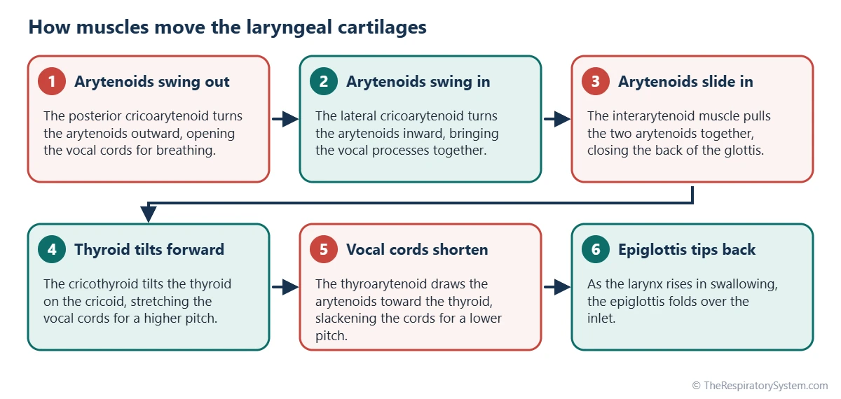
Its pointed apex curves backward and carries the corniculate cartilage. Most of each arytenoid is hyaline cartilage, but the tip of the vocal process is elastic cartilage, which bends without breaking during the constant motion of speech. When both arytenoids swing outward, the gap between the vocal cords, the glottis, opens for breathing; when they swing inward and slide together, it closes.
Epiglottis, Corniculate and Cuneiform Cartilages
The epiglottis, corniculate and cuneiform cartilages are the three elastic cartilages of the larynx: the epiglottis forms the flexible lid over the laryngeal inlet, while the tiny corniculate and cuneiform cartilages stiffen the folds that form its sides. Elastic cartilage bends and springs back, which suits parts that must flex with every swallow.
Epiglottis
The epiglottis is a leaf-shaped plate whose narrow stalk is fixed to the inside of the thyroid angle by the thyroepiglottic ligament. Its broad upper part rises behind the root of the tongue, where small hollows called the valleculae separate the two. When the larynx rises during swallowing, the epiglottis tips backward over the inlet and steers food toward the esophagus. Its full anatomy is on our epiglottis page.
Corniculate cartilages
The two corniculate cartilages, also called the cartilages of Santorini, are small horn-shaped nodules perched on the apex of each arytenoid. They lie in the back part of the aryepiglottic folds, the mucosal folds that run from the sides of the epiglottis to the arytenoids, and lengthen the arytenoids upward.
Cuneiform cartilages
The two cuneiform cartilages, or cartilages of Wrisberg, are small rod- or club-shaped pieces inside the aryepiglottic folds, just in front of the corniculates. They are not attached to any other cartilage, and in some people they are tiny or absent. Through a laryngoscope they show as small pale bumps on each side of the inlet.
Small extra cartilages
Some people also have a triticeal cartilage, a grain-sized nodule inside each lateral thyrohyoid ligament between the hyoid bone and the thyroid cartilage. These are not counted among the nine, but they can show up on neck X-rays once they calcify.
How Are the Laryngeal Cartilages Connected and Moved?
The laryngeal cartilages are connected by two pairs of synovial joints, several ligaments and membranes, and are moved by the small intrinsic muscles of the larynx, which change the width and tension of the vocal cords. Larger muscles of the neck move the whole larynx up and down, for example during swallowing.
The two key joints
- Cricothyroid joints: the inferior horns of the thyroid cartilage hinge on the sides of the cricoid. When the cricothyroid muscle contracts, the thyroid tilts forward and down, stretching the vocal cords and raising the pitch of the voice.
- Cricoarytenoid joints: each arytenoid rocks, rotates and glides on the cricoid lamina. The posterior cricoarytenoid muscle is the only muscle that opens the vocal cords, while the lateral cricoarytenoid and the interarytenoid muscles close them.
Nerves
All the intrinsic muscles that move the cartilages are supplied by the recurrent laryngeal nerve, a branch of the vagus nerve, except the cricothyroid muscle, which is supplied by the external branch of the superior laryngeal nerve. These movements are what turn airflow into voice, a process covered on our page on how the voice is produced.
| Connection | What it joins | What it does |
|---|---|---|
| Cricothyroid joint | Inferior horns of the thyroid to the sides of the cricoid | Lets the thyroid tilt to stretch the vocal cords |
| Cricoarytenoid joint | Base of each arytenoid to the cricoid lamina | Lets the arytenoids rotate and glide to open or close the glottis |
| Thyrohyoid membrane | Upper edge of the thyroid cartilage to the hyoid bone | Hangs the larynx from the hyoid |
| Cricothyroid ligament | Cricoid arch to the lower edge of the thyroid cartilage | Closes the front gap; part of the conus elasticus, whose upper edges are the vocal ligaments |
| Cricotracheal ligament | Lower edge of the cricoid to the first tracheal ring | Joins the larynx to the trachea |
| Thyroepiglottic ligament | Stalk of the epiglottis to the inside of the thyroid angle | Anchors the epiglottis |
| Hyoepiglottic ligament | Front of the epiglottis to the hyoid bone | Steadies the epiglottis and forms the floor of the valleculae |
| Vocal ligament | Inside of the thyroid angle to the vocal process of the arytenoid | Core of each true vocal cord |
| Vestibular ligament | Thyroid angle to the front of the arytenoid, above the vocal ligament | Core of each false vocal cord |
| Source: Typical adult anatomy, our editors | ||
How Do the Laryngeal Cartilages Develop and Change With Age?
The laryngeal cartilages develop from tissue of the fourth and sixth pharyngeal arches in the embryo, grow sharply at puberty, and the hyaline ones gradually turn partly to bone during adult life. The epiglottis forms separately, from a swelling at the base of the developing tongue, as described on our respiratory system development page.
In a newborn the larynx sits high in the neck and its cartilages are small and soft, so the airway is easily narrowed. During childhood the larynx slowly descends. At puberty the thyroid cartilage grows much more in boys than in girls, the angle between its laminae narrows, the Adam's apple appears and the lengthening vocal cords deepen the voice.
From early or middle adulthood, the hyaline cartilages, mainly the thyroid, cricoid and arytenoids, begin to calcify and ossify, usually earlier and more extensively in men. The elastic cartilages, including the epiglottis, normally stay flexible. Ossified laryngeal cartilages are visible on X-rays and CT scans and are a normal finding with age.
Common Conditions That Affect the Laryngeal Cartilages
Conditions of the laryngeal cartilages mostly involve cartilage that is too soft, narrowed, injured or inflamed, and they usually show up as noisy breathing, hoarseness or difficulty breathing. Our respiratory diseases section covers airway conditions in more depth, and breathing difficulty is always a reason to see a doctor.
- Laryngomalacia: the soft cartilages and folds above the vocal cords, mainly the epiglottis and aryepiglottic folds, fall inward when a baby breathes in. It is the most common cause of noisy breathing (stridor) in infants and usually improves as the cartilage firms up.
- Subglottic stenosis: narrowing of the airway inside the cricoid ring, present from birth or caused by scarring, for example after long use of a breathing tube.
- Croup: a viral infection in young children in which the lining inside the cricoid ring swells, producing a barking cough.
- Laryngeal fracture: a rare injury from a hard blow to the front of the neck, most often affecting the thyroid cartilage.
- Arytenoid dislocation: a rare displacement of an arytenoid from its joint, usually after intubation or trauma, causing hoarseness.
- Relapsing polychondritis: a rare autoimmune disease that inflames cartilage in the ears, nose and airway and can weaken the laryngeal frame.
Inflammation of the lining over the cartilages, as in laryngitis, affects the voice but does not usually damage the cartilage itself.
Key Facts
- The larynx has 9 cartilages: 3 unpaired (thyroid, cricoid, epiglottis) and 3 paired (arytenoid, corniculate, cuneiform).
- The thyroid cartilage is the largest, and its laryngeal prominence is the Adam's apple.
- The cricoid cartilage is the only complete ring of cartilage in the airway and sits just above the trachea.
- The arytenoid cartilages hold the back ends of the vocal cords and open or close the glottis.
- The thyroid, cricoid and most of the arytenoids are hyaline cartilage; the epiglottis, corniculate and cuneiform cartilages are elastic.
- The posterior cricoarytenoid is the only muscle that opens the vocal cords.
- Hyaline laryngeal cartilages commonly calcify and partly turn to bone during adult life, while elastic ones stay flexible.
Frequently Asked Questions
What is the largest cartilage of the larynx?
The thyroid cartilage is the largest cartilage of the larynx. Its two broad plates form most of the front and side walls of the voice box and meet in front to create the laryngeal prominence, or Adam's apple. It is made of hyaline cartilage and connects to the hyoid bone above and the cricoid cartilage below.
Which laryngeal cartilage is a complete ring?
The cricoid cartilage is the only laryngeal cartilage, and the only cartilage anywhere in the airway, that forms a complete ring. It is narrow in front and tall at the back, like a signet ring. The cartilages of the trachea below are C-shaped and open at the back, where the esophagus lies against them.
Do women have an Adam's apple?
Yes. Everyone has a thyroid cartilage with a laryngeal prominence, but in women the two plates meet at a wider angle, about 120 degrees compared with about 90 degrees in men, so the prominence is flatter and usually hidden under the skin. The male larynx also grows more at puberty, which makes the Adam's apple easier to see.
Which laryngeal cartilages are paired?
The paired laryngeal cartilages are the arytenoid, corniculate and cuneiform cartilages, one of each on the left and the right, for six in total. The arytenoids are the largest and most important of these because they move the vocal cords. The corniculate and cuneiform cartilages are small elastic pieces that support the aryepiglottic folds.
Is the hyoid bone part of the larynx?
No. The hyoid is a U-shaped bone in the upper neck that sits above the larynx and is not counted among its cartilages. The larynx hangs from it by the thyrohyoid membrane, and the epiglottis is tied to it by the hyoepiglottic ligament. Because of these links, the larynx rises with the hyoid bone every time you swallow.
Can you feel the laryngeal cartilages from outside?
Yes, two of them. Running a finger down the front of the neck, you first meet the thyroid cartilage with its notch and Adam's apple. Just below it is a small soft dip, the cricothyroid ligament, and below that a firm narrow band, the arch of the cricoid cartilage. The arytenoids and the other small cartilages lie too deep to feel.
Why do the laryngeal cartilages turn to bone?
The thyroid, cricoid and arytenoid cartilages are made of hyaline cartilage, the same type that forms much of the growing skeleton, and with age it tends to calcify and be replaced in part by bone. This change usually starts in early or middle adulthood and goes further in men. It is a normal part of aging, and elastic cartilages such as the epiglottis are not affected.
What holds the laryngeal cartilages together?
The cartilages are held together by synovial joints, ligaments and sheets of connective tissue. The cricothyroid and cricoarytenoid joints let the cartilages move, while the thyrohyoid membrane, the cricothyroid ligament and the cricotracheal ligament tie the larynx to the hyoid bone above and the trachea below. Inside, the quadrangular membrane and the conus elasticus form the vestibular and vocal ligaments.
Keep Exploring


