At a Glance
The maxillary sinus is a pyramid-shaped air space in the body of the maxilla, one in each cheek. It is the largest paranasal sinus, holding roughly 15 milliliters of air in an adult, and it drains into the middle meatus of the nose through an opening near its roof, so its cilia must sweep mucus upward to empty it. Its floor lies just above the roots of the upper back teeth.
- Location
- Body of the maxilla in each cheek, below the eye socket and above the upper back teeth
- Shape
- Pyramid, with its base on the side wall of the nose and its tip toward the cheekbone
- Drains into
- Middle meatus, through the maxillary ostium and the ethmoidal infundibulum
- Lining
- Thin ciliated respiratory mucoperiosteum (Schneiderian membrane)
- Nerve supply
- Infraorbital and superior alveolar branches of the maxillary nerve
- Part of
- Paranasal sinuses, upper respiratory tract
On this page
- What Is the Maxillary Sinus?
- Where Is the Maxillary Sinus Located?
- Maxillary Sinus Anatomy: Walls, Opening and Lining
- Where Does the Maxillary Sinus Drain?
- What Is the Function of the Maxillary Sinus?
- How Is the Maxillary Sinus Related to the Upper Teeth?
- Maxillary Sinus vs Sphenoid Sinus: How Do They Differ?
- Common Conditions That Affect the Maxillary Sinus
- Key Facts
- Quiz
- FAQ
What Is the Maxillary Sinus?
The maxillary sinus is a paired, air-filled cavity in the maxilla, the bone that forms the upper jaw, most of the cheek and part of the floor of the eye socket. It is the largest of the paranasal sinuses, the group of air spaces around the nasal cavity that also includes the frontal, ethmoid and sphenoid sinuses.
Its older name is the maxillary antrum, or antrum of Highmore, after the English physician Nathaniel Highmore, who described it in the seventeenth century. Antrum simply means cave. Like the other sinuses, it is lined by mucous membrane, is normally filled with air and connects to the nose through a narrow opening.
Explained simply: behind each cheek is a small hollow room filled with air. A tiny doorway near the top of that room leads into the side of your nose, and the mucus made inside the room leaves through that doorway.
Where Is the Maxillary Sinus Located?
The maxillary sinus is located in the cheek on each side of the nose, directly below the eye socket and above the roots of the upper back teeth. There is one on the right and one on the left, and each fills most of the body of its maxilla.
To find it on your own face, place a fingertip on the cheek just below the lower rim of the eye and beside the nose: the sinus lies under the bone there. Its inner wall is the outer wall of the nasal cavity, its roof is the floor of the orbit, and its floor sits over the tooth-bearing ridge of the upper jaw. Toward the outside it narrows into the zygomatic process of the maxilla, under the cheekbone.
The right and left maxillary sinuses are mirror images, though they are rarely exactly the same size. Each one drains into its own side of the nose, lateral to the nasal septum.
Maxillary Sinus Anatomy: Walls, Opening and Lining
The maxillary sinus has the shape of a pyramid lying on its side, with its base forming part of the lateral wall of the nasal cavity and its apex pointing toward the cheekbone. In an adult it holds roughly 15 milliliters on average, though size varies widely from person to person.
Walls
The sinus has a roof, a floor and front, back and inner walls, each facing a different neighbor, as the table below shows. The roof is thin and carries the infraorbital nerve and vessels in a groove or canal on their way to the face. The back wall separates the sinus from the infratemporal and pterygopalatine fossae, deep spaces that hold branches of the maxillary artery and nerve.
Opening
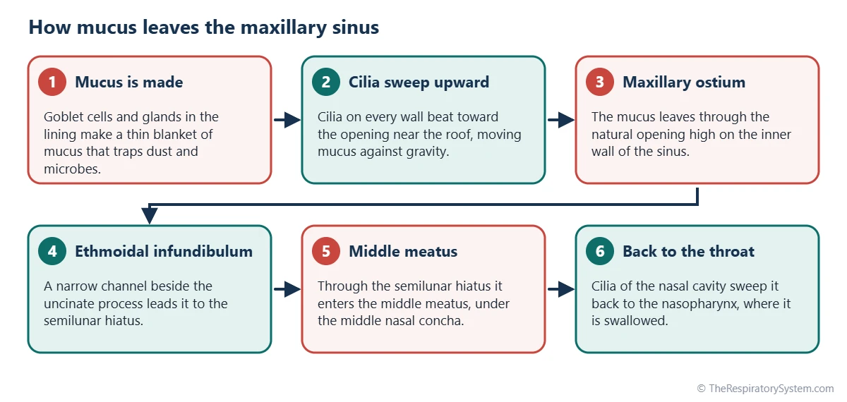
The natural opening, the maxillary ostium, lies high on the inner wall, close to the roof. It leads into a narrow channel called the ethmoidal infundibulum, which empties through the semilunar hiatus into the middle nasal meatus, under the middle nasal concha. Some people also have one or more small accessory openings.
Lining, vessels and nerves
The sinus is lined by a thin mucoperiosteum, often called the Schneiderian membrane, made of ciliated pseudostratified columnar respiratory epithelium with goblet cells. Blood comes from branches of the maxillary artery, including the infraorbital and superior alveolar arteries. Sensation travels in the infraorbital nerve and the superior alveolar nerves, all branches of the maxillary division of the trigeminal nerve.
| Wall | Formed by | What lies on the other side |
|---|---|---|
| Roof | Orbital surface of the maxilla (floor of the eye socket) | The orbit and eye; the infraorbital nerve and vessels run in the roof |
| Floor | Alveolar process and part of the palatine process of the maxilla | Roots of the upper molars and premolars |
| Medial wall (base) | Maxilla, completed by parts of the palatine, ethmoid, lacrimal and inferior concha bones | Middle and inferior meatuses of the nasal cavity |
| Anterior wall | Facial surface of the maxilla, including the canine fossa | Soft tissues of the cheek; infraorbital foramen near the top |
| Posterior wall | Infratemporal surface of the maxilla | Infratemporal and pterygopalatine fossae |
| Apex | Zygomatic process of the maxilla | Zygomatic bone (cheekbone) |
| Opening (ostium) | High on the medial wall, close to the roof | Ethmoidal infundibulum, then the middle meatus |
| Source: Typical adult anatomy, our editors | ||
Where Does the Maxillary Sinus Drain?
The maxillary sinus drains into the middle meatus of the nasal cavity, through the maxillary ostium, the ethmoidal infundibulum and the semilunar hiatus. Because the ostium sits near the roof rather than the floor, gravity cannot empty the sinus while you stand or sit upright.
Instead, the cilia of the lining beat in a coordinated pattern that moves the mucus blanket upward and inward, converging on the ostium from every wall. This is the mucociliary escalator at work. In the middle meatus the mucus joins secretions from the frontal sinus and the anterior ethmoid cells and is swept back toward the nasopharynx, where it is swallowed.
The narrow region where these three sinuses drain is called the ostiomeatal complex. Swelling there can block several sinuses at once, which helps explain why the maxillary sinus is so often involved when the sinuses become inflamed.
What Is the Function of the Maxillary Sinus?
The maxillary sinus shares the general jobs of all paranasal sinuses: it lightens the facial skeleton, adds resonance to the voice and supplies mucus that keeps the nasal lining moist. The full list, including theories still debated, is on the paranasal sinuses page; here are the points that apply most to this sinus.
- Lightening the midface. As the largest sinus, it accounts for much of the weight saved in the middle of the face.
- Shaping the face. It grows together with the upper teeth, helping give the cheeks and upper jaw their adult form.
- Moistening the airway. Its mucus flows into the busy middle meatus and adds to the film that conditions air on its way to the pharynx.
- Voice resonance. The air in the cheeks adds to the resonating spaces that color the sound of the voice, alongside the nose, mouth and throat.
Some researchers also describe the air-filled cheek as a crumple zone that absorbs force from a blow to the face and protects the eye, though this idea is harder to test.
How Is the Maxillary Sinus Related to the Upper Teeth?
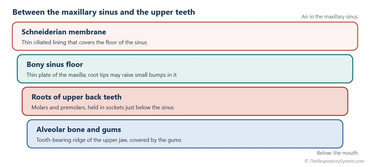
The floor of the maxillary sinus lies just above the roots of the upper back teeth, usually the first and second molars and often the premolars. In many adults the root tips raise small bumps in the sinus floor, and sometimes only a thin plate of bone or the lining itself separates a root from the air space.
This closeness has well-known effects. The teeth and the sinus share the superior alveolar nerves, so pain from an inflamed sinus can feel like an upper toothache, and dental pain can feel like sinus pressure. Infection from an upper back tooth can spread upward into the sinus, and removing an upper molar occasionally leaves an opening between the mouth and the sinus, called an oroantral communication.
In adults the floor of the sinus usually lies lower than the floor of the nasal cavity. After upper back teeth are lost, the sinus can expand downward into the empty bone, a process called pneumatization.
Maxillary Sinus vs Sphenoid Sinus: How Do They Differ?
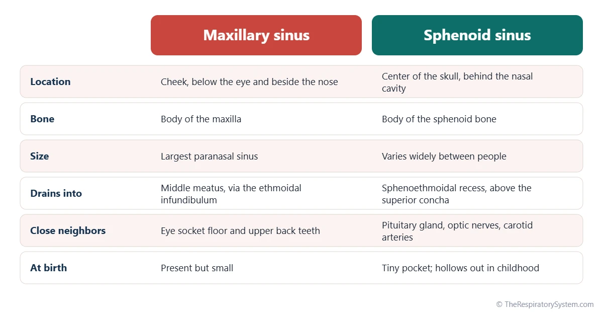
The maxillary sinus is the large sinus in the cheek at the front of the face, while the sphenoid sinus is a deep sinus in the center of the skull, behind the nasal cavity. They are the two sinuses farthest apart, and their neighbors are very different.
The maxillary sinus borders the eye socket and the upper teeth. The sphenoid sinus borders the pituitary gland, the optic nerves and the internal carotid arteries, and it drains into the sphenoethmoidal recess above the superior nasal concha rather than into the middle meatus. The maxillary sinus is present, though small, at birth, while the sphenoid sinus is only a tiny pocket at birth and hollows out its bone during childhood.
Common Conditions That Affect the Maxillary Sinus
The maxillary sinus is the sinus most often involved in sinus inflammation, and it can also be affected by dental problems, injuries and growths. These are listed for education only; the respiratory diseases page gives wider context.
- Maxillary sinusitis: inflammation of the lining, often after a cold, felt as pressure in the cheek. See sinusitis.
- Odontogenic sinusitis: sinus inflammation that starts from an upper tooth or a dental procedure.
- Retention cysts and polyps: small fluid-filled swellings of the lining are common and often found by chance on scans; an antrochoanal polyp grows from the maxillary sinus back toward the nasopharynx.
- Fractures: blows to the cheek can break the sinus walls, and a blowout fracture of the eye socket floor pushes into the sinus roof.
- Tumors: rare growths of the sinus lining, which can press on the eye, teeth or nose.
Lasting facial pain or swelling, or symptoms on only one side of the nose, are reasons to see a doctor.
Key Facts
- The maxillary sinus is the largest paranasal sinus, with one in each maxilla below the eye.
- It is shaped like a pyramid, with its base on the lateral wall of the nasal cavity and its apex toward the cheekbone.
- It drains into the middle meatus through the maxillary ostium, which lies high on its inner wall.
- Cilia must carry mucus upward to the ostium, because gravity cannot empty the sinus when the head is upright.
- Its roof is the floor of the orbit, and its floor lies above the roots of the upper molars and premolars.
- It is the first paranasal sinus to form and is already present, though small, at birth.
- Its older name is the antrum of Highmore.
Quick Quiz: Test What You Know About the Maxillary Sinus
6 questions. Tap an answer to check it.
Which bone contains the maxillary sinus?
The maxillary sinus fills most of the body of the maxilla, the upper jaw bone in each cheek.
Where does the maxillary sinus drain?
It drains through the maxillary ostium and the ethmoidal infundibulum into the middle meatus, under the middle nasal concha.
Which is the largest paranasal sinus?
The maxillary sinus is the largest of the four pairs of paranasal sinuses.
What forms the roof of the maxillary sinus?
The roof of the maxillary sinus is the thin floor of the eye socket, which carries the infraorbital nerve.
Why can gravity not drain the maxillary sinus when you are upright?
The maxillary ostium lies near the roof of the sinus, so cilia must sweep mucus upward to it.
The roots of which teeth lie closest to the maxillary sinus floor?
The roots of the upper molars and premolars sit just below the sinus floor and can even bulge into it.
Frequently Asked Questions
Where is the right maxillary sinus?
The right maxillary sinus is inside the right maxilla, in the right cheek. It lies below the right eye socket, beside the right side of the nose and above the roots of the upper right back teeth. It opens into the right half of the nasal cavity, in the middle meatus. The left maxillary sinus is its mirror image on the other side of the face.
Are babies born with maxillary sinuses?
Yes. The maxillary sinus is the first paranasal sinus to begin forming before birth, so a newborn already has one on each side, though they are only small slits beside the nose. The ethmoid cells are also present at birth, while the frontal and sphenoid sinuses develop mostly during childhood.
When does the maxillary sinus reach adult size?
The maxillary sinus grows in spurts during childhood, especially as the permanent teeth form and come in. It expands downward into the bone that the developing teeth leave behind, and usually reaches close to its adult size in the teenage years, once the permanent teeth have erupted. Its floor then ends up lower than the floor of the nasal cavity.
Why can sinus problems make the upper teeth hurt?
The upper back teeth and the lining of the maxillary sinus are supplied by the same superior alveolar nerves, branches of the maxillary nerve. When the sinus lining is swollen or under pressure, the brain can read the signal as coming from the teeth. The roots of these teeth also sit right below the sinus floor, so the two areas are close physically as well.
Can the maxillary sinus be seen on an X-ray?
Yes. On a plain X-ray of the face, the maxillary sinuses appear as two dark, air-filled areas in the cheeks on either side of the nose. A view taken with the chin raised, called the occipitomental or Waters view, shows them especially well. Computed tomography (CT) gives a much more detailed picture of the walls and the drainage pathway.
What does a normal maxillary sinus look like on a CT scan?
On a CT scan, a normal maxillary sinus looks black, because air blocks almost no X-rays, surrounded by a thin white outline of bone. The healthy lining is too thin to see clearly. A coronal CT, taken in slices from front to back, also shows the ostium and the ethmoidal infundibulum leading up to the middle meatus.
Why is the maxillary sinus opening at the top?
The reason is not certain. The opening forms where the sinus first buds out from the middle meatus in the embryo, and the sinus then grows downward and outward into the maxilla, especially as the teeth develop. The space ends up hanging below its own doorway, which is why the maxillary sinus depends on cilia rather than gravity to drain.
Is the maxillary sinus part of the respiratory system?
Yes. The maxillary sinuses are part of the paranasal sinuses, which belong to the upper respiratory tract. They do not carry the main airflow and take no part in gas exchange, but they open into the nasal cavity, share its lining and add mucus to the air passages that lead to the pharynx, larynx and lungs.
Keep Exploring


