At a Glance
The frontal sinus is the paranasal sinus of the forehead: a pair of air spaces in the frontal bone above the eye sockets, usually unequal in size and separated by a thin bony wall. It drains downward through the frontal sinus ostium and the frontal recess into the middle meatus of the nose. It is absent at birth, grows during childhood and reaches adult size after puberty.
- Location
- Frontal bone, behind the brow ridges, above the eye sockets and the root of the nose
- Shape
- Two funnel-shaped spaces, usually unequal, split by an off-center bony septum
- Drains into
- Middle meatus, through the frontal sinus ostium and the frontal recess
- Neighbors
- Forehead in front, frontal lobes of the brain behind, orbits below
- Nerve supply
- Supraorbital nerve, a branch of the ophthalmic nerve
- Part of
- Paranasal sinuses, upper respiratory tract
On this page
- What Is the Frontal Sinus?
- Where Is the Frontal Sinus Located?
- Frontal Sinus Anatomy: Walls, Septum and Lining
- Where Does the Frontal Sinus Drain?
- What Is the Function of the Frontal Sinuses?
- Frontal Sinus Anatomy on CT and X-Ray
- How Does the Frontal Sinus Develop?
- Common Conditions That Affect the Frontal Sinus
- Key Facts
- Quiz
- FAQ
What Is the Frontal Sinus?
The frontal sinus is a pair of air-filled cavities in the frontal bone, the bone of the forehead. It is one of the four pairs of paranasal sinuses, together with the maxillary, ethmoid and sphenoid sinuses, and like them it is lined with mucous membrane and opens into the nasal cavity.
The frontal sinus is often described as an ethmoid air cell that has grown upward. It begins in early childhood as an outpouching from the front of the ethmoid region and only later expands into the frontal bone, which is why its drainage pathway stays so closely tied to the ethmoid cells below it.
Explained simply: right behind your eyebrows, the forehead bone is not solid. It holds two small air pockets, one over each eye, with a drain at the bottom of each that empties into your nose.
Where Is the Frontal Sinus Located?
The frontal sinus is located in the lower part of the forehead, inside the frontal bone, behind the brow ridges and above the eye sockets and the root of the nose. On the skull it lies in the flat, upright part of the frontal bone and can extend backward into the roof of each orbit.
The two frontal sinuses sit side by side, separated by a thin bony partition called the intersinus septum. This septum is rarely in the exact midline, so one sinus is usually larger and may even cross over to the other side. Their size varies more than that of any other sinus:
- some reach high into the forehead and spread out over the eyes;
- some stay small, just above the root of the nose;
- a small minority of people have one frontal sinus missing, or both.
Below and behind each frontal sinus lie the anterior ethmoid air cells, and its drainage channel runs down past them to the front of the middle nasal meatus.
Frontal Sinus Anatomy: Walls, Septum and Lining
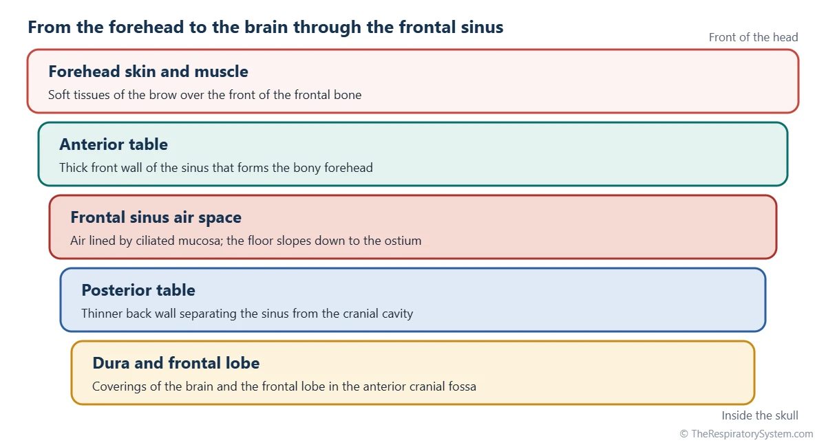
The frontal sinus has a thick front wall, a thinner back wall, a floor that forms part of the orbital roof and a bony septum between the two sides. Each wall faces a different neighbor, which matters in injuries and in the rare spread of infection.
Anterior and posterior tables
The front wall, called the anterior table, is the thickest wall and forms the bony forehead under the brow. The back wall, the posterior table, is thinner and separates the sinus from the anterior cranial fossa, where the dura mater and the frontal lobes of the brain lie. The floor is the thinnest part; it forms the inner roof of the orbit and slopes down toward the drainage opening.
Lining, vessels and nerves
Like the other sinuses, it is lined by thin ciliated respiratory epithelium with goblet cells, attached directly to the bone as a mucoperiosteum. Blood comes from the supraorbital and anterior ethmoidal arteries, both branches of the ophthalmic artery. Veins drain into the supraorbital and superior ophthalmic veins and into small diploic veins inside the bone, which connect with veins around the brain. Sensation travels in the supraorbital nerve, a branch of the ophthalmic division of the trigeminal nerve.
| Part | What it is like | What lies beyond it |
|---|---|---|
| Anterior table (front wall) | Thickest wall, under the brow ridge | Forehead skin, muscle and eyebrows |
| Posterior table (back wall) | Thinner plate of bone | Dura mater and frontal lobes in the anterior cranial fossa |
| Floor | Thinnest wall, sloping down toward the ostium | Roof of the orbit and, toward the midline, the anterior ethmoid cells |
| Intersinus septum | Thin bony partition, usually off center | The opposite frontal sinus |
| Ostium | Narrow opening in the back inner part of the floor | Frontal recess |
| Frontal recess | Narrow space bordered by anterior ethmoid cells | Front of the middle meatus of the nasal cavity |
| Lining | Thin ciliated mucoperiosteum with goblet cells | Continuous with the lining of the nasal cavity |
| Source: Typical adult anatomy, our editors | ||
Where Does the Frontal Sinus Drain?
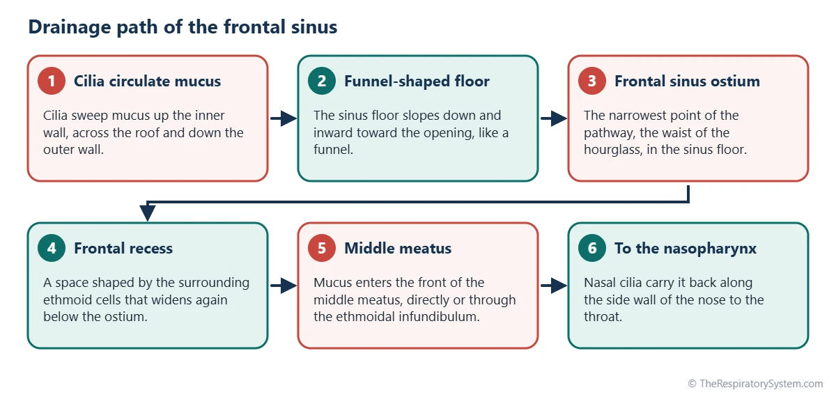
The frontal sinus drains into the middle meatus of the nasal cavity, through an opening in its floor called the frontal sinus ostium and a narrow passage below it called the frontal recess. Older textbooks call this passage the frontonasal duct, but in most people it is not a true tube.
The pathway is often compared to an hourglass: the funnel-shaped sinus narrows to the ostium, then widens again into the frontal recess, which opens into the front of the middle meatus. Depending on how a thin ridge of bone called the uncinate process attaches, the recess empties either into the ethmoidal infundibulum or directly into the middle meatus. Cilia move mucus toward the exit by sweeping it up the inner wall, across the roof and down the outer wall to the ostium.
In the middle meatus, frontal mucus joins drainage from the maxillary sinus and the anterior ethmoid cells and is carried back to the nasopharynx under the middle nasal concha.
Frontal sinus anatomy classification
The frontal recess is crowded by small anterior ethmoid cells, and their pattern differs from person to person. Surgeons and radiologists describe them with the International Frontal Sinus Anatomy Classification (IFAC), which sorts them into anterior, posterior and medial cells around the drainage pathway, as the second table shows. An older system, the Kuhn classification, grouped frontal cells into four types.
| Cell | Group | Where it lies |
|---|---|---|
| Agger nasi cell | Anterior | In front of the attachment of the middle concha, below the frontal sinus |
| Supra agger cell | Anterior | Above the agger nasi cell, staying below the frontal sinus |
| Supra agger frontal cell | Anterior | Above the agger nasi cell, reaching up into the frontal sinus |
| Supra bulla cell | Posterior | Above the ethmoid bulla, below the frontal sinus |
| Supra bulla frontal cell | Posterior | Above the bulla, extending along the skull base into the frontal sinus |
| Supraorbital ethmoid cell | Posterior | Over the roof of the orbit, behind the frontal sinus |
| Frontal septal cell | Medial | Against the intersinus septum, pushing the drainage pathway sideways |
| Source: Typical adult anatomy, our editors | ||
What Is the Function of the Frontal Sinuses?
The frontal sinuses lighten the front of the skull, add mucus to the nasal lining and form part of the resonating spaces of the voice, the same broad jobs shared by all paranasal sinuses. Their exact purpose is still debated, partly because people with very small or absent frontal sinuses live normal lives.
Frontal sinus function in the respiratory system
In the respiratory system, the frontal sinus is a side chamber of the airway rather than part of the main air path. Air inside it exchanges slowly through the ostium, while its mucus drains into the middle meatus and joins the moist film that warms, humidifies and cleans inhaled air before it reaches the pharynx and the lungs. The cilia that clear it belong to the same mucociliary escalator that protects the whole upper airway.
Other proposed roles are specific to the forehead. The air-filled sinus may act as a crumple zone that absorbs some of the force of a blow to the forehead before it reaches the brain, and its growth helps shape the adult brow ridges, which are usually more prominent in men than in women.
Frontal Sinus Anatomy on CT and X-Ray
On imaging, the frontal sinus appears as a dark, air-filled space in the lower forehead, outlined by the white bone of its anterior and posterior tables. On a plain X-ray of the skull taken from the front, such as the occipitofrontal or Caldwell view, the two sinuses show up above the orbits with scalloped upper edges and the off-center septum between them.
Computed tomography (CT) is the standard way to study frontal sinus anatomy in detail. Coronal and sagittal slices show the thickness of each table, the narrow ostium and the cells crowding the frontal recess, which are named with the classification above. On sagittal images the hourglass shape of the drainage pathway is especially clear.
The outline of the frontal sinuses is so variable that it is considered unique to each person, somewhat like a fingerprint. Forensic experts have compared frontal sinus X-rays taken in life and after death to help identify human remains.
How Does the Frontal Sinus Develop?
The frontal sinus is absent at birth and develops during childhood, making it one of the last paranasal sinuses to form. It starts as a small outgrowth from the front of the ethmoid region, near the future frontal recess, and slowly pushes up into the frontal bone.
It usually becomes visible on X-rays during the early school years and keeps enlarging through adolescence, reaching adult size after puberty. Because it develops late, frontal sinus infections are uncommon in young children, whose ethmoid and maxillary sinuses are already present. How far the sinus grows differs widely between people, which explains the large range of adult sizes and shapes. Timing for all four sinuses is outlined on the development of the respiratory system page.
Common Conditions That Affect the Frontal Sinus
The frontal sinus can be affected by inflammation, blocked drainage, injuries and growths. These are described for education only; the respiratory diseases page covers related conditions.
- Frontal sinusitis: inflammation of the lining, often felt as pressure or pain in the forehead above the eyes. See sinusitis.
- Mucocele: a slowly growing, mucus-filled sac that forms when the drainage pathway is blocked. The frontal sinus is its most common site, and it can thin the surrounding bone and push on the eye.
- Osteoma: a benign bony growth found most often in the frontal sinus, usually by chance on a scan.
- Fractures: a hard blow to the forehead can break the anterior table, and more severe injuries can involve the posterior table or the drainage pathway.
- Rare spread of infection: because its veins connect with veins inside the skull and its back wall borders the brain, a frontal sinus infection can, rarely, spread to the bone or to the coverings of the brain.
Marked forehead swelling, changes around the eye, or headache with fever are reasons to see a doctor promptly.
Key Facts
- The frontal sinus is a paired air space in the frontal bone, behind the brow ridges and above the eye sockets.
- The two frontal sinuses are separated by an intersinus septum that is usually off center, so they are rarely the same size.
- Its thick anterior table forms the forehead, while the thinner posterior table separates it from the frontal lobes of the brain.
- It drains through the frontal sinus ostium and the frontal recess into the middle meatus of the nasal cavity.
- It is absent at birth, starts growing from the ethmoid region in early childhood and reaches adult size after puberty.
- The supraorbital nerve, a branch of the ophthalmic nerve, carries sensation from its lining.
- Its outline is so variable that frontal sinus X-rays have been used to identify people.
Quick Quiz: Test What You Know About the Frontal Sinus
6 questions. Tap an answer to check it.
Which bone contains the frontal sinus?
The frontal sinus lies in the frontal bone of the forehead, behind the brow ridges.
Where does the frontal sinus drain?
It drains through the frontal sinus ostium and the frontal recess into the front of the middle meatus.
Which wall separates the frontal sinus from the brain?
The thinner posterior table lies between the sinus and the anterior cranial fossa, where the frontal lobes sit.
When does the frontal sinus develop?
The frontal sinus is absent at birth, grows up from the ethmoid region in childhood and reaches adult size after puberty.
Which nerve carries sensation from the frontal sinus?
The supraorbital nerve, a branch of the ophthalmic division of the trigeminal nerve, supplies the frontal sinus.
Which system names the cells around the frontal recess?
The IFAC sorts the cells around the frontal recess into anterior, posterior and medial groups. Keros grades the depth of the olfactory fossa instead.
Frequently Asked Questions
Is it normal to have no frontal sinus?
Yes. A small minority of people have no frontal sinus on one side, and some have none on either side. In these people the frontal bone simply stays solid where the air space would be. This is usually found by chance on an X-ray or CT scan and is considered a normal variation rather than a disease.
Why are my two frontal sinuses different sizes?
The left and right frontal sinuses grow independently during childhood, each pushing up into the frontal bone at its own pace. The thin septum between them usually ends up off center, so one side is larger and may even extend over the other. Uneven frontal sinuses are the rule rather than the exception in adults.
Where is frontal sinus pain felt?
Discomfort from the frontal sinus is usually felt in the lower forehead, above the eyes and between the eyebrows, because that is where the sinus lies. Pressing on the forehead or the inner part of the upper eye socket may make it more noticeable. Pain in the same area can also come from other causes, such as tension headaches.
Is the frontal sinus part of the skull?
Yes. The frontal sinus is a hollow inside the frontal bone, which is one of the bones of the cranium. On a dried skull it can be seen where the bone is cut or broken above the eye sockets, as a cavity between the outer and inner plates of the forehead bone, often divided into two by a thin bony wall.
What is inside the frontal sinus?
Normally the frontal sinus contains only air and a thin layer of mucus spread over its lining. The air exchanges slowly with the nasal cavity through the ostium, and the mucus is constantly moved toward the drainage pathway by cilia. Fluid, thickened lining or a growth inside the sinus is not normal and can show up on a CT scan.
Are frontal sinuses larger in men than in women?
On average, yes. Men tend to have larger frontal sinuses and more prominent brow ridges than women, although there is a lot of overlap between individuals. Because of this, the size and shape of the frontal sinus are sometimes considered, together with other features of the skull, when experts estimate sex from skeletal remains.
Do animals have frontal sinuses?
Many mammals do. Dogs, horses, cattle, goats and many other species have frontal sinuses, and in some they are very large. In cattle and goats, the frontal sinus even extends into the bony core of the horns. Their shape and size vary with the build of the skull, and they connect to the nasal cavity just as in humans.
How big is the frontal sinus?
The frontal sinus varies in size more than any other paranasal sinus. In some adults it is a small pocket just above the root of the nose, while in others it spreads high into the forehead and back over the roofs of both orbits. On average it is smaller than the maxillary sinus, and the two sides are rarely equal.
Keep Exploring


