At a Glance
The ethmoid sinus is not a single cavity but a group of small air cells, called ethmoid air cells, inside each side of the ethmoid bone between the nose and the eye socket. The anterior cells drain into the middle meatus and the posterior cells into the superior meatus. They are present at birth and are separated from the orbit only by a paper-thin plate of bone, the lamina papyracea.
- Location
- Ethmoid labyrinth on each side, between the upper nasal cavity and the orbit
- Structure
- A variable number of small, thin-walled air cells in anterior and posterior groups
- Drains into
- Middle meatus (anterior cells) and superior meatus (posterior cells)
- Borders
- Lamina papyracea to the orbit, skull base above, sphenoid sinus behind
- Nerve supply
- Anterior and posterior ethmoidal nerves, with small branches linked to the maxillary nerve
- Part of
- Paranasal sinuses, upper respiratory tract
On this page
- What Is the Ethmoid Sinus?
- Where Is the Ethmoid Sinus Located?
- Ethmoid Sinus Anatomy: Anterior and Posterior Ethmoid Cells
- Where Do the Ethmoid Air Cells Drain?
- What Is the Function of the Ethmoid Sinuses?
- Ethmoid Sinus Anatomy on CT and Radiology
- How Do the Ethmoid Sinuses Develop?
- Common Conditions That Affect the Ethmoid Sinus
- Key Facts
- Quiz
- FAQ
What Is the Ethmoid Sinus?
The ethmoid sinus is the collective name for the ethmoid air cells, many small air-filled spaces inside the ethmoid bone, a light, box-shaped bone at the center of the face behind the bridge of the nose. Instead of one large cavity, each side holds a cluster of cells separated by very thin bony walls, so the region is often called the ethmoid labyrinth.
It is one of the four pairs of paranasal sinuses, along with the frontal, maxillary and sphenoid sinuses. It also sits at the crossroads of the others: the frontal and maxillary sinuses drain through channels that pass among the anterior ethmoid cells, so the ethmoid region acts as the gateway for sinus drainage.
Explained simply: between your eyes, behind the top of your nose, the bone looks like a tiny sponge or honeycomb. Each little hole in that honeycomb is an air cell, and all of them together make up the ethmoid sinus.
Where Is the Ethmoid Sinus Located?
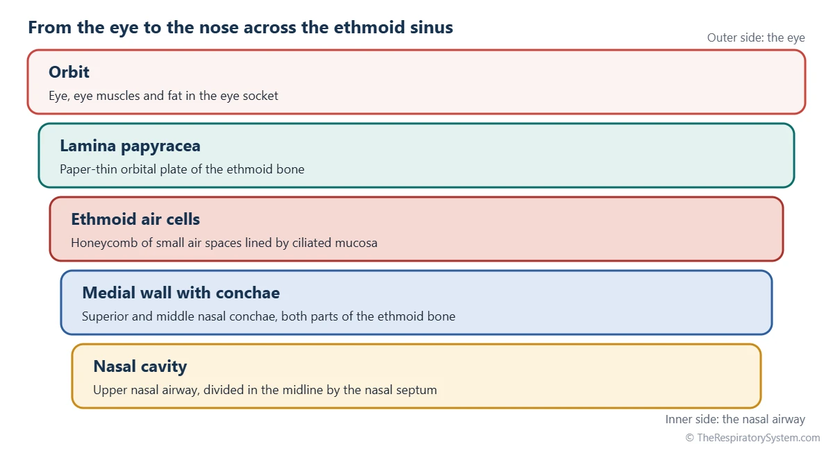
The ethmoid sinus is located between the eyes, in the two lateral masses of the ethmoid bone on either side of the upper nasal cavity. Each cluster of cells lies between the nasal cavity on its inner side and the orbit on its outer side, just below the floor of the front part of the skull.
The bone around the cells is thin on every side, as the table below shows. The outer wall, the lamina papyracea or orbital plate, is so thin that its name means paper layer. The roof, the fovea ethmoidalis, is formed mainly by the frontal bone and separates the cells from the anterior cranial fossa. The inner wall carries the superior and middle nasal conchae, which are themselves parts of the ethmoid bone.
Between the two ethmoid labyrinths lie the upper nasal septum and, above it, the cribriform plate, which carries the nerve fibers serving the sense of smell.
| Border | Formed by | What lies beyond it |
|---|---|---|
| Lateral (outer) | Lamina papyracea, the paper-thin orbital plate of the ethmoid | Orbit: eye, eye muscles and fat |
| Roof | Fovea ethmoidalis, mainly of the frontal bone | Anterior cranial fossa and the frontal lobes |
| Medial (inner) | Ethmoid bone carrying the superior and middle conchae | Upper nasal cavity |
| Floor | Maxilla, at the inner edge of the orbital floor | Maxillary sinus and middle meatus |
| Front | Frontal process of the maxilla and the lacrimal bone | Lacrimal sac and frontal recess |
| Back | Body of the sphenoid bone | Sphenoid sinus |
| Source: Typical adult anatomy, our editors | ||
Ethmoid Sinus Anatomy: Anterior and Posterior Ethmoid Cells
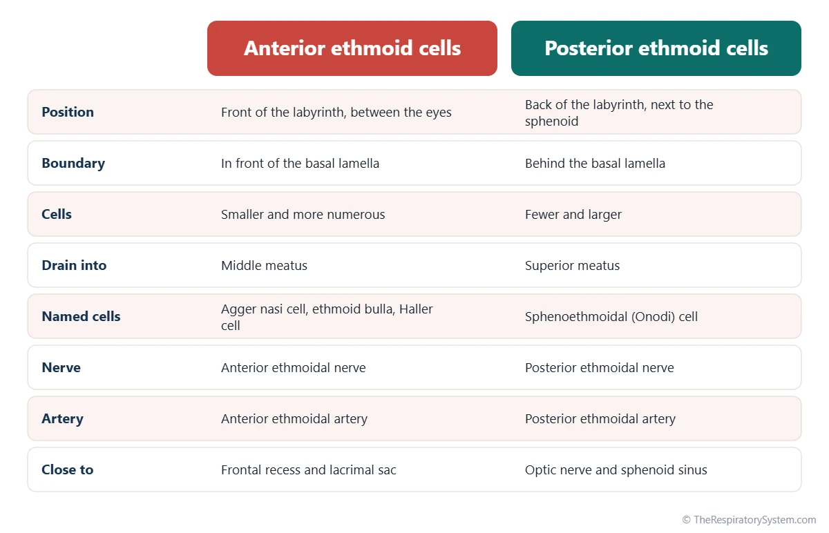
The ethmoid air cells are divided into anterior and posterior groups by the basal lamella of the middle concha, a thin sheet of bone where the middle turbinate attaches to the side wall of the nose. Some textbooks also describe a middle group, which today is usually counted with the anterior cells.
The number and size of the cells vary widely between people. Anterior cells tend to be smaller and more numerous, while posterior cells are fewer and larger. Several cells are distinctive enough to have their own names, such as the agger nasi cell at the front, the ethmoid bulla in the middle meatus and the Onodi cell at the back; the second table lists them with their positions.
Lining, vessels and nerves
The cells are lined by thin ciliated respiratory epithelium, continuous with the lining of the nose. Blood comes mainly from the anterior and posterior ethmoidal arteries, branches of the ophthalmic artery that cross the roof, with help from the sphenopalatine artery. Sensation travels in the anterior and posterior ethmoidal nerves, from the ophthalmic division of the trigeminal nerve, and in small orbital branches linked to the maxillary nerve.
| Cell | Group | Where it lies | Why it matters |
|---|---|---|---|
| Agger nasi cell | Anterior | Most forward cell, in front of the middle concha attachment | Landmark for the frontal sinus drainage pathway |
| Ethmoid bulla | Anterior | Behind the uncinate process, bulging into the middle meatus | Usually the largest anterior cell; borders the semilunar hiatus |
| Supraorbital ethmoid cell | Anterior | Over the roof of the orbit, behind the frontal sinus | Can look like a divided frontal sinus on scans |
| Infraorbital ethmoid cell (Haller cell) | Anterior | In the floor of the orbit, above the maxillary sinus | Can narrow the drainage of the maxillary sinus |
| Concha bullosa | Anterior | An air cell inside the middle concha | Common variant that can crowd the middle meatus |
| Posterior ethmoid cells | Posterior | Behind the basal lamella, above the superior meatus | Fewer, larger cells near the optic nerve |
| Sphenoethmoidal cell (Onodi cell) | Posterior | Above or beside the sphenoid sinus | Lies close to the optic nerve and sometimes the internal carotid artery |
| Source: Typical adult anatomy, our editors | |||
Where Do the Ethmoid Air Cells Drain?
The anterior ethmoid cells drain into the middle meatus, and the posterior ethmoid cells drain into the superior meatus. Each cell opens into a neighbor or directly into the nose through small openings, rather than through one large ostium.
- Anterior cells: into the ethmoidal infundibulum and semilunar hiatus, or onto the ethmoid bulla, in the middle meatus.
- Middle cells, when described: on or above the ethmoid bulla, also in the middle meatus.
- Posterior cells: into the superior meatus under the superior concha, and sometimes into a small supreme meatus above it.
The anterior ethmoid region is part of the ostiomeatal complex, the narrow area of the middle meatus where the frontal, maxillary and anterior ethmoid sinuses all empty. Because these channels are so close together, swelling of the ethmoid lining can block the drainage of the larger sinuses as well. Mucus from every cell is swept by the mucociliary escalator toward the nasopharynx; the nasal meatus page shows where each channel opens.
What Is the Function of the Ethmoid Sinuses?
The ethmoid sinuses lighten the center of the face, add mucus and moisture to the nasal lining and help form the resonating spaces of the voice, the general roles of all paranasal sinuses described on the paranasal sinuses page.
Ethmoid sinus function in the respiratory system
In the respiratory system, the ethmoid cells provide a large lined surface right beside the upper nasal airway. Their mucus drains into the middle and superior meatuses, adding to the film that traps particles and moistens inhaled air on its way to the pharynx and the lungs. The honeycomb of thin walls also keeps the bone between the eyes light while bracing it, much like the cells of a beehive.
The ethmoid bone that holds the cells also carries the superior and middle conchae, which shape airflow through the upper nose, and it lies just below the olfactory area in the roof of the nose. The air cells themselves do not detect odors.
Ethmoid Sinus Anatomy on CT and Radiology
On CT, the ethmoid sinus appears as a honeycomb of small dark air cells separated by thin white bony walls, lying between the orbits on coronal images. CT is the standard way to study ethmoid anatomy, because plain X-rays show the overlapping cells poorly; on a front-facing Caldwell view they look like a mottled dark area between the eyes.
Radiologists check several landmarks on a sinus CT:
- whether the lamina papyracea is intact and in its usual position;
- the course of the anterior ethmoidal artery across the roof;
- Haller cells, Onodi cells and concha bullosa, which narrow drainage paths or lie near the optic nerve;
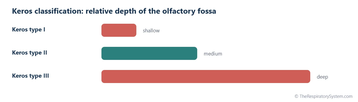
- the depth of the olfactory fossa, graded with the Keros classification.
Keros classification
The Keros classification describes how far the cribriform plate sits below the ethmoid roof, measured as the height of the thin lateral lamella of the cribriform plate. Type I is shallow, about 1 to 3 millimeters; type II is medium, about 4 to 7 millimeters; and type III is deep, about 8 to 16 millimeters. A deeper olfactory fossa means a longer stretch of very thin bone at the skull base, which surgeons take into account.
How Do the Ethmoid Sinuses Develop?
The ethmoid sinuses are present at birth, making them, with the maxillary sinuses, the earliest paranasal sinuses to form. They begin before birth as outpouchings of the nasal lining into the developing ethmoid bone, and a newborn already has a set of small cells on each side.
The cells enlarge and multiply through childhood and reach close to their adult size around the early teens. Their early presence is one reason the ethmoid sinuses are so often involved when young children have sinus inflammation, while the frontal sinus, which grows up from the anterior ethmoid region, appears only later. The overall timeline is covered on the development of the respiratory system page.
Common Conditions That Affect the Ethmoid Sinus
The ethmoid sinus is involved in many sinus conditions because it sits at the center of sinus drainage. These are listed for education only; the respiratory diseases page gives wider context.
- Ethmoid sinusitis: inflammation of the ethmoid lining, often felt as pressure between the eyes or at the bridge of the nose. See sinusitis.
- Chronic rhinosinusitis and nasal polyps: long-lasting inflammation often centers on the ethmoid cells, and nasal polyps commonly arise from the ethmoid region.
- Orbital complications: because the lamina papyracea is so thin, ethmoid inflammation can, uncommonly, spread to the tissues around the eye, especially in children.
- Mucocele: a mucus-filled sac from a blocked cell; the ethmoid cells are the second most common site after the frontal sinus.
- Tumors: rare growths of the ethmoid lining, which can involve the orbit or the skull base.
Swelling or redness around the eye, changes in vision or a severe headache are reasons to see a doctor without delay.
Key Facts
- The ethmoid sinus is a group of small air cells in the ethmoid bone, between the nasal cavity and the eye sockets.
- The basal lamella of the middle concha divides the cells into anterior and posterior groups.
- Anterior ethmoid cells drain into the middle meatus; posterior ethmoid cells drain into the superior meatus.
- The lamina papyracea, the outer wall of the cells, is a paper-thin plate of bone that separates them from the orbit.
- The ethmoid sinuses are present at birth and reach near-adult size around the early teens.
- The anterior ethmoid region forms part of the ostiomeatal complex, where the frontal and maxillary sinuses also drain.
- The Keros classification grades the depth of the olfactory fossa beside the ethmoid roof as type I, II or III.
Quick Quiz: Test What You Know About the Ethmoid Sinus
6 questions. Tap an answer to check it.
Which bone contains the ethmoid air cells?
The ethmoid air cells fill the lateral masses, or labyrinths, of the ethmoid bone between the eyes.
What thin plate separates the ethmoid air cells from the orbit?
The lamina papyracea, or orbital plate of the ethmoid, is a paper-thin wall between the cells and the eye socket.
Where do the posterior ethmoid cells drain?
Posterior ethmoid cells open into the superior meatus, under the superior concha. Anterior cells drain into the middle meatus.
What divides the anterior ethmoid cells from the posterior ones?
The basal lamella, where the middle concha attaches to the side wall of the nose, separates the two groups.
Which ethmoid cell can lie close to the optic nerve?
The Onodi, or sphenoethmoidal, cell is a posterior ethmoid cell that extends beside or above the sphenoid sinus, near the optic nerve.
Which paranasal sinuses are already present at birth?
The ethmoid and maxillary sinuses are present at birth; the frontal and sphenoid sinuses develop mainly during childhood.
Frequently Asked Questions
How many ethmoid air cells are there?
There is no fixed number. Each side of the ethmoid bone holds anywhere from a few to well over a dozen cells, and the count, size and shape differ from person to person and even between the left and right sides. Because the cells vary so much, anatomists describe them by group and position rather than by a set number.
Which sinus is between the eyes?
The ethmoid sinus is the sinus between the eyes. Its air cells fill the space between the two eye sockets, behind the bridge of the nose and on either side of the upper nasal cavity. The frontal sinus lies higher, in the forehead, and the sphenoid sinus lies further back, deep behind the ethmoid cells.
Where is ethmoid sinus pain felt?
Discomfort from the ethmoid sinus is usually felt between the eyes, at the bridge of the nose or around the inner corners of the eyes, because that is where the cells lie. It may feel stronger when touching the sides of the nose near the eyes. Similar pain can also come from other causes, so the location alone does not identify the source.
What is the difference between the ethmoid sinus and the ethmoid bone?
The ethmoid bone is a whole skull bone at the center of the face. It includes the cribriform plate, the perpendicular plate in the nasal septum, the superior and middle conchae and two lateral masses. The ethmoid sinus is the set of air cells hollowed out inside those lateral masses, so it is a space within the bone.
Are the ethmoid sinuses connected to the eyes?
No, there is no opening between the ethmoid sinus and the eye socket. The two are separated by the lamina papyracea, a very thin plate of bone, and by the lining and connective tissue around the eye. Because this wall is so thin, and small natural gaps can occur in it, inflammation can occasionally spread across it.
Can ethmoid air cells grow into other bones?
Yes. During growth, ethmoid cells often expand into neighboring bones. They can reach into the frontal bone over the orbit, into the floor of the orbit above the maxillary sinus as Haller cells, into the sphenoid region as Onodi cells and into the middle concha as a concha bullosa. These extensions are common variations.
What is a concha bullosa?
A concha bullosa is an air cell inside a nasal concha, most often the middle concha. It forms when ethmoid air cells extend into the bone of the concha during growth. It is a common anatomical variation that usually causes no problems, but a large one can crowd the middle meatus, where the frontal, maxillary and anterior ethmoid sinuses drain.
Is the ethmoid sinus part of the respiratory system?
Yes. The ethmoid air cells belong to the paranasal sinuses, which are part of the upper respiratory tract. They do not carry the main stream of air and take no part in gas exchange, but they open into the nasal cavity, share its ciliated lining and add mucus to the airway that leads to the pharynx, larynx and lungs.
Keep Exploring


