At a Glance
Lung development passes through five stages: embryonic (about weeks 4 to 7), pseudoglandular (about weeks 5 to 17), canalicular (about weeks 16 to 26), saccular (about weeks 26 to 36) and alveolar (from about 36 weeks into childhood). The airway lining comes from endoderm, while cartilage, muscle and blood vessels come from mesoderm, and most alveoli form after birth.
- Begins
- About week 4, as the respiratory diverticulum (lung bud) from the foregut
- Germ layers
- Endoderm forms the lining and glands; splanchnic mesoderm forms cartilage, muscle, connective tissue and vessels
- Stages
- Embryonic, pseudoglandular, canalicular, saccular and alveolar
- Surfactant
- Made by type II pneumocytes; amounts rise sharply in the final weeks of pregnancy
- Before birth
- Lungs are filled with fluid; the placenta handles gas exchange
- Continues
- Alveoli keep forming after birth, through early childhood and possibly into adolescence
On this page
- What Is the Development of the Respiratory System?
- How Does the Respiratory System Begin in the Embryo?
- What Are the Stages of Lung Development?
- When Do the Lungs Start Making Surfactant?
- What Is Fetal Lung Development, and How Do the Lungs Work Before Birth?
- What Is Lung Development Like at 36 and 37 Weeks?
- What Happens to the Lungs at Birth?
- How Do the Lungs Keep Developing After Birth?
- How Do the Nose, Sinuses and Diaphragm Develop?
- Common Conditions Linked to Respiratory Development
- Key Facts
- Quiz
- FAQ
What Is the Development of the Respiratory System?
The development of the respiratory system is the process by which the airways, lungs, chest structures and breathing muscles form, from a simple outgrowth of the embryonic gut to a working organ that can breathe air at birth. In embryology it is studied in two parts: the early formation of the airways, and the later maturation of the lungs for gas exchange.
Two germ layers build the system. The endoderm, the inner layer that also forms the gut, gives rise to the lining epithelium of the larynx, trachea, bronchi and alveoli, along with their glands. The surrounding splanchnic mesoderm forms the cartilage, smooth muscle, connective tissue, blood vessels and the visceral pleura. Signals passed back and forth between these two tissues control where and how often each airway branches.
Explained simply
The lungs start as a tiny pouch budding off the tube that later becomes the throat and food pipe. The pouch splits in two, and each half keeps splitting, like a tree growing more and more branches. Near the end of pregnancy, the tips of the smallest branches turn into tiny, thin-walled bubbles, and after the baby is born many more bubbles keep forming for years.
How weeks are counted
Embryology texts usually count weeks from fertilization, while pregnancy weeks are counted from the first day of the last menstrual period, about two weeks earlier. Stage timings therefore differ by a week or two between sources, and the stages also overlap, because different parts of the lung mature at slightly different times.
How Does the Respiratory System Begin in the Embryo?
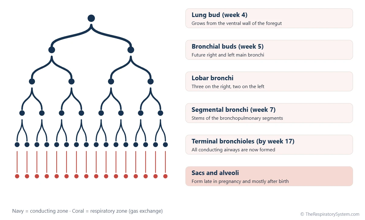
The respiratory system begins in the embryo as the respiratory diverticulum, or lung bud, which grows out of the front (ventral) wall of the foregut around the fourth week after fertilization. This bud becomes the larynx, trachea, bronchi and lungs, so in the earliest weeks the airway and the digestive tract share a common origin.
Separating the airway from the esophagus
As the lung bud grows downward, ridges along its sides grow toward each other and fuse into a wall called the tracheoesophageal septum. This wall divides the foregut into a front tube, the trachea with its lung buds, and a back tube, the esophagus. The two stay connected only at the top, through the laryngeal inlet, which is why food and air still share the pharynx, as the trachea vs esophagus comparison explains.
Larynx and cartilages
The opening of the lung bud becomes the larynx. Its lining comes from endoderm, but the laryngeal cartilages and muscles come from the fourth and sixth pharyngeal arches, which is why the larynx is supplied by branches of the vagus nerve.
Bronchial buds and lobes
During the fifth week the lung bud splits into two bronchial buds, the future main bronchi. The right bud then divides into three secondary bronchi and the left into two, foreshadowing the three lobes of the right lung and the two lobes of the left lung. By about the seventh week, segmental bronchi have formed, ten on the right and eight to ten on the left, each the stem of a future bronchopulmonary segment.
What Are the Stages of Lung Development?
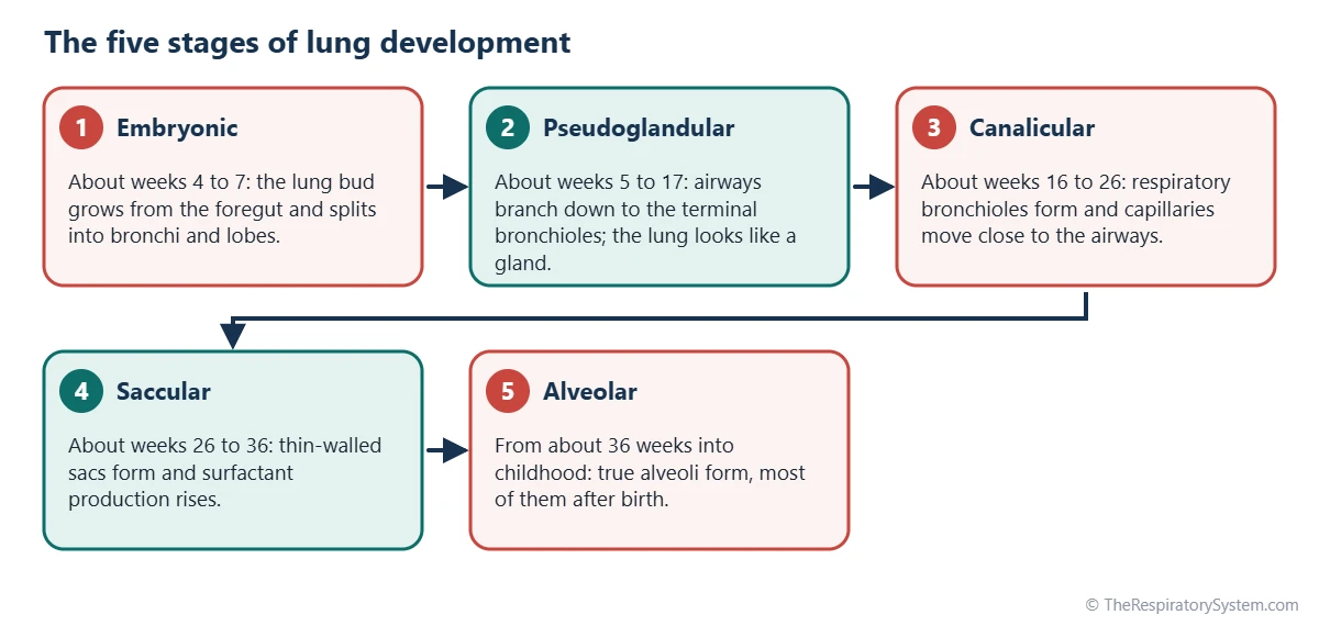
The five stages of lung development are the embryonic, pseudoglandular, canalicular, saccular and alveolar stages, each named after what the developing lung looks like under a microscope. The first two build the airways, and the last three build the gas-exchange surface.
1. Embryonic stage (about weeks 4 to 7)
The lung bud appears, separates from the esophagus and branches into the main, lobar and segmental bronchi. The developing lungs grow into the pericardioperitoneal canals, spaces that later become the pleural cavities.
2. Pseudoglandular stage (about weeks 5 to 17)
The airways branch again and again until all the conducting airways down to the terminal bronchioles are formed. In sections the lung looks like a gland, with many small tubes in loose tissue, which gives the stage its name. Cartilage, smooth muscle, glands and the first ciliated cells of the airway lining appear, but there are no air sacs yet, so gas exchange is impossible.
3. Canalicular stage (about weeks 16 to 26)
The terminal bronchioles give rise to respiratory bronchioles, the airways widen, and the lung tissue becomes rich in blood vessels. Capillaries move up against the thinning airway lining, and the first type I and type II pneumocytes appear. By the end of this stage the air-blood barrier first becomes thin enough for limited gas exchange.
4. Saccular stage (about weeks 26 to 36)
The ends of the respiratory bronchioles widen into thin-walled terminal sacs, or saccules, the forerunners of alveoli. The tissue between them thins, capillaries bulge into the sacs, and surfactant production by type II cells rises.
5. Alveolar stage (from about 36 weeks into childhood)
True alveoli form as small walls called secondary septa grow up from the sac walls and subdivide them. Only a fraction of the adult number of alveoli exist at birth; most form after birth, rapidly in the first years and continuing to about age 8, and possibly into adolescence.
| Timing (approximate) | Stage | What develops |
|---|---|---|
| About week 4 | Embryonic | Lung bud (respiratory diverticulum) grows from the ventral foregut |
| About week 5 | Embryonic | Two bronchial buds form; the tracheoesophageal septum separates trachea and esophagus |
| About weeks 6 to 7 | Embryonic | Lobar bronchi (three right, two left), then segmental bronchi |
| About weeks 5 to 17 | Pseudoglandular | Branching down to the terminal bronchioles; cartilage, glands and cilia appear |
| About weeks 16 to 26 | Canalicular | Respiratory bronchioles; capillaries approach the airways; type I and II cells appear |
| About weeks 26 to 36 | Saccular | Thin-walled terminal sacs; surfactant production rises |
| About 36 weeks to birth | Alveolar | First true alveoli form by secondary septation |
| Birth | Transition | Lung fluid cleared, first breaths, pulmonary vessels widen, fetal shunts close |
| Birth to about age 8 | Alveolar (continued) | Most alveoli form; capillary network matures |
| Childhood to early adulthood | Growth | Airways and air sacs enlarge; lung function peaks in the early to mid twenties |
| Source: Typical textbook timings, counted from fertilization; sources differ by a week or two. Our editors | ||
When Do the Lungs Start Making Surfactant?
The fetal lungs start making pulmonary surfactant when type II pneumocytes appear, from around the 20th to 24th week, and the amount rises sharply during the last weeks of pregnancy, usually reaching mature levels by about 34 to 36 weeks. This timing is one of the main reasons why babies born early may have trouble breathing.
Surfactant is a film of lipids and proteins that lowers the surface tension of the fluid lining the air sacs. Without it, the smallest sacs tend to collapse at the end of every breath out, and each new breath has to reopen them, which takes much more effort. Surfactant production is boosted by hormones, especially cortisol, which rises naturally in the fetus toward the end of pregnancy.
Some surfactant leaves the lungs with lung fluid and mixes into the amniotic fluid, which is why it can be detected there late in pregnancy. Our respiratory physiology section explains how surface tension affects breathing in more detail.
What Is Fetal Lung Development, and How Do the Lungs Work Before Birth?
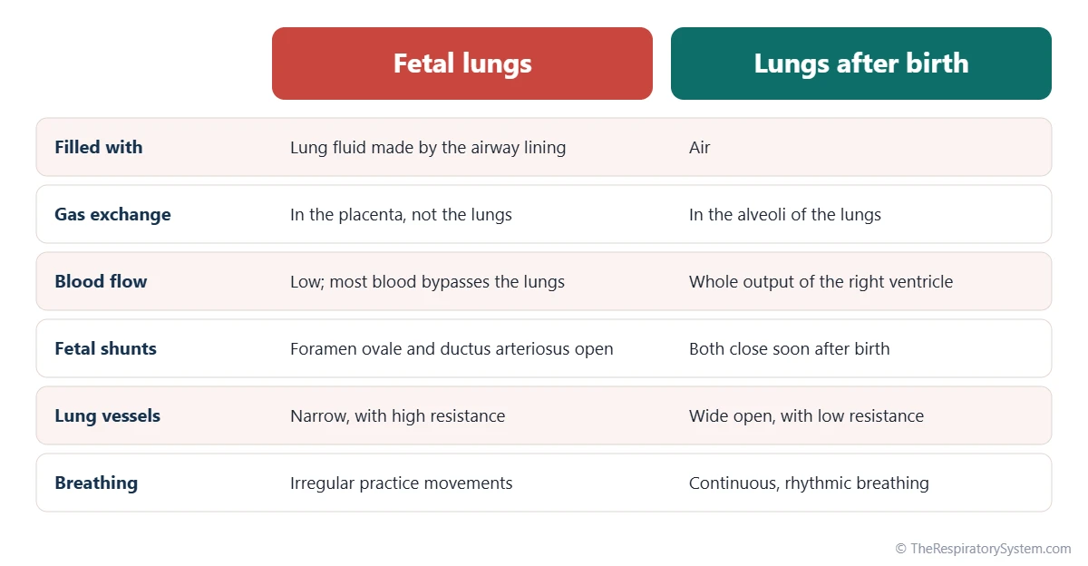
Fetal lung development is the growth and maturation of the lungs before birth, during a time when the lungs are filled with fluid and do no breathing at all, because the placenta supplies oxygen and removes carbon dioxide. The lungs are preparing for a job they will only start at the first breath.
Lung fluid
The lining of the fetal lungs constantly secretes fluid into the airways. This fluid keeps the lungs gently expanded, which stretches the tissue and is an important signal for growth. Part of it flows up the trachea and is either swallowed or added to the amniotic fluid. When too little amniotic fluid surrounds the fetus, or when something takes up space in the chest, the lungs may not grow to their full size.
Fetal breathing movements
From around the end of the first trimester, the fetus makes irregular breathing movements, moving the diaphragm and shifting small amounts of fluid in and out of the airways. These movements do not exchange gas, but they train the breathing muscles and help the lungs grow.
Blood flow before birth
Because the fetal lungs are not used for gas exchange, their blood vessels are narrow and resist flow, and only a small share of the blood reaches them. Most of the blood is diverted through two shortcuts: the foramen ovale, an opening between the atria, and the ductus arteriosus, a vessel linking the pulmonary trunk to the aorta. The adult route is described on the pulmonary circulation page.
What Is Lung Development Like at 36 and 37 Weeks?
At 36 to 37 weeks of pregnancy, the lungs are at the end of the saccular stage and entering the alveolar stage, and in most babies they produce enough surfactant to breathe air. They are close to ready for birth, but they are still not finished.
Around 36 weeks the terminal sacs have thin walls covered by type I pneumocytes, the capillaries lie close against them, and the first true alveoli are forming. Thirty-seven weeks marks the start of the term period, and at this point lung development is advanced enough that most newborns adapt to breathing well. Even so, studies show that babies born before 39 weeks have slightly higher rates of breathing problems than those born a little later, as the last weeks still add alveoli and surfactant.
Every pregnancy is different, and questions about an individual baby's development belong with the doctor or midwife caring for the pregnancy.
What Happens to the Lungs at Birth?
At birth the lungs switch from fluid-filled organs to air-filled ones within minutes: the lung fluid is cleared, the first breaths inflate the air sacs, and blood flow through the lungs rises sharply. It is one of the fastest and most dramatic changes the body ever makes.
- Fluid is cleared. Late in pregnancy and during labor, the lining stops secreting fluid and starts absorbing it, pumping sodium and water out of the airways into the lung tissue, where blood and lymph vessels remove it. Some fluid is also squeezed out during a vaginal birth.
- The first breaths. The first breaths need a strong pull from the diaphragm to open the air sacs for the first time. Once surfactant spreads over the new air-liquid surface, the sacs stay partly open and later breaths become much easier.
- The blood vessels open. Rising oxygen and the stretch of inflation relax the pulmonary arteries, so resistance falls and the whole output of the right ventricle begins to flow through the lungs.
- The shortcuts close. The foramen ovale closes as pressure in the left atrium rises, and the ductus arteriosus constricts within the first days, completing the adult pattern of circulation.
From then on the lungs and heart work as the linked system described on the respiratory and circulatory system page.
How Do the Lungs Keep Developing After Birth?
After birth the lungs keep developing by adding many more alveoli, mostly in the first few years of life, and then by growing in size until early adulthood. A newborn's lungs are not just a small copy of an adult's; they still have most of their gas-exchange surface left to build.
The number of alveoli rises quickly in infancy and early childhood, and alveolar formation continues to about age 8, with evidence that it may continue at a slower rate into adolescence. An adult ends up with about 300 to 500 million alveoli. At the same time the capillary network matures from a double layer into the single sheet seen in adult alveolar walls.
After the alveoli are in place, the lungs grow mainly by enlarging the existing airways and air sacs. Lung function keeps rising through the teenage years and usually peaks in the early to mid twenties, then slowly declines with age, as measured by the lung volumes and capacities used in breathing tests. The breathing rate also changes, falling from fast newborn rates toward the adult respiratory rate.
How Do the Nose, Sinuses and Diaphragm Develop?
The nose, sinuses and diaphragm develop separately from the lungs: the nose from thickened patches of surface tissue on the face, the sinuses as outgrowths of the nasal cavity, and the diaphragm from four tissues that fuse across the body cavity.
Nose and nasal cavity
During the fifth week, two thickened patches called nasal placodes appear on the front of the head and sink inward to form nasal pits. These deepen into the nasal cavities, separated from the mouth at first by a thin membrane. When that membrane breaks down, openings form at the back of the nose, the future choanae, and the later fusion of the palate separates the nasal cavity from the mouth.
Paranasal sinuses
The paranasal sinuses begin as small pockets of the nasal lining that grow into the surrounding bones. The maxillary and ethmoid sinuses are present but small at birth, while the frontal and sphenoid sinuses develop mainly during childhood, and all of them keep growing until the teenage years.
Diaphragm
The diaphragm forms from four parts: the septum transversum, the pleuroperitoneal membranes, the mesentery of the esophagus and muscle from the body wall. It starts high, at the level of the neck, and descends as the embryo grows, which is why it is supplied by the phrenic nerve from the cervical spinal nerves C3 to C5.
Common Conditions Linked to Respiratory Development
Most conditions linked to respiratory development arise when a step of formation does not finish normally, such as the separation of the trachea from the esophagus, the closure of the diaphragm or the production of surfactant. Our respiratory diseases section covers lung conditions in more depth.
- Esophageal atresia and tracheoesophageal fistula: the tracheoesophageal septum forms incompletely, so the esophagus ends blindly and/or an abnormal channel links it to the trachea. The most common form combines a blind upper esophagus with a lower esophagus connected to the trachea.
- Congenital diaphragmatic hernia: a gap in the diaphragm, usually at the back on the left side, lets abdominal organs move into the chest and limits lung growth.
- Pulmonary hypoplasia: underdeveloped lungs, often linked to too little amniotic fluid or lack of space in the chest.
- Neonatal respiratory distress syndrome: breathing difficulty in newborns, mostly preterm, because the lungs have not yet made enough surfactant.
- Bronchopulmonary dysplasia: a long-term lung condition that can develop in babies born very early, when the lungs are still in the canalicular or saccular stage.
- Choanal atresia: the openings at the back of the nose fail to form, blocking the nasal airway.
Key Facts
- The respiratory system begins around the fourth week as a lung bud growing from the ventral wall of the foregut.
- Endoderm forms the lining of the airways and alveoli, while mesoderm forms the cartilage, muscle and blood vessels.
- The tracheoesophageal septum separates the trachea from the esophagus early in development.
- Lung development has five stages: embryonic, pseudoglandular, canalicular, saccular and alveolar.
- Type II pneumocytes start making surfactant around weeks 20 to 24, and levels usually mature by about 34 to 36 weeks.
- Before birth the lungs are filled with fluid, and the placenta handles gas exchange.
- Most alveoli form after birth, with alveolar formation continuing to about age 8.
Quick Quiz: Test What You Know About the Development of the Respiratory System
6 questions. Tap an answer to check it.
From which embryonic structure does the lung bud grow?
The respiratory diverticulum grows from the ventral (front) wall of the foregut around the fourth week.
Which germ layer forms the epithelial lining of the airways and alveoli?
The airway lining comes from endoderm; cartilage, muscle and vessels come from splanchnic mesoderm.
During which stage are all the conducting airways down to the terminal bronchioles formed?
By the end of the pseudoglandular stage, branching has produced every conducting airway, but no air sacs yet.
Which cells produce pulmonary surfactant?
Type II pneumocytes appear in the canalicular stage and make surfactant, which rises sharply late in pregnancy.
When do most alveoli form?
Only a fraction of the adult number of alveoli exist at birth; most form in infancy and early childhood.
What separates the trachea from the esophagus during development?
Ridges on either side of the lung bud fuse into the tracheoesophageal septum, dividing the foregut into two tubes.
Frequently Asked Questions
When are a baby's lungs fully developed?
A baby's lungs are mature enough to breathe air for most babies by about 36 to 37 weeks of pregnancy, but they are not fully developed at birth. Most alveoli form after birth, rapidly during infancy and continuing to about age 8, and the lungs keep growing in size until early adulthood. Lung function usually peaks in the early to mid twenties.
Do babies breathe in the womb?
Not in the sense of breathing air. The fetal lungs are filled with fluid, and the placenta supplies oxygen and removes carbon dioxide through the umbilical cord. However, the fetus makes intermittent practice breathing movements with the diaphragm, moving small amounts of fluid in and out of the airways. These movements train the breathing muscles and help the lungs grow.
Why are the stages of lung development named the way they are?
Each stage is named after what the developing lung looks like under a microscope. In the pseudoglandular stage it resembles a gland, with many small tubes. In the canalicular stage, wide channels and vessels appear. In the saccular stage, thin-walled sacs form. In the alveolar stage, true alveoli appear. The embryonic stage is named for the period of the embryo itself.
What germ layers form the respiratory system?
The endoderm forms the epithelial lining of the larynx, trachea, bronchi and alveoli, plus the airway glands. The splanchnic mesoderm around the lung bud forms the cartilage, smooth muscle, connective tissue, blood vessels and visceral pleura. The nasal cavity lining comes from surface ectoderm, and the laryngeal cartilages come from the fourth and sixth pharyngeal arches.
Why does the right lung have three lobes and the left two?
The difference begins in the fifth to sixth week, when the right bronchial bud divides into three secondary bronchi and the left into two. Each secondary bronchus becomes the stem of one lobe. The left lung also has to share space with the heart, which sits slightly to the left in the chest, and it has a notch where the heart presses against it.
Why do babies born early often have breathing problems?
Babies born early may arrive before their lungs have made enough surfactant, and before the air sacs and capillaries are fully formed. Without enough surfactant the tiny air sacs tend to collapse after each breath, which makes breathing hard work. The earlier the birth, the less mature the lungs are, which is why breathing support is a common part of newborn intensive care.
Is lung development affected by smoking during pregnancy?
Yes. Research links smoking during pregnancy with smaller airways and lower lung function in the child, along with a higher risk of wheezing illnesses in early childhood. Nicotine crosses the placenta and can affect how the developing lungs grow. Smoke exposure after birth, including secondhand smoke, also affects the lungs while they are still forming alveoli.
Keep Exploring


