At a Glance
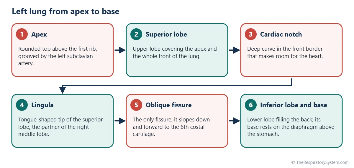
The left lung is the lung in the left side of the chest. It has 2 lobes (superior and inferior) separated by one oblique fissure, 8 to 10 bronchopulmonary segments, a cardiac notch on its front border and a lingula on its superior lobe. Because the heart lies toward the left, it is narrower and holds roughly 45 percent of total lung volume, but it is slightly longer than the right lung.
- Location
- Left side of the thoracic cavity, beside the heart, resting on the left dome of the diaphragm
- Lobes
- Two: superior (upper), including the lingula, and inferior (lower)
- Fissures
- One: the oblique fissure
- Segments
- 8 to 10, depending on how combined segments are counted
- Connects to
- Trachea by the left main bronchus; heart by the left pulmonary artery and two left pulmonary veins
- Part of
- Lower respiratory tract
On this page
What Is the Left Lung?
The left lung is the cone-shaped, spongy organ in the left half of the chest that exchanges oxygen and carbon dioxide between the air and the blood, shaped around the heart that sits beside it. It belongs to the lower respiratory tract and has the same tissue and job as the right lung, but a different outline and fewer lobes. The lungs page covers what both lungs share; this page covers only the left one.
Left lung definition
In anatomy, the left lung is the respiratory organ in the left pleural cavity, supplied with air by the left main bronchus and with blood by the left pulmonary artery. It is wrapped in its own pleura, so it can keep working even if the right lung collapses, and the reverse is also true.
Explained simply
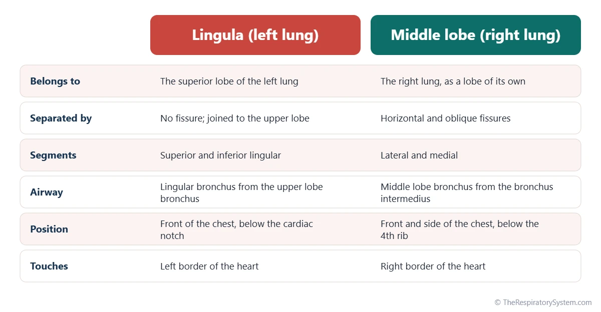
The heart is not in the exact middle of the chest; it leans to the left. The left lung has to wrap around it, so it is slimmer than the right lung and has a scooped-out bite in its front edge where the heart pokes forward. It is split into only two big lobes, and the small flap just below the bite, called the lingula, does the job that the middle lobe does on the right.
Where Is the Left Lung Located?
The left lung is located in the left side of the thoracic cavity, between the left ribs and the mediastinum, with its apex in the root of the neck and its base resting on the left dome of the diaphragm. The heart, inside its pericardium, lies against its inner surface and pushes deeply into it.
Where is the left lung apex?
The apex of the left lung is its rounded top, rising through the upper opening of the rib cage to about 2 to 3 cm above the inner third of the left collarbone. It is capped by the cervical pleura and is grooved by the left subclavian artery, which arches over it on its way to the arm.
Surfaces, borders and base
- Costal surface: large and convex, facing the ribs and intercostal muscles.
- Mediastinal surface: deeply hollowed by the heart and grooved by the aorta, with the hilum near its middle.
- Anterior border: straight near the top, then curving sharply outward as the cardiac notch before turning back in at the lingula.
- Base: sits on the left dome of the diaphragm, which is lower than the right dome. Under it lie the stomach, the spleen and part of the left lobe of the liver.
Because the left dome of the diaphragm sits lower, the left lung reaches a little further down than the right. From the back, almost everything below the shoulder blade belongs to the inferior lobe.
What Are the Lobes of the Left Lung?
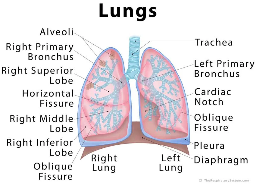
The lobes of the left lung are the superior (upper) lobe and the inferior (lower) lobe, separated by a single oblique fissure; there is no middle lobe and no horizontal fissure. The superior lobe instead carries the lingula, a thin extension that sits in the same place as the right middle lobe. The lung lobes page compares the lobes of both lungs side by side.
The oblique fissure
The oblique fissure of the left lung starts at the back at about the level of the 4th thoracic vertebra and slopes downward and forward to the 6th costal cartilage at the front. Like the right oblique fissure, it can be incomplete, leaving the two lobes partly joined at their deep ends. See lung fissures for its full course.
Superior lobe, cardiac notch and lingula
The superior lobe forms the apex, the whole front of the lung and much of its upper back. On its anterior border, a deep curved cut called the cardiac notch makes room for the apex of the heart, and just below the notch the lobe ends in the lingula, Latin for "little tongue". The cardiac notch and lingula page describes both in detail.
Inferior lobe
The inferior lobe lies behind and below the oblique fissure. It fills most of the back of the lung and the whole base on the diaphragm, and in many people it is about as large as the superior lobe.
Bronchopulmonary segments
The left lung has 8 to 10 bronchopulmonary segments. Counted as 10, it mirrors the right lung, but in most people two pairs share a single segmental bronchus: the apical and posterior segments of the superior lobe form one apicoposterior segment, and the anterior and medial basal segments of the inferior lobe form one anteromedial basal segment. The table below uses the common count of 8.
| Segment | Lobe | Where it lies |
|---|---|---|
| Apicoposterior | Superior lobe | Top and upper back of the lung, in the dome of pleura |
| Anterior | Superior lobe | Upper front of the lung, above the cardiac notch |
| Superior lingular | Superior lobe (lingula) | Upper part of the lingula, beside the cardiac notch |
| Inferior lingular | Superior lobe (lingula) | Lower tip of the lingula, against the left border of the heart |
| Superior | Inferior lobe | Top of the inferior lobe, at the back below the oblique fissure |
| Anteromedial basal | Inferior lobe | Front and inner part of the base, behind the heart |
| Lateral basal | Inferior lobe | Outer part of the base, toward the side of the chest |
| Posterior basal | Inferior lobe | Back part of the base, lowest part of the lung beside the spine |
| Source: Typical adult anatomy, our editors | ||
Left Lung Anatomy: Hilum, Bronchus and Impressions
The anatomy of the left lung is shaped by the heart and the aorta: the heart carves a deep cardiac impression into its inner surface, and the aorta arches over its root and runs down behind it. Its airway and hilum also follow their own pattern, different from the right.
The left main bronchus
The left main bronchus leaves the trachea at the carina and is about twice as long as the right, roughly 5 cm. It is narrower and more horizontal, passing below the arch of the aorta and in front of the esophagus and descending aorta before it enters the lung. Inside the hilum it divides into a superior lobe bronchus, which gives off the lingular bronchus, and an inferior lobe bronchus. The bronchi page follows the whole airway.
What passes through the left hilum?
At the left hilum, the left pulmonary artery lies highest, the main bronchus lies behind and below it, and the two left pulmonary veins sit lowest and most anterior. Unlike the right side, there is no eparterial bronchus: every lobar bronchus on the left branches below the pulmonary artery. The left phrenic nerve passes in front of the hilum and the left vagus nerve behind it, while the left recurrent laryngeal nerve hooks under the aortic arch just above.
Impressions on the mediastinal surface
On a preserved left lung, the heart, mainly its left ventricle, leaves a deep cardiac impression in front of the hilum. The aortic arch makes a curved groove over the hilum, the descending aorta a broad vertical groove behind it, and the left subclavian artery a groove near the apex. The second table lists these marks with the structures that make them.
| Structure | Where it touches the left lung | What it leaves or does |
|---|---|---|
| Heart (mainly left ventricle) | Front of the mediastinal surface | Deep cardiac impression and the cardiac notch |
| Arch of the aorta | Curving over the top of the hilum | Curved groove above the root of the lung |
| Descending aorta | Behind the hilum and pulmonary ligament | Broad vertical groove down the back of the mediastinal surface |
| Left subclavian artery | Upper mediastinal surface and apex | Groove running up over the apex |
| Left brachiocephalic vein | Upper front of the mediastinal surface | Shallow groove near the apex |
| Esophagus | Lower mediastinal surface, in front of the descending aorta | Short groove just above the diaphragm |
| Left phrenic and vagus nerves | In front of and behind the hilum | Pass the root on their way to the diaphragm and abdomen |
| Left dome of the diaphragm | Under the base, above the stomach and spleen | Concave base that sits a little lower than the right |
| Source: Typical adult anatomy, our editors | ||
What Is the Function of the Left Lung?
The function of the left lung is to load the blood with oxygen and unload carbon dioxide into the air, and because it gives up space to the heart, it handles roughly 45 percent of this work. Its role in the respiratory system is the same as the right lung's; it simply has less tissue.
Air reaches it through the left main bronchus and the branching bronchial tree, ending in the alveoli where gas exchange happens. Oxygen-poor blood arrives from the right ventricle through the left pulmonary artery, and oxygen-rich blood returns to the left atrium through the two left pulmonary veins. Our how do the lungs work page explains the full process.
Left lung lobes and their function
The superior and inferior lobes, and the lingula, all perform the same gas exchange; none has a separate job. Like any lung, the left lung also cleans inhaled air with its cilia and mucus, filters small clots out of the venous blood, and helps keep the blood's pH stable by breathing out carbon dioxide. Its division into lobes and segments keeps many infections or blockages confined to one part of the lung.
Left Lung Anatomy on CT and Bronchoscopy
On imaging and bronchoscopy, the left lung is recognized by its single oblique fissure, its long, horizontal main bronchus, and its close contact with the heart and aorta. Doctors use these landmarks to name exactly which lobe or segment a finding is in.
- Chest X-ray: the lingula touches the left border of the heart, while the inferior lobe touches the left dome of the diaphragm and the descending aorta. When one of these regions fills with fluid, the edge it touches becomes blurred, which helps locate the problem.
- CT scan: slices show the oblique fissure as a thin line, the aortic arch and descending aorta curving around the root, and the lingula wrapping the front of the heart.
- Bronchoscopy: a thin camera passed down the airway enters the left main bronchus after a longer, more angled run than on the right. At its end it reaches the split into the upper and lower lobe bronchi, with the lingular bronchus branching off the upper one.
Part of the left inferior lobe sits behind the heart, so on a frontal X-ray it is partly hidden by the heart's shadow, and a side view or CT is often used to see it clearly. The same anatomy appears on our respiratory system diagram.
Common Conditions That Affect the Left Lung
The left lung can develop any lung disease, and a few problems are linked to its closeness to the heart or to its lingula and lower lobe. The respiratory diseases section explains each condition, and anyone with breathing symptoms should see a doctor.
- Left lower lobe collapse: part of the lower lobe loses its air, sometimes after surgery or when an enlarged heart presses on it, and it can hide behind the heart shadow on X-rays.
- Lingular pneumonia: infection of the lingula, which blurs the left border of the heart on a chest X-ray.
- Pleural effusion: fluid in the left pleural cavity pressing on the lung from outside.
- Pneumothorax: air in the left pleural cavity allowing the lung to partly or fully collapse.
- Aspiration: less common on the left than on the right, because the left main bronchus is narrower and leaves the trachea at a sharper angle.
Key Facts
- The left lung has 2 lobes (superior and inferior), a single oblique fissure and 8 to 10 bronchopulmonary segments.
- It holds roughly 45 percent of total lung volume, because the heart leans to the left and takes up part of its space.
- The cardiac notch on its anterior border and the deep cardiac impression on its inner surface both make room for the heart.
- The lingula is a tongue-shaped part of the superior lobe and is the counterpart of the right lung's middle lobe.
- The left main bronchus is about 5 cm long, roughly twice the length of the right, and runs more horizontally under the aortic arch.
- The arch of the aorta curves over the root of the left lung, and the descending aorta runs down behind it.
- The left lung is narrower than the right lung but slightly longer, because the left dome of the diaphragm sits lower.
Quick Quiz: Test What You Know About the Left Lung
6 questions. Tap an answer to check it.
How many lobes does the left lung have?
The left lung has two lobes, superior and inferior, separated by the oblique fissure.
Which part of the left lung is the counterpart of the right middle lobe?
The lingula, part of the left superior lobe, lies in the same position as the right middle lobe.
What makes the cardiac notch in the left lung?
The heart leans to the left, and the cardiac notch in the anterior border of the left lung makes room for it.
Which fissure does the left lung have?
The left lung has only an oblique fissure. The horizontal fissure is found in the right lung.
Which large vessel arches over the root of the left lung?
The arch of the aorta curves over the left lung root; the azygos vein does the same on the right.
Compared with the right main bronchus, the left main bronchus is:
The left main bronchus is about 5 cm long and more horizontal, passing under the aortic arch.
Frequently Asked Questions
Why does the left lung have only 2 lobes?
During development, the left lung bud branches into only two lobar bronchi, while the right lung bud branches into three. This matches the body's left-right plan, in which the heart leans to the left and takes up space on that side. The part that would form a middle lobe stays attached to the upper lobe as the lingula, so the left lung ends up with two lobes.
Is the left lung smaller than the right lung?
Yes. The left lung is smaller overall, holding roughly 45 percent of total lung volume against about 55 percent for the right, and it is usually a little lighter. The difference comes mainly from the heart, which leans into the left side of the chest. The left lung is narrower but slightly longer than the right, as our which lung is bigger page explains.
Does the left lung have a horizontal fissure?
No. The left lung normally has only one fissure, the oblique fissure, which separates its superior and inferior lobes. The horizontal fissure is found only in the right lung. In a small number of people, an extra accessory fissure partly separates the lingula from the rest of the superior lobe, but this is a normal variation and not a true horizontal fissure.
Why do some books say the left lung has 8 segments and others 10?
Both counts describe the same lung. In most people, the apical and posterior segments of the left superior lobe share one segmental bronchus, and so do the anterior and medial basal segments of the inferior lobe. Books that count each shared airway as one segment list 8; books that keep the right lung's pattern list 10, and some list 9 by combining only one of the pairs.
Can a person live with only the left lung?
Yes. People can live for many years after the right lung is removed in an operation called a pneumonectomy. The left lung then performs all gas exchange and is usually enough for everyday activity at rest, although hard exercise becomes more limited. Losing the larger right lung removes more lung tissue than losing the left, so the reserve left afterward is smaller.
Why is the left lung longer than the right lung?
The left lung is slightly longer because the left dome of the diaphragm sits lower than the right dome. On the right, the large liver pushes the diaphragm up and shortens the right lung. On the left, the stomach and spleen lie under the diaphragm and allow it to sit lower, so the left lung's base reaches a little further down.
How much of the heart lies on the left side of the chest?
About two-thirds of the heart lies to the left of the midline of the body, and its apex points down, forward and to the left. This is why the left lung has a deep cardiac impression and a cardiac notch, while the right lung has only a shallow impression. The heart and lungs share the chest tightly, as our mediastinum page shows.
Keep Exploring


