At a Glance
The hilum of the lung is the depression on the medial (mediastinal) surface of each lung through which the structures of the lung root pass: a main bronchus, one pulmonary artery, two pulmonary veins, bronchial vessels, nerves of the pulmonary plexus, lymph vessels and hilar lymph nodes. It lies roughly opposite the fifth to seventh thoracic vertebrae, and in both lungs the pulmonary veins are the most anterior and inferior structures.
- Location
- Middle of the medial (mediastinal) surface of each lung, opposite about the T5 to T7 vertebrae
- What it is
- The doorway on the lung surface; the root is the bundle of structures passing through it
- Main contents
- Main bronchus, pulmonary artery, two pulmonary veins, bronchial vessels, nerves, lymph vessels and nodes
- Covering
- A sleeve of pleura where visceral and parietal pleura meet, extending down as the pulmonary ligament
- Function
- Connects each lung to the heart, trachea and mediastinum and anchors it in place
- Part of
- Lungs and mediastinum, lower respiratory tract
On this page
- What Is a Lung Hilum?
- Where Is the Hilum of the Lung?
- Hilum of Lung Anatomy: What Passes Through It?
- Hilum of Lung vs Root of Lung: What Is the Difference?
- What Is the Function of the Hilum of the Lung?
- What Does the Hilum of the Lung Look Like on an X-Ray?
- Common Conditions That Affect the Hilum of the Lung
- Key Facts
- Quiz
- FAQ
What Is a Lung Hilum?
A lung hilum is the opening on the inner surface of each lung where the airway, blood vessels, nerves and lymph vessels pass between the lung and the center of the chest. Each of the two lungs has one hilum, and it is the only place where the lung is attached to anything; everywhere else the lung moves freely inside its pleural sac.
The word hilum, plural hila, is used in anatomy for the notch or depression where vessels and ducts enter an organ; the kidneys and spleen have hila too. In the lung, the hilum is a roughly wedge-shaped or racquet-shaped area on the mediastinal surface, outlined by the line where the pleura covering the lung turns back to line the chest.
Explained simply
Think of each lung as a large, soft house with only one doorway, on the side facing the heart. Every pipe and cable the house needs, the air pipe, the blood pipes in and out, the wires and the drains, comes in through that single doorway. The doorway is the hilum, and the bundle of pipes and cables running through it is the root of the lung.
Where Is the Hilum of the Lung?
The hilum of the lung is on the medial surface of each lung, the side that faces the mediastinum, a little above the middle of that surface and slightly toward the back. The root of the lung lies roughly opposite the bodies of the fifth to seventh thoracic vertebrae.
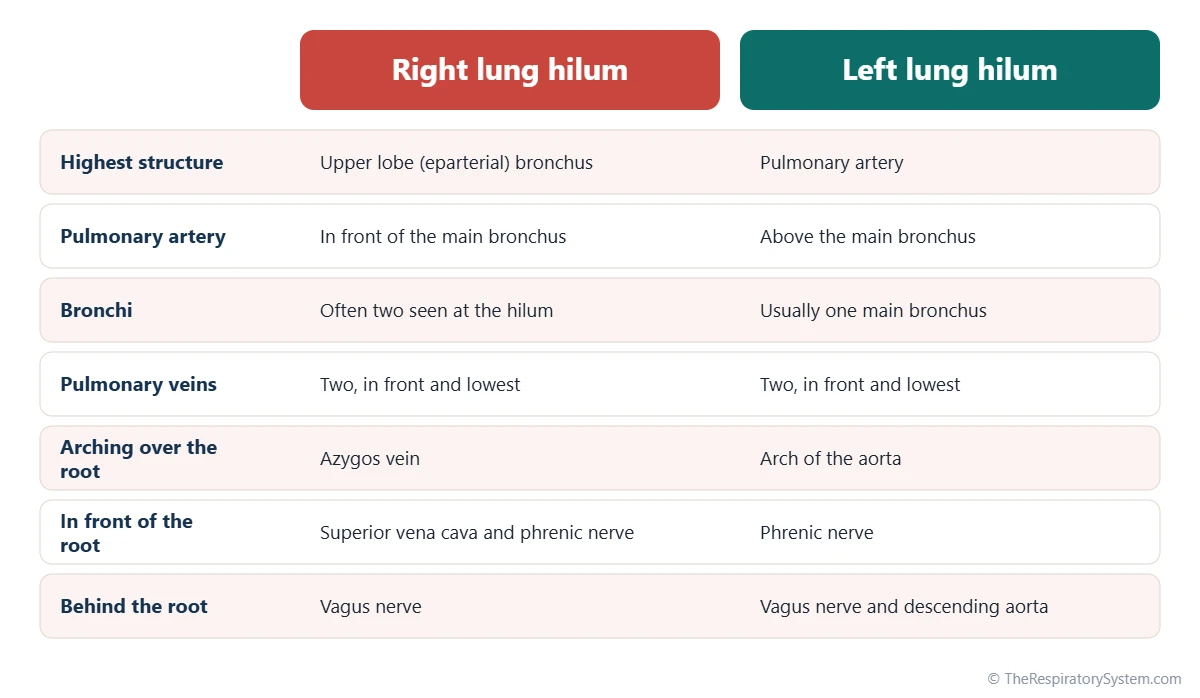
On the right lung, the hilum sits just behind the shallow cardiac impression made by the right atrium and beneath the groove for the arch of the azygos vein. On the left lung, it lies behind and above the deeper impression made by the heart, which continues forward as the cardiac notch, and beneath the groove for the arch of the aorta. Each hilum connects the lung to the structures in the middle of the thoracic cavity.
Relations of the root of the lung
- In front: the phrenic nerve on both sides; the superior vena cava and right atrium on the right.
- Behind: the vagus nerve on both sides; the descending aorta on the left.
- Above: the arch of the azygos vein on the right and the arch of the aorta on the left.
- Below: the pulmonary ligament, a fold of pleura hanging down from the root.
Hilum of Lung Anatomy: What Passes Through It?
Structures passing through the hilum of each lung include one main bronchus, one pulmonary artery, two pulmonary veins, the bronchial arteries and veins, the pulmonary nerve plexus, lymph vessels and the bronchopulmonary (hilar) lymph nodes. Together these form the root of the lung.
- Main bronchus. Carries air from the trachea into the lung, where it begins the branching of the bronchial tree. On the right, the upper lobe bronchus often branches off before the hilum, so two bronchi may be seen. See the bronchi page.
- Pulmonary artery. Brings oxygen-poor blood from the right ventricle into the lung for gas exchange.
- Pulmonary veins. Usually two on each side, a superior and an inferior, carrying oxygen-rich blood back to the left atrium.
- Bronchial arteries and veins. Small vessels that supply the walls of the airways and lung tissue.
- Pulmonary plexus. A network of autonomic nerve fibers from the vagus nerve and the sympathetic trunks that controls airway width, glands and blood vessels.
- Lymph vessels and hilar lymph nodes. Drain lymph from the lung toward the tracheobronchial nodes around the carina.
How the structures are arranged
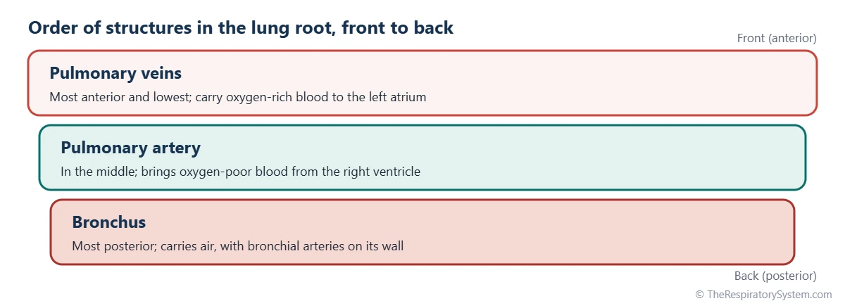
In both hila, the pulmonary veins lie in front and lowest, the pulmonary artery lies in the middle and the bronchus lies at the back. The main difference between the two sides is the position of the artery relative to the bronchus. In the left hilum, the pulmonary artery is the highest structure, lying above the bronchus. In the right hilum, the upper lobe bronchus is the highest structure, sitting above the pulmonary artery, which is why it is called the eparterial bronchus. Students often remember this as RALS: Right, Anterior; Left, Superior, describing where the artery lies relative to the bronchus.
| Structure | Right root | Left root | What it does |
|---|---|---|---|
| Bronchus | Often two: upper lobe (eparterial) bronchus and the main bronchus below it | One main bronchus, below the artery | Carries air into and out of the lung |
| Pulmonary artery | One, in front of the bronchus | One, the highest structure, arching over the bronchus | Brings oxygen-poor blood for gas exchange |
| Pulmonary veins | Two, superior and inferior, front and lowest | Two, superior and inferior, front and lowest | Return oxygen-rich blood to the left atrium |
| Bronchial arteries | Usually one | Usually two, from the thoracic aorta | Nourish the airway walls and lung tissue |
| Bronchial veins | Drain toward the azygos vein | Drain toward the hemiazygos system | Return part of the blood from the airway walls |
| Pulmonary nerve plexus | Vagus and sympathetic fibers | Vagus and sympathetic fibers | Controls airway muscle, glands and vessels |
| Lymph vessels | Present | Present | Drain lymph from the lung toward the mediastinum |
| Hilar lymph nodes | Bronchopulmonary nodes at the hilum | Bronchopulmonary nodes at the hilum | Filter lymph and trap microbes and particles |
| Source: Typical adult anatomy, our editors | |||
Hilum of Lung vs Root of Lung: What Is the Difference?
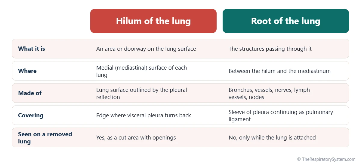
The difference between the hilum and the root of the lung is that the hilum is the place, an area on the lung surface, while the root is the content, the bundle of structures that passes through that area. The two terms are often used as if they meant the same thing, which is why this page covers both.
The root of the lung runs a short distance from the hilum to the mediastinum and is wrapped in a sleeve of pleura. At the hilum, the visceral pleura covering the lung folds back on itself and becomes continuous with the parietal pleura lining the mediastinum, enclosing the root like a cuff. Below the root, this sleeve hangs down as a double fold called the pulmonary ligament, which gives the vessels room to expand and lets the root move as the lung inflates. The pleural cavity page shows how the two pleural layers form one closed sac.
A useful way to keep the two apart: you can see the hilum on a lung removed from the body, as a cut area with vessel and airway openings, but the root only exists while the lung is still attached to the heart and trachea.
What Is the Function of the Hilum of the Lung?
The function of the hilum of the lung is to act as the single gateway through which air, blood, nerve signals and lymph travel between each lung and the rest of the body, while also anchoring the lung to the mediastinum. Without it, the lung could not connect to the airways and the heart.
- Air in and out: every breath passes through the main bronchus at the hilum on its pathway to the alveoli.
- Blood for gas exchange: the pulmonary artery and veins at the hilum form the link between the lung and the pulmonary circulation.
- Nourishment of the lung: the bronchial arteries carry oxygen-rich blood to the airway walls.
- Nerve control: nerves of the pulmonary plexus pass through the hilum to the airways and vessels.
- Immune filtering: lymph from the lung passes through hilar lymph nodes, which trap microbes and particles.
- Anchoring: the root holds each lung in position while the rest of its surface slides freely during breathing.
What Does the Hilum of the Lung Look Like on an X-Ray?
On a chest X-ray, each hilum appears as a branching, slightly denser shadow beside the heart, formed mainly by the pulmonary arteries and veins. The air-filled bronchi and normal-sized lymph nodes add little to the shadow.
In most people the left hilum sits slightly higher than the right, because the left pulmonary artery arches over the left main bronchus while the right pulmonary artery runs lower, in front of the right bronchus. Radiologists compare the size, shape, density and height of the two hila on every chest film. A hilum that looks larger or denser than expected can reflect enlarged lymph nodes or enlarged pulmonary arteries, and a hilum pulled up or down can point to shrinkage or collapse of part of a lung. A CT scan shows the separate structures of the root in much more detail.
Medical students also study the hilum on anatomy models and preserved specimens, where its arrangement of veins in front, artery in the middle and bronchus behind is easy to trace.
Common Conditions That Affect the Hilum of the Lung
Most conditions involving the hilum cause the hilar lymph nodes or the pulmonary vessels to enlarge, or involve a growth near the main bronchus. Our respiratory diseases section has more background, and anyone with symptoms should see a doctor.
- Hilar lymph node enlargement (hilar lymphadenopathy): swelling of the nodes at one or both hila, seen in infections such as tuberculosis, in sarcoidosis, in lymphoma and when cancer spreads to the nodes.
- Lung cancer: tumors starting in or near the main bronchi, often called central tumors, can make one hilum look larger.
- Pulmonary hypertension: high pressure in the pulmonary arteries can widen them, enlarging both hilar shadows.
- Pulmonary embolism: a large clot may lodge in a pulmonary artery near the hilum.
- Lung collapse: loss of air in a lobe, for example from a blocked bronchus, can pull the hilum up or down on an X-ray.
Key Facts
- The hilum is the area on the medial surface of each lung where the root structures enter and leave.
- The root of the lung contains a main bronchus, one pulmonary artery, two pulmonary veins, bronchial vessels, nerves, lymph vessels and hilar lymph nodes.
- The root of each lung lies roughly opposite the fifth to seventh thoracic vertebrae.
- In both hila the pulmonary veins are the most anterior and lowest structures, and the bronchus is the most posterior.
- In the left hilum the pulmonary artery is the highest structure; in the right hilum the upper lobe bronchus lies above the artery.
- The pleura forms a sleeve around the root and hangs below it as the pulmonary ligament.
- On a chest X-ray the left hilum usually sits slightly higher than the right.
Quick Quiz: Test What You Know About the Hilum of the Lung
6 questions. Tap an answer to check it.
On which surface of the lung is the hilum found?
The hilum is on the medial surface of each lung, the side facing the mediastinum and heart.
Which structure is the most anterior in the root of both lungs?
In both roots the pulmonary veins lie in front and lowest, the artery in the middle and the bronchus at the back.
In the left hilum, which structure is usually the highest?
On the left, the pulmonary artery arches over the main bronchus and is the highest structure in the hilum.
What is the difference between the hilum and the root of the lung?
The hilum is the doorway on the lung surface, and the root is the bundle of bronchus, vessels, nerves and lymphatics running through it.
What fold of pleura hangs down below the root of the lung?
The pleural sleeve around the root continues downward as a double fold called the pulmonary ligament.
Which vessel arches over the root of the right lung?
The azygos vein arches over the right lung root to join the superior vena cava; the aortic arch crosses over the left root.
Frequently Asked Questions
What is the right lung hilum?
The right lung hilum is the entry point on the medial surface of the right lung. Through it pass the right main bronchus, usually with its upper lobe branch already separate, the right pulmonary artery, two right pulmonary veins, bronchial vessels, nerves and lymphatics. The superior vena cava lies in front of it, and the azygos vein arches over its top to join the superior vena cava.
What are the hilar lymph nodes?
Hilar lymph nodes, also called bronchopulmonary nodes, are small bean-shaped nodes clustered around the main bronchi and vessels at each hilum. They receive lymph from the lung tissue and filter out microbes, dust and other particles before the lymph moves on to the tracheobronchial nodes near the carina. They are often checked on imaging because many lung conditions make them swell.
Is the hilum part of the mediastinum?
The hilum belongs to the lung, since it is an area on the lung's own surface. The root of the lung, however, runs from the hilum into the mediastinum, where its bronchus joins the trachea and its vessels join the heart. So the hilum marks the boundary between the lung and the mediastinum, and the root bridges the two.
Why is the left hilum higher than the right?
The left hilum is usually higher on a chest X-ray because of the course of the left pulmonary artery. It arches over the left main bronchus, so the main vascular shadow on the left sits higher. On the right, the pulmonary artery runs lower, in front of the bronchus. A clear change from this usual pattern can be a sign that part of a lung has shrunk or collapsed.
Do the pulmonary veins always number two on each side?
Usually, yes. Most people have a superior and an inferior pulmonary vein on each side, four in total, entering the left atrium. Variations are common, however. On the right, a separate middle lobe vein is sometimes present, and on the left the two veins sometimes join into one short trunk before reaching the heart. These variants are normal and usually cause no problems.
Can you see the hilum of the lung on an anatomy model?
Yes. On most anatomy models the lungs can be removed from the chest, and the hilum is shown on the medial surface of each lung as a cut area with the openings of the bronchus, pulmonary artery and pulmonary veins. Models are often colored with arteries in blue and veins in red, because in the lungs the arteries carry oxygen-poor blood and the veins carry oxygen-rich blood.
What is the pulmonary ligament?
The pulmonary ligament is a double fold of pleura that hangs down from the root of each lung toward the diaphragm. It forms where the pleural sleeve around the root extends below it. It contains no important vessels, but it gives the pulmonary veins room to expand and lets the root of the lung move up and down slightly as the lung fills and empties.
Does the hilum move during breathing?
Yes, a little. As the diaphragm contracts and the lungs expand, the root of the lung moves slightly downward, and it rises again on breathing out. The movement is small compared with that of the lung base, because the root is held by its connections to the heart and trachea, and the pulmonary ligament below gives it room to shift.
Keep Exploring


