At a Glance
The phrenic nerve is a mixed motor and sensory nerve that arises from the C3, C4 and C5 spinal nerve roots, mainly C4, in the cervical plexus of the neck. There is one on each side: each runs down over the anterior scalene muscle, through the mediastinum in front of the root of the lung, and is the only motor nerve to its half of the diaphragm.
- Origin
- Cervical plexus in the neck, spinal nerve roots C3, C4 and C5 (mainly C4)
- Course
- Down the front of the anterior scalene muscle, into the chest, through the mediastinum to the diaphragm
- Type
- Mixed nerve: motor, sensory and a few sympathetic fibers
- Function
- Only motor supply of the diaphragm; feeling from the central diaphragm, pericardium and mediastinal pleura
- Number
- Two, a right and a left phrenic nerve, each serving its own half of the diaphragm
- Part of
- Cervical plexus; carries breathing commands from the brainstem to the diaphragm
On this page
- What Is the Phrenic Nerve?
- Phrenic Nerve Anatomy: Roots and Fibers
- Phrenic Nerve Location and Course: From the Neck to the Diaphragm
- How Do the Right and Left Phrenic Nerves Differ?
- What Is the Phrenic Nerve Function?
- Which Structures Work With the Phrenic Nerve?
- Common Conditions That Affect the Phrenic Nerve
- Key Facts
- Quiz
- FAQ
What Is the Phrenic Nerve?
The phrenic nerve is a paired nerve from the neck that carries the signals that make the diaphragm contract, and it is the only motor nerve that muscle has. Because the diaphragm does most of the work of quiet breathing, the phrenic nerves are among the most important nerves for staying alive. The name comes from the Greek word phren, an old term for the diaphragm.
Medical students remember its roots with a short rhyme: "C3, 4 and 5 keep the diaphragm alive." The nerve belongs to the cervical plexus, a network of nerves formed by the upper neck spinal nerves, yet it travels all the way down through the thoracic cavity to reach a muscle that sits at the bottom of the chest.
Explained simply
Think of the phrenic nerve as a long cable running from the neck to the floor of the chest. Your brainstem sends a pulse down the cable with every breath, the diaphragm tightens and pulls down, and air rushes into your lungs. When the pulse stops, the muscle relaxes and you breathe out.
Phrenic Nerve Anatomy: Roots and Fibers
Each phrenic nerve forms in the neck from the front branches (ventral rami) of the third, fourth and fifth cervical spinal nerves, with the fourth giving the largest share. The cell bodies of its motor fibers sit in a column of the spinal cord called the phrenic motor nucleus, which spans the C3 to C5 segments.
What kinds of fibers does it carry?
- Motor fibers to the muscle of its own half of the diaphragm, including the crus on that side.
- Sensory fibers that carry pain and touch from the central part of the diaphragm, the mediastinal pleura, the fibrous and parietal pericardium, and the membranes just above and below the central diaphragm.
- A few sympathetic fibers, picked up from the cervical sympathetic ganglia, that travel along with it.
The accessory phrenic nerve
In some people, a small accessory phrenic nerve adds extra fibers, usually from C5 through the nerve to the subclavius. It runs a separate short course and joins the main phrenic nerve near the first rib or in the upper chest. This normal variation matters to surgeons, because cutting the main trunk may not fully stop the diaphragm on that side.
Phrenic Nerve Location and Course: From the Neck to the Diaphragm
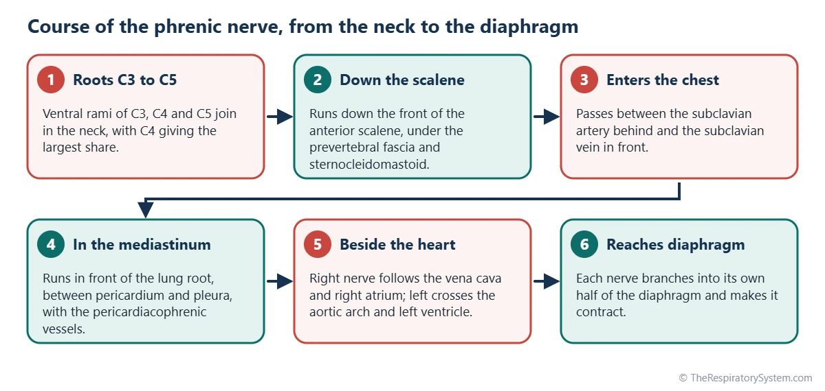
The phrenic nerve runs from the side of the neck, down the front of the anterior scalene muscle, into the chest between the subclavian artery and vein, and then through the mediastinum in front of the root of the lung to the diaphragm. Its path can be followed in three parts.
Phrenic nerve anatomy in the neck
The nerve forms at the upper outer edge of the anterior scalene muscle and slides down and inward across its front surface, so it is the only nerve that runs straight down on that muscle. It lies under the prevertebral layer of deep fascia, behind the sternocleidomastoid muscle and the internal jugular vein, and the transverse cervical and suprascapular arteries cross in front of it.
Entering the chest
At the root of the neck the nerve passes in front of the subclavian artery and behind the subclavian vein, then crosses the internal thoracic artery as it enters the thorax. From here the right and left nerves follow different routes because the large vessels and the heart are not symmetrical.
In the mediastinum
Inside the mediastinum, both nerves run in front of the root of the lung, squeezed between the fibrous pericardium around the heart and the mediastinal layer of the pleura. A small artery and vein, the pericardiacophrenic vessels, travel with each nerve. At the diaphragm, each nerve divides into several branches that spread through its half of the muscle, mostly from the underside.
| Segment | Where the nerve runs | Main neighbors |
|---|---|---|
| Origin | Ventral rami of C3, C4 and C5 join at the upper outer edge of the anterior scalene | Cervical plexus, upper neck spinal nerves |
| Neck | Straight down the front of the anterior scalene, from its outer to its inner edge | Prevertebral fascia, sternocleidomastoid, internal jugular vein |
| Root of the neck | Between the subclavian artery behind and the subclavian vein in front | Internal thoracic artery, first rib |
| Upper chest, right | Along the right brachiocephalic vein and superior vena cava | Right mediastinal pleura |
| Upper chest, left | Between the left common carotid and left subclavian arteries, then over the aortic arch | Left vagus nerve, which lies behind it |
| Middle mediastinum | In front of the lung root, between fibrous pericardium and mediastinal pleura | Pericardiacophrenic artery and vein |
| Beside the heart | Right over the right atrium; left over the left ventricle | Pericardium, which it supplies with sensory fibers |
| Diaphragm | Right at or near the caval opening; left through the muscle of the left dome | Branches spread through its own half of the diaphragm |
| Source: Standard adult anatomy, our editors | ||
How Do the Right and Left Phrenic Nerves Differ?
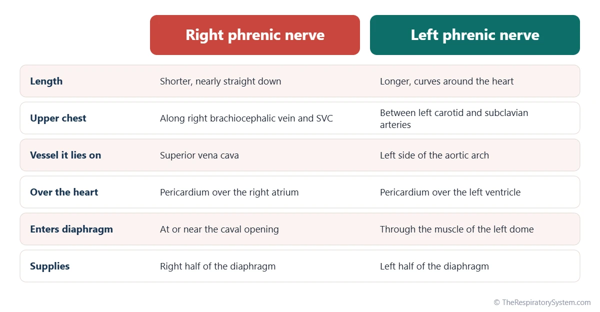
The right phrenic nerve is shorter and runs almost straight down along the great veins, while the left phrenic nerve is longer because it curves over the aortic arch and the bulge of the left side of the heart. Both still end in the same way, by supplying their own half of the diaphragm.
Phrenic nerve anatomy and the heart
The right nerve lies on the right brachiocephalic vein and the superior vena cava, then on the pericardium over the right atrium, and finally beside the inferior vena cava. It reaches the diaphragm at or near the caval opening, described on our page about the openings of the diaphragm. The left nerve descends between the left common carotid and left subclavian arteries, crosses the left side of the aortic arch in front of the left vagus nerve, and runs over the pericardium of the left ventricle before piercing the muscular left dome.
Neither nerve supplies the heart muscle itself. They only pass over the pericardium, the sac around the heart, and carry pain sensation from it.
What Is the Phrenic Nerve Function?
The main function of the phrenic nerve is to carry the motor signals that make the diaphragm contract during inhalation, and its second function is to carry sensation from the central diaphragm, pericardium and nearby pleura. Without it, the diaphragm on that side cannot contract at all.
Motor function in breathing
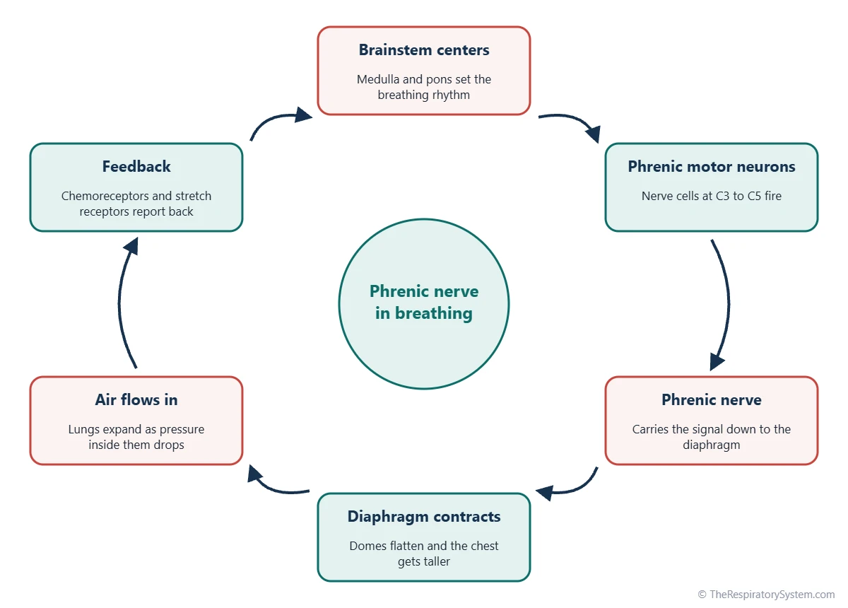
Groups of nerve cells in the brainstem set the breathing rhythm and send bursts of signals to the phrenic motor neurons in the spinal cord, as described on our control of breathing page. Each burst travels down the phrenic nerves, the diaphragm contracts and flattens, and the chest gets taller, which pulls air in during inhalation. Between bursts the muscle relaxes and quiet exhalation happens passively. The same pathway also produces deeper breaths during exercise, plus sudden diaphragm actions such as the spasm of a hiccup and the build-up of pressure before a cough or sneeze.
Sensory function and referred pain
Because the phrenic nerve enters the spinal cord at the C3 to C5 level, the brain can mistake irritation of the central diaphragm for pain in the skin of the same segments, especially the top of the shoulder. This referred shoulder tip pain is a classic clue that something is irritating the diaphragm from above or below. The outer rim of the diaphragm is different: its feeling travels in the lower intercostal nerves instead.
Common Conditions That Affect the Phrenic Nerve
The phrenic nerve can be stretched, compressed, inflamed or cut anywhere along its long path, and the main result is weakness or paralysis of the diaphragm on the affected side. Our respiratory diseases section covers lung conditions more broadly, and anyone with unexplained breathlessness should see a doctor.
- Phrenic nerve injury: may follow surgery on the heart, chest or neck, a neck injury, or a nerve block given near the neck, and is often temporary.
- Compression by a mass: a tumor or enlarged lymph nodes in the chest, for example from lung cancer, can press on the nerve.
- Nerve inflammation: some inflammatory nerve disorders and viral illnesses can weaken one or both phrenic nerves.
- High spinal cord injury: damage at or above the C3 to C5 segments can cut the brainstem off from the phrenic motor neurons.
When one side stops working, a chest X-ray often shows that half of the diaphragm sitting higher than normal, and a fluoroscopy "sniff test" can show it moving the wrong way. Many people with one paralyzed side have few symptoms at rest, while loss of both sides causes marked breathlessness, especially when lying flat. Long-lasting hiccups and diaphragmatic pleurisy with shoulder tip pain also involve this nerve.
Key Facts
- The phrenic nerve arises from the C3, C4 and C5 spinal nerve roots, mainly C4, and is the only motor nerve of the diaphragm.
- There are two phrenic nerves, and each one controls only its own half of the diaphragm.
- In the neck, the phrenic nerve runs straight down the front of the anterior scalene muscle.
- Both phrenic nerves pass in front of the root of the lung, while the vagus nerves pass behind it.
- The left phrenic nerve is longer than the right because it curves around the left side of the heart.
- Irritation of the central diaphragm can be felt as shoulder tip pain because both share the C3 to C5 spinal segments.
- Damage to one phrenic nerve often shows on a chest X-ray as a raised half of the diaphragm, a sign explained alongside the mechanics of breathing.
Quick Quiz: Test What You Know About the Phrenic Nerve
6 questions. Tap an answer to check it.
Which spinal nerve roots form the phrenic nerve?
The phrenic nerve comes from C3, C4 and C5, with C4 giving the largest share: "C3, 4 and 5 keep the diaphragm alive."
On which muscle does the phrenic nerve run down in the neck?
It runs straight down the front surface of the anterior scalene muscle, under the prevertebral fascia.
Where do the phrenic nerves pass in relation to the root of the lung?
Both phrenic nerves run in front of the lung root; the vagus nerves run behind it.
What is the main motor function of the phrenic nerve?
The phrenic nerve is the only motor nerve of the diaphragm, the main muscle of inhalation.
Why is the left phrenic nerve longer than the right?
The left nerve crosses the aortic arch and runs over the left ventricle, which lengthens its path.
Pain from irritation of the central diaphragm is often felt where?
Phrenic sensory fibers enter the spinal cord at C3 to C5, the same segments that supply the skin over the shoulder.
Frequently Asked Questions
What does the word phrenic mean?
Phrenic comes from the Greek word phren, which ancient writers used for the diaphragm and also for the mind, because they linked feelings and thought with the middle of the body. In anatomy, phrenic simply means related to the diaphragm, so the phrenic nerve is the diaphragm nerve, and the phrenic arteries and veins are the vessels of the diaphragm.
Can you breathe with one paralyzed phrenic nerve?
Yes. If one phrenic nerve stops working, the other half of the diaphragm and the other breathing muscles can usually keep breathing going, and many people notice little at rest. Shortness of breath tends to show during exercise or when lying flat. If both phrenic nerves fail, breathing becomes much harder, even at rest.
Can you see the phrenic nerve on a CT scan?
Usually not directly. The phrenic nerve is only a few millimeters thick, so on CT and in chest radiology its course is traced from landmarks such as the anterior scalene muscle and the pericardiacophrenic vessels beside it. High-resolution ultrasound can often show the nerve in the neck, and its effect is judged by watching how the diaphragm moves.
Does the phrenic nerve supply the heart?
No. The phrenic nerve runs over the heart but does not supply the heart muscle. It lies on the pericardium, the protective sac around the heart, and carries pain sensation from that sac. The heartbeat itself is set by the heart's own pacemaker cells and adjusted by the vagus nerve and sympathetic nerves, not by the phrenic nerve.
Is the phrenic nerve under voluntary control?
Both. Most of the time the phrenic nerve is driven automatically by breathing centers in the brainstem, so breathing continues during sleep. You can also take over for a while, for example to take a deep breath, talk, sing or hold your breath, because signals from the brain's motor cortex reach the same phrenic motor neurons in the spinal cord.
How is the phrenic nerve involved in hiccups?
A hiccup is a sudden, involuntary contraction of the diaphragm, and the phrenic nerve carries the signal that makes the muscle jerk. Right after the contraction starts, the vocal cords snap shut and make the hic sound. Most hiccup bouts are short. Very long-lasting hiccups can occasionally point to irritation of the phrenic or vagus nerve somewhere along their path.
What is the difference between the phrenic nerve and the vagus nerve?
The phrenic nerve is a spinal nerve from C3 to C5 that mainly moves the diaphragm. The vagus nerve is a cranial nerve from the brainstem that controls the larynx, slows the heart and serves the airways and gut. In the chest, the phrenic nerves pass in front of the lung roots, while the vagus nerves pass behind them.
Why does a problem under the diaphragm cause shoulder pain?
The central diaphragm sends its pain signals through the phrenic nerve into the C3 to C5 spinal segments. The same segments receive touch and pain from the skin over the top of the shoulder. The brain cannot easily tell the two sources apart, so irritation of the diaphragm, from above or below, is often felt as an ache at the shoulder tip.
Keep Exploring


