At a Glance
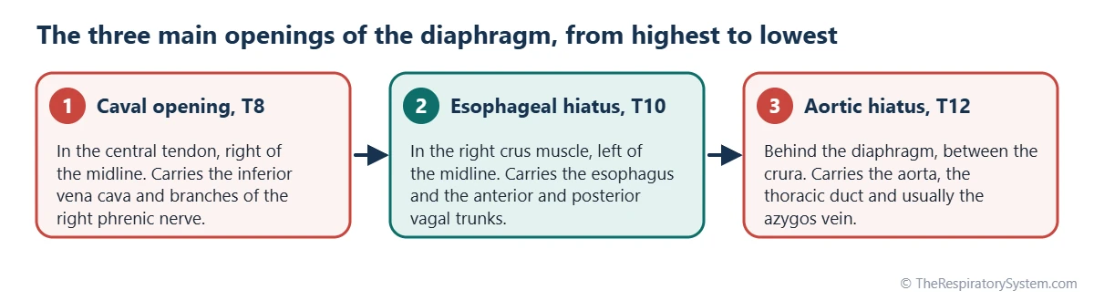
The diaphragm has three main openings. The caval opening, at the level of the T8 vertebra, carries the inferior vena cava and branches of the right phrenic nerve; the esophageal hiatus, at T10, carries the esophagus and the vagal trunks; and the aortic hiatus, at T12, carries the aorta, the thoracic duct and usually the azygos vein.
- Main openings
- Caval opening, esophageal hiatus and aortic hiatus
- Vertebral levels
- About T8, T10 and T12, from front and highest to back and lowest
- Caval opening
- Inferior vena cava and branches of the right phrenic nerve, in the central tendon
- Esophageal hiatus
- Esophagus, anterior and posterior vagal trunks, esophageal branches of the left gastric vessels
- Aortic hiatus
- Aorta, thoracic duct and usually the azygos vein, behind the median arcuate ligament
- Connects
- Thoracic cavity above with the abdominal cavity below
On this page
- What Are the Openings of the Diaphragm?
- Diaphragm Openings Levels: Where Is Each Opening?
- Diaphragm Openings and Contents: What Passes Through Each?
- What Is the Mnemonic for the Diaphragm Openings and Contents?
- How Do the Openings Behave When You Breathe?
- What Are the Smaller Openings of the Diaphragm?
- Common Conditions That Affect the Diaphragm Openings
- Key Facts
- Quiz
- FAQ
What Are the Openings of the Diaphragm?
The openings of the diaphragm are passages through or behind the muscle that let large blood vessels, the esophagus, nerves and lymph vessels travel between the chest and the abdomen. The diaphragm forms a complete wall between the thoracic cavity and the abdominal cavity, so anything that must cross it needs its own doorway.
Anatomists divide them into three main openings, each linked to a typical vertebral level, and a group of smaller passages for nerves and small vessels. The three main openings carry the body's largest vein, its largest artery and the food tube, so their position and shape matter every time you breathe, swallow or stand up.
Explained simply
Imagine the diaphragm as a trampoline stretched across the bottom of the rib cage. To get from the chest to the belly, the big pipes of the body have to pass through it, so the trampoline has three main holes, one for a vein, one for the food pipe and one for an artery, plus a few small slits for nerves.
Diaphragm Openings Levels: Where Is Each Opening?
The three main openings lie at about the levels of the T8, T10 and T12 vertebrae, and they step down and backward as they go: the caval opening is highest and most anterior, the esophageal hiatus is in the middle, and the aortic hiatus is lowest and furthest back. This order follows the shape of the diaphragm, whose tendon sits high in the front while its crura reach far down the spine at the back.
- Caval opening (vena caval foramen), T8: in the central tendon, just to the right of the midline.
- Esophageal hiatus, T10: in the muscular part, formed by a sling of fibers from the right crus, just to the left of the midline.
- Aortic hiatus, T12: in the midline, behind the diaphragm, between the right and left crura and under the median arcuate ligament.
These levels are typical values. In real people they can sit about one vertebra higher or lower, and they shift slightly with posture and with each breath, because the whole diaphragm moves up and down during inhalation and exhalation.
Diaphragm Openings and Contents: What Passes Through Each?
The caval opening carries the inferior vena cava, the esophageal hiatus carries the esophagus with the vagal trunks, and the aortic hiatus carries the aorta, the thoracic duct and usually the azygos vein. Each opening also lets small lymph vessels through.
Caval opening contents
The inferior vena cava, the large vein that returns blood from the legs and abdomen to the heart, passes through the caval opening and almost immediately enters the right atrium. Branches of the right phrenic nerve pass through the same opening or the tendon right beside it to reach the underside of the diaphragm. Its edges are firmly fused with the wall of the vein.
Esophageal hiatus contents
The esophagus passes from the posterior mediastinum into the abdomen, where it joins the stomach only a short distance below the diaphragm. It travels with the anterior and posterior vagal trunks, which are formed mainly from the left and right vagus nerves, and with the esophageal branches of the left gastric artery and vein. The trachea vs esophagus page shows how the food tube lies behind the windpipe higher in the chest.
Aortic hiatus contents
The descending aorta passes behind the diaphragm and becomes the abdominal aorta. The thoracic duct, the main lymph vessel of the body, runs upward through the same gap from its start in the abdomen, and the azygos vein usually passes with them, carrying blood up toward the superior vena cava. Because the aortic hiatus lies behind the muscle rather than through it, it is more a gap than a true hole.
| Opening | Typical level | Where it is | What passes through |
|---|---|---|---|
| Caval opening (vena caval foramen) | About T8 | Central tendon, just right of the midline | Inferior vena cava, branches of the right phrenic nerve, lymph vessels |
| Esophageal hiatus | About T10 | Muscular part, sling of right crus fibers, just left of the midline | Esophagus, anterior and posterior vagal trunks, esophageal branches of left gastric vessels, lymph vessels |
| Aortic hiatus | About T12 | Behind the diaphragm, between the crura, under the median arcuate ligament | Aorta, thoracic duct, usually the azygos vein |
| Openings in the crura | Close to the aortic hiatus | Through the right and left crura | Greater and lesser splanchnic nerves; hemiazygos vein through the left crus |
| Behind the medial arcuate ligament | Upper lumbar region | Over the psoas major muscle | Sympathetic trunk |
| Behind the lateral arcuate ligament | Upper lumbar region | Over the quadratus lumborum muscle | Subcostal nerve and vessels |
| Left dome | Moves with breathing | Muscular part of the left dome | Left phrenic nerve |
| Sternocostal gap | Front of the diaphragm | Between the sternal and costal parts | Superior epigastric vessels, lymph vessels |
| Source: Standard adult anatomy; levels are typical and vary by about one vertebra, our editors | |||
What Is the Mnemonic for the Diaphragm Openings and Contents?
The most popular mnemonic for the diaphragm openings and their levels is "I 8 10 Eggs At 12": the Inferior vena cava passes at T8, the Esophagus at T10 and the Aorta at T12. Read aloud, it sounds like "I ate ten eggs at twelve."
Other memory aids
- Count the letters: "vena cava" has 8 letters (T8), the British spelling "oesophagus" has 10 (T10) and "aortic hiatus" has 12 (T12).
- Red, white and blue: the aortic hiatus carries the aorta (red blood), the thoracic duct (white, milky lymph) and the azygos vein (blue, oxygen-poor blood).
- Vagus at ten: the vagus nerve is cranial nerve X, the Roman numeral for 10, and its trunks pass with the esophagus at T10.
- Front to back, top to bottom: as the numbers rise, the openings move further back and lower down.
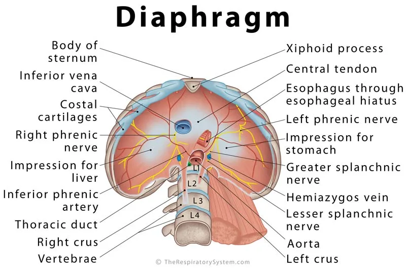
These aids help with exams, but the structures are easier to remember once you can picture them on the labeled diagram below, which shows the diaphragm from underneath.
How Do the Openings Behave When You Breathe?
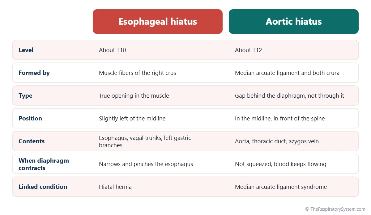
When the diaphragm contracts during inhalation, the caval opening is pulled wider, the esophageal hiatus is squeezed narrower, and the aortic hiatus is left unaffected. Each response suits the structure passing through.
Caval opening: helping blood return
Because the caval opening lies in the central tendon and its edges are fused to the vein, contraction of the muscle around the tendon tugs the opening and the vein wall open. At the same time, pressure in the chest falls during inhalation, so blood is drawn up the inferior vena cava toward the heart, an effect explained further on our mechanics of breathing page.
Esophageal hiatus function
The fibers of the right crus loop around the esophagus like a sling, and when they contract they pinch it closed from the outside. This works together with the muscle of the lower esophagus as part of the barrier that keeps stomach contents from flowing back up, and it is strongest during inhalation, exactly when pressure in the abdomen rises and pressure in the chest falls.
Aortic hiatus function
The aorta passes behind the diaphragm, under the arching median arcuate ligament, so the muscle never squeezes it. Blood can therefore flow to the abdomen and legs at full force no matter how hard you are breathing, whether during sleep or during exercise.
What Are the Smaller Openings of the Diaphragm?
Besides the three main openings, several nerves and small vessels pierce the diaphragm or slip behind its edges through smaller gaps. They are easy to miss but complete the list of diaphragm openings and structures.
- Through the crura: the greater and lesser splanchnic nerves pierce each crus, and the hemiazygos vein usually pierces the left crus.
- Behind the medial arcuate ligament: the sympathetic trunk passes into the abdomen over the psoas major muscle.
- Behind the lateral arcuate ligament: the subcostal nerve and vessels pass over the quadratus lumborum muscle.
- Through the left dome: the left phrenic nerve pierces the muscle on its own, rather than using one of the main openings.
- Between the sternal and costal parts: the superior epigastric vessels, the continuation of the internal thoracic vessels, pass down into the front of the abdominal wall.
- Between the costal slips: the lower intercostal nerves and vessels run from the rib cage into the abdominal wall.
These passages also mark weak points, which is why some hernias appear in the same places, as the next section shows.
Common Conditions That Affect the Diaphragm Openings
The most common problem at the diaphragm openings is a hiatal hernia, in which part of the stomach pushes up through the esophageal hiatus into the chest; other conditions involve weak spots in the diaphragm or compression near the aortic hiatus. Our respiratory diseases section covers conditions of the breathing organs, and anyone with symptoms should see a doctor.
- Sliding hiatal hernia: the most common type, in which the junction of the esophagus and stomach slides up through a widened hiatus. It is often linked with acid reflux.
- Paraesophageal hiatal hernia: part of the stomach rolls up beside the esophagus while the junction stays in place.
- Congenital diaphragmatic hernia: a birth defect in which abdominal organs move into the chest through a gap in the diaphragm, most often at the back on the left side (Bochdalek type), less often at the front near the sternum (Morgagni type). It can crowd the developing lungs.
- Median arcuate ligament syndrome: a rare condition in which the ligament that arches over the aortic hiatus presses on the celiac artery just below it.
Because the diaphragm is the main muscle of breathing, large hernias can also reduce how far the lungs can expand, a link explored on our muscles of respiration page.
Key Facts
- The diaphragm has three main openings, at about the T8, T10 and T12 vertebral levels.
- The caval opening at T8 lies in the central tendon and carries the inferior vena cava.
- The esophageal hiatus at T10 carries the esophagus and the anterior and posterior vagal trunks.
- The aortic hiatus at T12 carries the aorta, the thoracic duct and usually the azygos vein.
- "I 8 10 Eggs At 12" is a popular mnemonic for the levels of the inferior vena cava, esophagus and aorta.
- When the diaphragm contracts, the caval opening widens, the esophageal hiatus narrows and the aortic hiatus is not squeezed.
- The left phrenic nerve pierces the left dome on its own, while the splanchnic nerves pass through the crura.
Quick Quiz: Test What You Know About the Openings of the Diaphragm
6 questions. Tap an answer to check it.
At about which vertebral level is the caval opening?
The caval opening, which carries the inferior vena cava, lies at about T8 in the central tendon.
Which structure passes through the esophageal hiatus together with the esophagus?
The anterior and posterior vagal trunks pass with the esophagus at about T10.
Which three structures pass through the aortic hiatus?
The aortic hiatus at T12 carries the aorta, the thoracic duct and usually the azygos vein, remembered as red, white and blue.
Which opening lies in the central tendon of the diaphragm?
The caval opening sits in the central tendon, just to the right of the midline.
What happens to the esophageal hiatus when the diaphragm contracts?
Fibers of the right crus loop around the esophagus and squeeze it when they contract, helping keep stomach contents down.
What is the most common condition involving the diaphragm openings?
In a hiatal hernia, part of the stomach pushes up through the esophageal hiatus into the chest.
Frequently Asked Questions
How many openings does the diaphragm have?
The diaphragm has three main openings: the caval opening, the esophageal hiatus and the aortic hiatus. In addition, several smaller passages let nerves and small vessels through, including gaps in the crura for the splanchnic nerves, spaces behind the arcuate ligaments, a gap at the front for the superior epigastric vessels and the spot where the left phrenic nerve pierces the muscle.
What is the difference between a hiatus and a foramen?
Both words mean an opening, but they are used a little differently. A foramen is usually a neat, well-defined hole, often with firm edges, like the caval opening in the tendon. A hiatus is a gap or cleft, often between muscle fibers or behind a ligament, like the esophageal and aortic hiatuses. In everyday anatomy the terms are partly interchangeable.
Does the vagus nerve pass through the diaphragm?
Yes. Below the lungs, the right and left vagus nerves form a network around the esophagus and regroup into the anterior and posterior vagal trunks. These trunks pass through the esophageal hiatus with the esophagus at about T10. The anterior trunk comes mainly from the left vagus and the posterior trunk mainly from the right vagus.
Why does the inferior vena cava pass through the tendon instead of the muscle?
Passing through the central tendon protects the inferior vena cava from being squeezed. If the vein crossed the muscle, every contraction during inhalation could narrow it and slow the return of blood to the heart. Instead, contraction pulls the tendon and widens the opening, and the vein, whose wall is fused to the edges, is held open at exactly the moment chest pressure falls.
Where does the thoracic duct pass through the diaphragm?
The thoracic duct passes through the aortic hiatus at about T12, between the aorta and the azygos vein. It begins in the upper abdomen as a small sac that collects lymph from the legs and abdomen, then runs up through the chest behind the esophagus and drains into the large veins at the base of the left side of the neck.
Are the diaphragm opening levels the same in everyone?
No. T8, T10 and T12 are typical textbook values. In real people each opening can sit about one vertebra higher or lower, and the diaphragm itself moves several centimeters between a full breath in and a full breath out. Body shape, posture and age also shift the levels, so on scans radiologists identify the openings by their contents rather than by vertebra alone.
What is a hiatal hernia?
A hiatal hernia is a condition in which part of the stomach pushes up through the esophageal hiatus into the chest. In the common sliding type, the junction between the esophagus and stomach moves up; in the paraesophageal type, part of the stomach bulges up beside the esophagus. Many small hiatal hernias cause no symptoms, while others are linked with acid reflux.
What is the median arcuate ligament?
The median arcuate ligament is a fibrous arch that joins the right and left crura of the diaphragm in front of the aorta, at about the T12 level. It forms the front edge of the aortic hiatus, so the aorta passes under it rather than through muscle. The celiac artery, which supplies the stomach, liver and spleen, branches off the aorta just below it.
Keep Exploring


