At a Glance
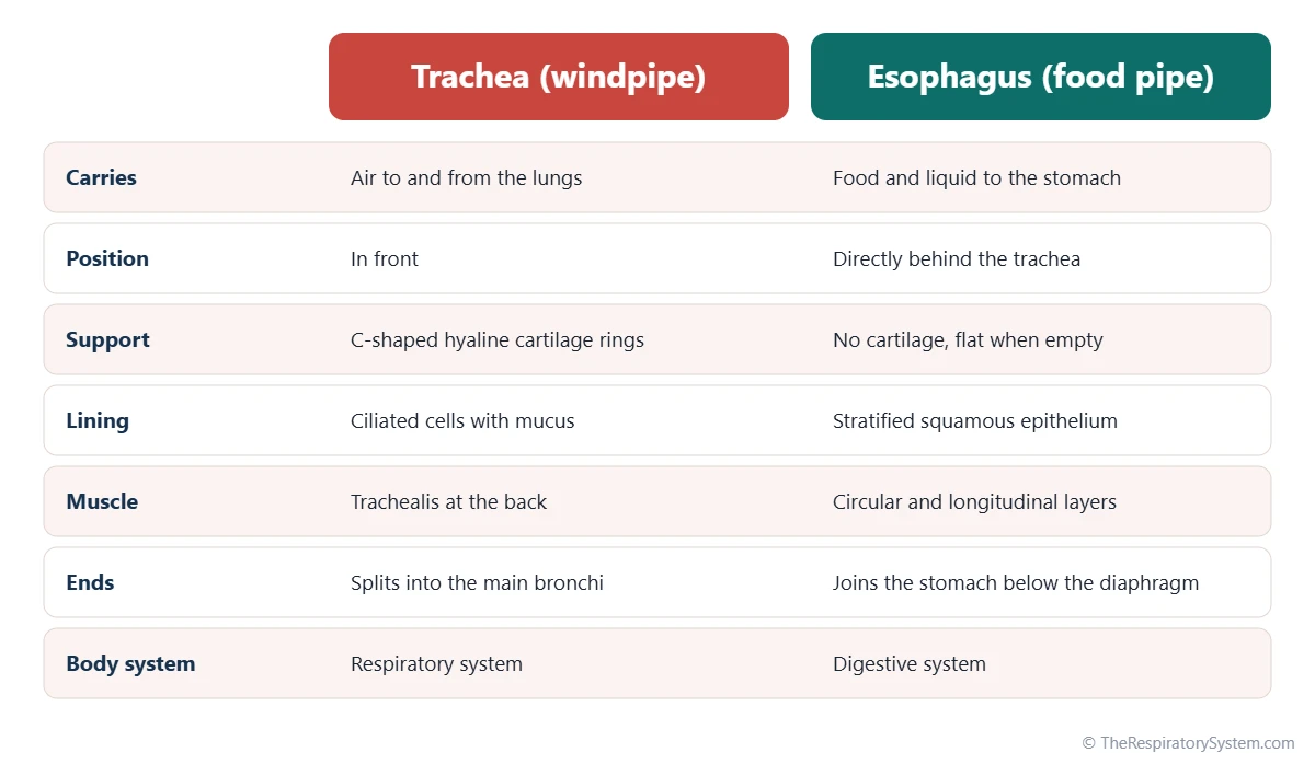
The trachea (windpipe) is an air tube about 10 to 12 cm long, kept open by 16 to 20 C-shaped rings of hyaline cartilage and lined by ciliated epithelium; it ends by splitting into the two main bronchi. The esophagus (food pipe) is a muscular tube about 25 cm long that lies directly behind the trachea, stays flat when empty, is lined by stratified squamous epithelium and moves food to the stomach by peristalsis.
- Trachea
- Windpipe: air from the larynx to the bronchi, about 10 to 12 cm long
- Esophagus
- Food pipe: food and liquid from the pharynx to the stomach, about 25 cm long
- Position
- Trachea in front, esophagus directly behind it, both starting at about the C6 vertebra
- Wall support
- Trachea: C-shaped cartilage rings. Esophagus: muscle only, no cartilage
- Lining
- Trachea: ciliated respiratory epithelium. Esophagus: stratified squamous epithelium
- Body system
- Trachea: respiratory system. Esophagus: digestive system
On this page
- What Is the Difference Between the Trachea and Esophagus?
- Where Are the Trachea and Esophagus Located?
- Trachea vs Esophagus Anatomy: How Are Their Walls Built?
- What Do the Trachea and Esophagus Do?
- What Do the Trachea and Esophagus Have in Common?
- How Do the Trachea and Esophagus Work Together When You Swallow?
- Pharynx vs Larynx vs Trachea vs Esophagus: How Do They Connect?
- Common Conditions That Affect the Trachea and Esophagus
- Key Facts
- Quiz
- FAQ
What Is the Difference Between the Trachea and Esophagus?
The main difference between the trachea and esophagus is what they carry: the trachea carries air to and from the lungs, and the esophagus carries food and drink to the stomach. Everything else about their build follows from that job.
An air tube must stay open all the time, even while you breathe in and the pressure inside it drops, so the trachea is stiffened by rings of cartilage. A food tube only needs to open when something passes through it, so the esophagus is a soft, collapsible muscle tube that squeezes each mouthful downward in a wave. The trachea is lined by a moist, ciliated layer that traps and clears dust, while the esophagus is lined by a tough, many-layered surface that resists scraping by food.
Windpipe vs food pipe in one line
The windpipe is the stiff tube in front that always holds air; the food pipe is the soft tube behind it that opens only when you swallow.
Explained simply
Imagine two hoses running down your neck. The front one is like a vacuum cleaner hose, ribbed so it never squashes flat, and it carries air. The back one is like an empty sock that only opens when food is pushed through it, and its muscles squeeze the food down to your stomach.
| Feature | Trachea (windpipe) | Esophagus (food pipe) |
|---|---|---|
| Carries | Air | Food and liquid |
| Body system | Respiratory system | Digestive system |
| Starts | Below the larynx, at about C6 | Below the laryngopharynx, at about C6 |
| Ends | Divides into the main bronchi at about T4 to T5 | Joins the stomach just below the diaphragm |
| Position | In front | Behind the trachea, in front of the spine |
| Typical adult length | About 10 to 12 cm | About 25 cm |
| Support | 16 to 20 C-shaped hyaline cartilage rings | No cartilage, collapses flat when empty |
| Lining | Pseudostratified ciliated columnar epithelium | Non-keratinized stratified squamous epithelium |
| Muscle | Trachealis smooth muscle at the back | Two layers, skeletal above and smooth below |
| How contents move | Air flow driven by breathing | Peristalsis, helped by gravity |
| Openings guarded by | Epiglottis and vocal folds | Upper and lower esophageal sphincters |
| Passes the diaphragm | No, ends in the chest | Yes, through the esophageal hiatus at about T10 |
| Source: Typical adult values, our editors | ||
Where Are the Trachea and Esophagus Located?
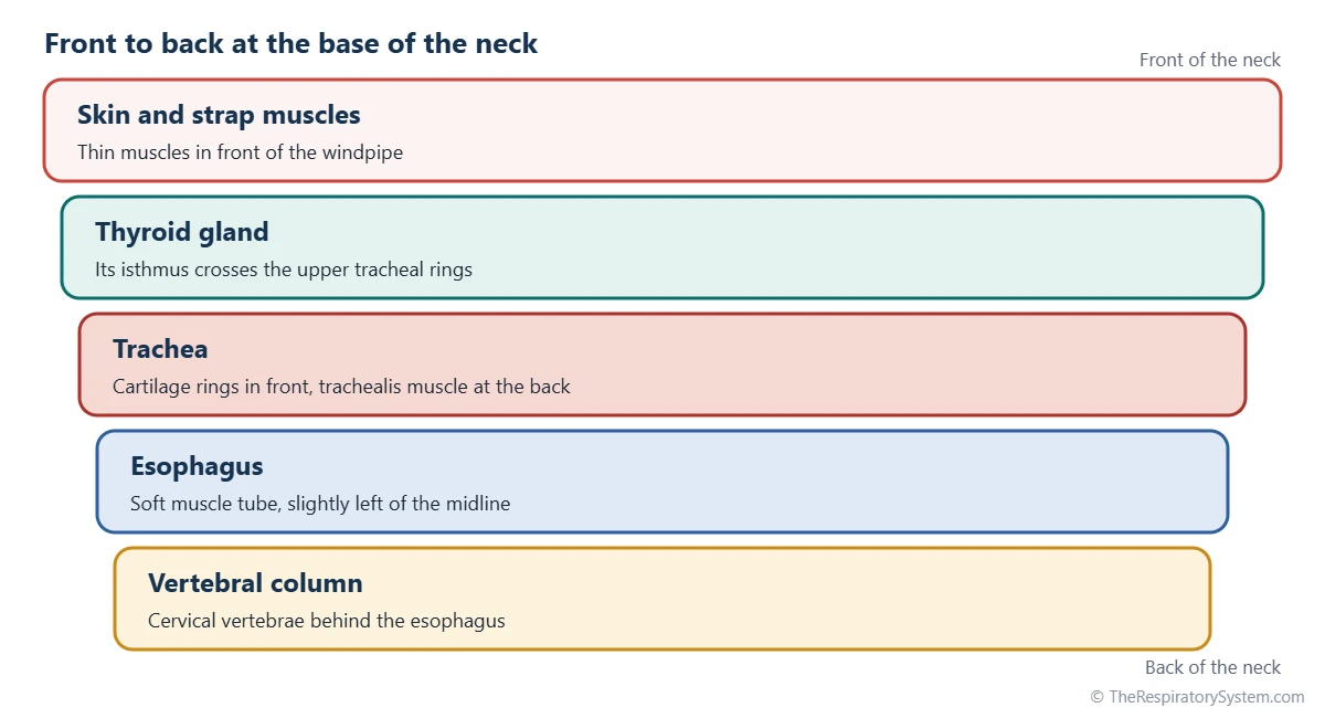
The trachea and esophagus both run down the middle of the neck into the chest, with the trachea in front and the esophagus directly behind it, pressed against the front of the spine.
Where they begin
Both tubes begin at about the level of the sixth cervical vertebra (C6), at the lower edge of the cricoid cartilage. Here the throat splits into two paths: the larynx continues as the trachea in front, and the laryngopharynx continues as the esophagus behind. In the neck the esophagus lies slightly to the left of the midline, which is why it peeks out to the left of the trachea in cross-sections.
Where they end
The trachea is the shorter tube. It travels into the superior mediastinum and ends at about the level of the sternal angle (T4 to T5), where it divides at the carina into the right and left main bronchi. The esophagus keeps going behind the heart through the posterior mediastinum, passes through the diaphragm at the esophageal hiatus at about T10, and joins the stomach just below it. Our page on the openings of the diaphragm shows that passage.
What lies between and around them
The narrow groove between the two tubes on each side is called the tracheoesophageal groove, and the recurrent laryngeal nerves run up inside it toward the larynx. In front of the trachea in the neck lies the thyroid gland, whose isthmus crosses the upper tracheal rings.
Trachea vs Esophagus Anatomy: How Are Their Walls Built?
The trachea has a wall built around cartilage rings and a ciliated lining, while the esophagus has a wall built around two thick layers of muscle and a stratified squamous lining.
Wall of the trachea
- Mucosa: pseudostratified ciliated columnar respiratory epithelium with goblet cells that make mucus.
- Submucosa: loose connective tissue with mucus and serous glands.
- Cartilage layer: 16 to 20 C-shaped rings of hyaline cartilage, open at the back.
- Trachealis muscle: smooth muscle that bridges the open ends of the rings, right against the esophagus.
- Adventitia: an outer layer of connective tissue.
Wall of the esophagus
- Mucosa: non-keratinized stratified squamous epithelium, built to withstand friction.
- Submucosa: connective tissue with mucous glands that lubricate the passage.
- Muscularis externa: an inner circular and an outer longitudinal layer. The upper third is skeletal muscle, the middle third is mixed, and the lower third is smooth muscle.
- Adventitia: an outer connective tissue layer; only the short part below the diaphragm has a serosa.
The open back of each tracheal ring is no accident: a large swallowed mouthful can bulge forward into the soft back wall of the trachea for a moment as it passes. Our respiratory system histology page shows the tracheal wall under the microscope.
What Do the Trachea and Esophagus Do?
The trachea conducts air between the larynx and the bronchi and cleans it on the way, while the esophagus transports swallowed food and liquid to the stomach and keeps stomach contents from flowing back.
Functions of the trachea
- Air passage: it is the only route for air between the throat and the lungs, part of the conducting zone described on our conducting zone vs respiratory zone page.
- Cleaning: mucus traps particles and cilia beat them upward toward the throat, the mucociliary escalator explained on our cilia and mucus page.
- Warming and humidifying: it finishes conditioning the air that the nose started.
- Coughing: sensitive nerve endings trigger the cough reflex, and the trachealis muscle narrows the tube to make a cough faster.
Functions of the esophagus
- Moving food: waves of muscle contraction called peristalsis push each swallowed bite down, even if you are lying down.
- Upper sphincter: a muscle ring at the top stays closed between swallows so air is not gulped into the stomach.
- Lower sphincter: a zone of muscle at the bottom relaxes to let food into the stomach and then closes to limit reflux.
The esophagus plays no part in breathing and the trachea plays no part in digestion, but the two must coordinate closely every time you swallow.
What Do the Trachea and Esophagus Have in Common?
The trachea and esophagus are both hollow tubes that start at the same level in the neck, run side by side through the neck and chest, and grow from the same part of the embryo.
- Common origin: both develop from the foregut. Early in development a groove in the front wall of the foregut becomes the trachea and lung buds, and a wall of tissue called the tracheoesophageal septum separates it from the esophagus behind, as described on our respiratory system development page.
- Shared starting point: both begin at about the C6 vertebra, below the pharynx.
- Shared neighbors: both lie in the neck and the superior mediastinum, near the thyroid gland, the great vessels and the vagus nerves.
- Similar wall plan: both have a mucosa, submucosa with glands, a muscle element and an outer adventitia.
- Same nerves: both receive branches of the vagus nerve, including the recurrent laryngeal nerves that run between them.
- Similar width: each is roughly 2 cm across in an adult, though the esophagus is flat when empty.
How Do the Trachea and Esophagus Work Together When You Swallow?
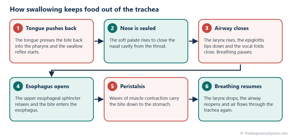
When you swallow, the airway is sealed for about a second so that food slides into the esophagus instead of the trachea, and breathing pauses briefly while it happens.
The tongue first pushes the bite back into the throat, and the soft palate rises to close off the nasal cavity. The larynx is then pulled up and forward, the epiglottis tips down over its entrance, and the vocal folds close tightly. At the same time, the upper esophageal sphincter relaxes and opens, and the bite drops into the esophagus, where peristalsis carries it to the stomach. A moment later the larynx drops, the airway reopens, and breathing usually resumes with a small breath out.
When food goes down the wrong pipe
If food or liquid slips past the closing larynx, sensory nerves in the larynx and trachea trigger strong coughing to push it back up. This is the familiar feeling of something going down the wrong pipe. Inhaling material into the airways is called aspiration, and in the lungs it can lead to infection.
Why the trachea stays out of the way
Because the back of the trachea is soft muscle rather than cartilage, the esophagus can expand forward as a bite passes, without squeezing the airway shut. Our glottis vs epiglottis page explains the two structures that guard the airway during this split second.
Pharynx vs Larynx vs Trachea vs Esophagus: How Do They Connect?
The pharynx is the shared throat for air and food, and below it the path splits in two: air goes forward through the larynx into the trachea, and food goes backward into the esophagus.
- Pharynx (throat): a muscular funnel behind the nose and mouth used by both air and food. Its lowest part, the laryngopharynx, leads to both the larynx and the esophagus.
- Larynx (voice box): the cartilage box at the top of the airway that guards it and makes the voice; compare the two on our larynx vs pharynx page.
- Trachea (windpipe): continues from the larynx down into the chest and splits into the bronchi.
- Esophagus (food pipe): continues from the laryngopharynx behind the trachea down to the stomach.
In short, air travels pharynx, larynx, trachea, while food travels pharynx, esophagus. The crossing of the two paths in the throat is the reason the swallowing reflex must close the airway so precisely. The full route of air is mapped on our pathway of air page.
Common Conditions That Affect the Trachea and Esophagus
Because the two tubes share a wall and a nerve supply, a problem in one can affect the other, but most conditions involve only the windpipe or only the food pipe. Our respiratory diseases section covers the airway conditions, and anyone with swallowing or breathing symptoms should see a doctor.
- Tracheoesophageal fistula and esophageal atresia: birth defects in which the two tubes fail to separate fully, leaving an abnormal connection or a blocked esophagus.
- Tracheitis and croup: infections that inflame the windpipe and the area just below the larynx, mainly in children.
- Tracheal stenosis: narrowing of the windpipe, sometimes after a long period with a breathing tube.
- Gastroesophageal reflux: stomach acid flows back into the esophagus; when acid reaches the throat it can irritate the larynx as well.
- Aspiration: food or liquid entering the trachea instead of the esophagus, which can lead to aspiration pneumonia.
Key Facts
- The trachea carries air to the lungs, while the esophagus carries food and liquid to the stomach.
- The trachea always lies in front of the esophagus, and both begin at about the level of the C6 vertebra.
- The trachea is about 10 to 12 cm long and ends at the carina; the esophagus is about 25 cm long and ends at the stomach.
- Sixteen to twenty C-shaped cartilage rings keep the trachea open, while the esophagus has no cartilage and lies flat when empty.
- The open back of each tracheal ring lets the esophagus bulge forward as a large bite passes.
- During swallowing the epiglottis and vocal folds seal the airway and breathing pauses for about a second.
- Both tubes grow from the embryonic foregut and are separated by the tracheoesophageal septum.
Quick Quiz: Test What You Know About the Trachea vs Esophagus
6 questions. Tap an answer to check it.
Which tube lies in front in the neck?
The trachea always lies in front of the esophagus, which lies between the trachea and the spine.
What keeps the trachea open?
Sixteen to twenty C-shaped hyaline cartilage rings hold the trachea open at all times.
What type of epithelium lines the esophagus?
Non-keratinized stratified squamous epithelium resists the friction of swallowed food.
Where does the trachea end?
The trachea divides at the carina, at about T4 to T5, into the right and left main bronchi.
Which structure tips down to cover the larynx during swallowing?
The epiglottis folds over the laryngeal inlet while the vocal folds close, guiding food into the esophagus.
Which tube passes through the diaphragm?
The esophagus passes through the esophageal hiatus at about T10 to reach the stomach. The trachea ends in the chest.
Frequently Asked Questions
Is the esophagus in front of or behind the trachea?
The esophagus is behind the trachea. In the neck and upper chest it lies between the back of the trachea and the front of the spine, slightly to the left of the midline. This is why the back wall of the trachea is soft muscle rather than cartilage: it lets the esophagus expand forward a little when a large mouthful passes through.
Can you feel your trachea and esophagus?
You can usually feel the trachea. Below the Adam's apple and the cricoid cartilage, the firm ridges of the tracheal rings can be felt in the front of the neck just above the breastbone. The esophagus cannot be felt from the outside, because it lies deep behind the trachea, against the spine, and is soft and flat when empty.
Why is the trachea rigid but the esophagus soft?
The trachea must stay open every moment, even when breathing in lowers the pressure inside it, so cartilage rings keep it from collapsing. The esophagus only needs to open when food passes, and a soft muscular tube can stretch around a bite and push it along by peristalsis. Each design fits what the tube carries, air in one case and solid food in the other.
Are the windpipe and esophagus connected?
In a healthy body they are not connected below the throat. They share a common entrance in the pharynx, and their walls touch along their whole length in the neck, but each is a separate tube. An abnormal opening between them, called a tracheoesophageal fistula, is a rare birth defect that is usually found in the first days of life.
Which is longer, the trachea or the esophagus?
The esophagus is longer. In an adult it is about 25 cm long because it continues through the whole chest and the diaphragm to reach the stomach. The trachea is only about 10 to 12 cm long, since it stops in the upper chest, at about the level of the sternal angle, where it divides into the two main bronchi.
Why can't you breathe and swallow at the same time?
The pharynx is shared by air and food, so the airway has to be closed while a bite or sip passes through. During each swallow, the larynx rises, the epiglottis covers its entrance, the vocal folds close and breathing stops for about a second. This short pause, sometimes called swallowing apnea, is controlled automatically by the brainstem.
Is the esophagus part of the respiratory system?
No. The esophagus belongs to the digestive system, even though it runs right beside the trachea and starts in the same part of the throat. It carries only food and liquid and plays no part in moving air. The trachea, by contrast, is part of the lower respiratory tract and carries only air between the larynx and the bronchi.
What happens when food goes into the trachea?
When food or liquid slips into the larynx or trachea, sensitive nerve endings there trigger a strong cough that usually pushes the material back up into the throat. Material that reaches deeper airways is called aspiration and can cause irritation or infection in the lungs.
Keep Exploring


