At a Glance
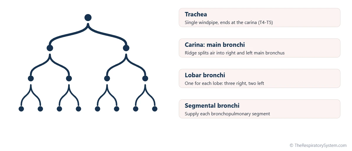
The tracheal carina is a ridge formed by the last cartilage ring of the trachea, projecting up between the openings of the right and left main bronchi at the point where the trachea splits. In adults it lies at about the level of the sternal angle and the disc between the T4 and T5 vertebrae, moving a little lower with a deep breath. Its lining is packed with sensory nerve endings that trigger a strong cough.
- Location
- Lower end of the trachea in the chest, behind the sternal angle, at about the T4-T5 level
- Type of tissue
- Hyaline cartilage of the last tracheal ring, lined by ciliated respiratory epithelium
- Function
- Divides airflow between the two main bronchi and triggers the cough reflex
- Nerve supply
- Vagus nerve branches through the pulmonary plexus
- Connects to
- Trachea above; right and left main bronchi below
- Part of
- The trachea, at the start of the bronchial tree in the lower respiratory tract
On this page
What Is the Carina of the Trachea?
The carina of the trachea is the ridge at the bottom of the windpipe that separates the openings of the two main bronchi, marking the exact point where one airway becomes two. Its name is Latin for "keel", because seen from inside, the ridge looks like the keel of a small boat pointing up into the trachea.
Carina definition in medicine
In medical terms, the carina (tracheal carina, or carina tracheae) is the internal ridge at the tracheal bifurcation, formed by the lowest tracheal cartilage, which curves down and back between the right and left main bronchi. The word carina is also used for a few other keel-shaped ridges in the body, so doctors usually say "main carina" or "tracheal carina" when they mean this one.
Explained simply
Imagine a garden hose that splits into a Y-shaped fitting so water can flow into two smaller hoses. Look into the fitting from the top and you see a sharp little divider in the middle, splitting the stream in two. The carina is that divider in your windpipe, sending air left and right into each lung.
Where Is the Carina Located?
The carina is located in the chest, in the upper part of the mediastinum, behind the sternal angle where the handle of the breastbone meets its body. This bony bump on the front of the chest is a handy surface landmark for finding it.
Carina of trachea vertebral level
In anatomy textbooks, the tracheal carina level is given as the T4-T5 intervertebral disc, the same horizontal plane as the sternal angle. This plane is measured in a person lying down. In an upright, living person the carina usually sits slightly lower, and during a deep breath in it can move down toward T6 as the diaphragm pulls the lungs and airways downward.
Neighbors of the carina
- Behind: the esophagus, which runs down just behind the trachea; see trachea vs esophagus.
- In front: the arch of the aorta and the split of the pulmonary trunk into the pulmonary arteries.
- Below: the subcarinal lymph nodes and, a little lower, the left atrium of the heart.
- To the left: the aortic arch, curving over the left main bronchus.
- To the right: the arch of the azygos vein, curving over the right main bronchus.
What Does the Carina Look Like Inside?
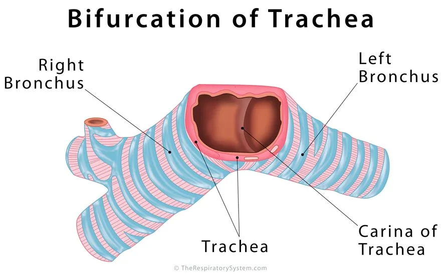
Inside the airway, the carina looks like a thin, sharp, slightly curved ridge running from front to back between two round openings, the larger right main bronchus and the narrower left main bronchus. It is built from the last C-shaped cartilage of the trachea, which is thicker in the middle and bends down into a hook-like point between the bronchi.
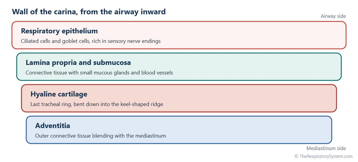
Wall of the carina
Like the rest of the trachea, the carina is covered by respiratory epithelium, a lining of ciliated cells and mucus-making goblet cells that keeps the mucociliary escalator moving. Beneath it lie connective tissue with small glands, then the hyaline cartilage, and an outer coat of connective tissue that blends with the tissues of the mediastinum.
The angle at the carina
The two main bronchi leave the carina at different angles. The right main bronchus is shorter, wider and runs more steeply downward, almost in line with the trachea, while the left main bronchus is longer, narrower and leaves at a wider angle because the heart and aortic arch push it sideways. The angle between them is called the carinal or subcarinal angle, and in adults it is usually less than a right angle.
| Feature | Typical description | Why it matters |
|---|---|---|
| Shape | Keel-shaped ridge running front to back | Divides the trachea's opening into two |
| Made of | Last tracheal cartilage ring, thick in the middle | Keeps the split rigid and open |
| Lining | Ciliated respiratory epithelium with goblet cells | Moves mucus up and away from the lungs |
| Vertebral level | T4-T5 intervertebral disc, lying down | Matches the sternal angle on the chest wall |
| Level in breathing | Moves lower on a deep breath in, toward T6 | Shows the airways stretch with the lungs |
| Right main bronchus | Shorter, wider, more vertical | Inhaled objects usually go right |
| Left main bronchus | Longer, narrower, more horizontal | Passes under the aortic arch |
| Nerve supply | Vagus nerve through the pulmonary plexus | Carries the signal for the cough reflex |
| Below it | Subcarinal lymph nodes, then the left atrium | Enlargement can widen the carina |
| Source: Standard adult anatomy, our editors | ||
What Is the Function of the Carina in the Respiratory System?
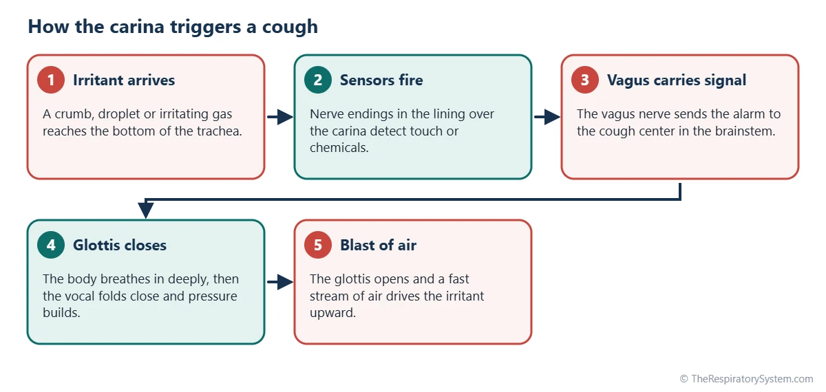
The carina has two main functions in the respiratory system: it divides inhaled air between the right and left lungs, and its very sensitive lining acts as an alarm that sets off a cough when anything foreign reaches the bottom of the trachea.
Splitting the airflow
As air flows down the windpipe on each breath, the sharp edge of the carina splits the stream into two, sending part to each of the lungs through its main bronchus. Because the right bronchus is wider and more vertical, inhaled objects such as food or small toys more often pass the carina into the right side, a fact covered on our bronchial tree page. From here the airways keep dividing on the pathway of air toward the alveoli.
The cough trigger
The mucous membrane over the carina holds many sensory nerve endings that respond to touch and irritating chemicals. When they fire, signals travel along the vagus nerve to the brainstem, which launches the cough reflex: a deep breath in, a brief closure of the glottis and a forceful blast of air that drives the irritant back up. This makes the carina the last checkpoint before particles can enter either lung.
A landmark for doctors
Because it is easy to see and lies at a known level, the carina is a key landmark in medicine. During bronchoscopy, doctors use it to orient themselves before entering the right or left bronchus. On a chest X-ray, the tip of a breathing tube placed in the trachea is checked against the carina, so that it sits a few centimeters above it and does not slip into one bronchus.
How Does the Carina Form?
The carina forms early in the embryo, at the spot where the single lung bud growing from the front of the foregut splits into two bronchial buds, one for each future lung. The trachea grows above that split, and the bronchial buds below it go on branching to build the bronchial tree.
Cartilage appears in the walls of the trachea and bronchi during later fetal weeks, and the last tracheal ring at the split becomes the cartilage of the carina. The steps of lung formation are described on our page about development of the respiratory system.
Common Conditions That Affect the Carina
Conditions that affect the carina usually involve the structures around it, such as enlarged lymph nodes or an enlarged heart chamber, or growths and inflammation in the airway itself. More information is in our respiratory diseases section.
- Widened carina: on imaging or bronchoscopy, a carina that looks splayed or blunted can mean that something below it is pushing the bronchi apart, such as enlarged subcarinal lymph nodes or an enlarged left atrium.
- Enlarged lymph nodes: the subcarinal nodes can swell with infections such as tuberculosis, with inflammatory diseases or when lung cancer spreads, which is why doctors examine this area when staging lung cancer.
- Tumors near the carina: cancers of the trachea or main bronchi that reach the carina affect both airways at once.
- Inhaled foreign bodies: objects that pass the carina usually lodge in a main bronchus, more often the right one, and the irritation triggers a strong cough.
- Inflammation: the lining over the carina becomes irritated in bronchitis and other airway infections, which helps explain why a dry, tickling cough can be felt deep behind the breastbone.
A cough that lasts for weeks or comes with coughing up blood is a reason to see a doctor.
Key Facts
- The carina is the keel-shaped ridge where the trachea divides into the right and left main bronchi.
- Its name comes from the Latin word for the keel of a boat.
- In textbooks it lies at the level of the sternal angle and the T4-T5 intervertebral disc, and it moves lower during a deep breath in.
- It is formed by the last tracheal cartilage, which bends down into a point between the two bronchi.
- The lining over the carina is one of the most cough-sensitive areas of the airway, linked to the brainstem by the vagus nerve.
- The right main bronchus leaves the carina more steeply than the left, so inhaled objects more often go to the right lung.
- Doctors use the carina as a landmark in bronchoscopy and to check the position of breathing tubes on X-rays.
Quick Quiz: Test What You Know About the Carina of the Trachea
6 questions. Tap an answer to check it.
What is the carina of the trachea?
The carina is the keel-shaped ridge at the tracheal bifurcation, between the right and left main bronchi.
At about which vertebral level is the carina found in textbooks?
The carina lies at about the T4-T5 intervertebral disc, the same level as the sternal angle.
Which surface landmark marks the level of the carina?
The sternal angle, where the manubrium meets the body of the sternum, lies in the same plane as the carina.
What kind of tissue forms the ridge of the carina?
The carina is formed by the last C-shaped ring of hyaline cartilage of the trachea.
Which nerve carries the cough signal from the carina to the brainstem?
Sensory fibers of the vagus nerve carry signals from the carina's lining to the cough center in the brainstem.
Why do inhaled objects more often enter the right main bronchus?
The right main bronchus is shorter, wider and more in line with the trachea, so objects passing the carina tend to drop into it.
Frequently Asked Questions
Can you feel your carina?
You cannot touch the carina from the outside, because it lies deep in the chest behind the breastbone, in front of the esophagus. Many people do feel it indirectly, though. A tickle or burning sensation deep behind the upper breastbone during a chest cold, or the sharp urge to cough when a crumb goes down the wrong way, often comes from irritation of the lining at or near the carina.
Is the carina part of the trachea or the bronchi?
The carina is usually counted as part of the trachea, because it is formed by the last tracheal cartilage ring at the lower end of the windpipe. It is also the starting point of the bronchi, since the right and left main bronchi begin on either side of it. In practice, it is the border between the two and is often described simply as the tracheal bifurcation.
What is the tracheal bifurcation?
The tracheal bifurcation is the place where the trachea divides into the two main bronchi, one for each lung. The carina is the ridge on the inside of this split. The two terms are closely related and often used together: bifurcation describes the branching of the tube as a whole, while carina names the specific keel-shaped ridge you would see looking down the airway.
What does a widened carina mean?
A widened carina means the angle between the two main bronchi is larger than usual, as seen on a chest X-ray or CT scan. It suggests that something below the carina is pushing the bronchi apart, most often enlarged subcarinal lymph nodes or an enlarged left atrium of the heart. It is a sign that prompts further tests rather than a diagnosis by itself.
Does the carina move when you breathe?
Yes. The trachea and bronchi are attached to the lungs, so they stretch and shift with each breath. When the diaphragm contracts and the lungs expand downward, the carina moves lower, by about one vertebral level on a deep breath. When you breathe out, it rises again. Its position also changes slightly between lying down and standing up.
Why is the carina important during intubation?
During intubation, a breathing tube is passed through the larynx into the trachea. If the tube is pushed too far, it can pass the carina and enter one main bronchus, usually the right, so only one lung receives air. For this reason, the tube's tip is checked against the carina on a chest X-ray, where it should sit a few centimeters above the bifurcation.
Are there other carinas in the lungs?
Yes. Every place where an airway divides has a small ridge between its two branches, and these are also called carinas, or secondary carinas. They are found at the splits of the main bronchi into lobar bronchi and at many smaller branch points further down the bronchial tree. The one at the bottom of the trachea is the largest and is called the main or tracheal carina.
What is the difference between the carina and the hilum of the lung?
The carina is inside the airway, at the point where the trachea divides into the two main bronchi in the mediastinum. The hilum of the lung is a region on the inner surface of each lung where the main bronchus, pulmonary vessels, nerves and lymphatics enter and leave. The main bronchi travel from the carina to the hilum of their own lung.
Keep Exploring


