At a Glance
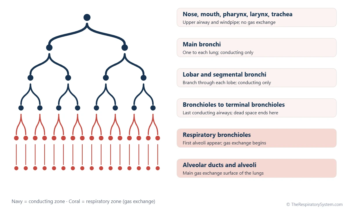
Dead space is inhaled air that takes no part in gas exchange. Anatomical dead space is the volume of the conducting airways, from the nose and mouth down to the terminal bronchioles, about 150 mL in a typical adult. Physiological (total) dead space is anatomical dead space plus alveolar dead space, and in healthy lungs the two are almost equal.
- Definition
- Air that is breathed in but takes no part in gas exchange
- Anatomical dead space
- Conducting airways, nose and mouth to terminal bronchioles; about 150 mL
- Alveolar dead space
- Alveoli that get air but little or no blood flow; close to zero in health
- Physiological dead space
- Anatomical plus alveolar dead space; about a quarter to a third of a resting breath
- Measured by
- Fowler's nitrogen washout (anatomical) and the Bohr equation (physiological)
- Linked to
- Tidal volume, alveolar ventilation and the ventilation-perfusion ratio
On this page
What Is Dead Space in the Lungs?
Dead space in the lungs is the volume of inhaled air that never takes part in gas exchange, either because it stays in the airways or because it reaches alveoli that have too little blood flowing past them. In medicine the term is used as a measure of wasted ventilation, the breathing effort that does not help load oxygen or remove carbon dioxide.
Dead space exists because we breathe in and out through the same set of tubes. At the end of each breath in, the last air drawn in is still sitting in the trachea and bronchi, and it is the first air to be breathed out again, unchanged. Only the air that travels beyond the conducting airways mixes with alveolar air and takes part in gas exchange.
Explained simply
Think of breathing through a long straw. Each time you breathe in, the first sip is the stale air left in the straw from your last breath out. Your airways are that straw: part of every breath only fills the tubes and never reaches the tiny air sacs where oxygen enters the blood.
What Is Anatomical Dead Space?
Anatomical dead space is the total volume of the conducting airways, the passages that carry air to and from the alveoli but have walls too thick for gas exchange, and it is about 150 mL in a typical adult.
It includes the nasal cavity or mouth, the pharynx, the larynx, the trachea, the bronchi and the bronchioles down to the terminal bronchioles. These airways make up the conducting zone, while the respiratory bronchioles, alveolar ducts and alveoli form the respiratory zone, as shown on our conducting zone vs respiratory zone page.
What changes anatomical dead space?
- Body size: larger people have longer and wider airways, so their dead space is bigger.
- Lung volume: deep breaths stretch the airways open, which slightly increases dead space.
- Head and neck position: extending the neck and pushing the jaw forward lengthens the upper airway; bending the head down shortens it.
- Airway tone: narrowing of the bronchi reduces dead space, and widening increases it.
- Bypassing the upper airway: a breathing tube or a tracheostomy skips the nose, mouth and pharynx, which reduces anatomical dead space.
What Is Physiological Dead Space?
Physiological dead space, also called total dead space, is the anatomical dead space plus the alveolar dead space: all the inhaled air that does not take part in gas exchange, wherever it ends up.
Alveolar dead space is the air that reaches alveoli but finds too little blood to exchange gases with. It arises when the ventilation of a lung region is much higher than its blood flow, a mismatch described by the ventilation-perfusion ratio. In an upright healthy person, the tops of the lungs receive relatively little blood, so a small amount of alveolar dead space exists even in normal lungs, but it is usually so small that physiological and anatomical dead space are nearly the same.
In lung disease the picture changes. When blood vessels are blocked or destroyed, many alveoli are ventilated without being perfused, and physiological dead space can grow far larger than anatomical dead space. This makes physiological dead space a useful measure of how well ventilation and blood flow are matched across the lungs.
Physiological Dead Space vs Anatomical Dead Space
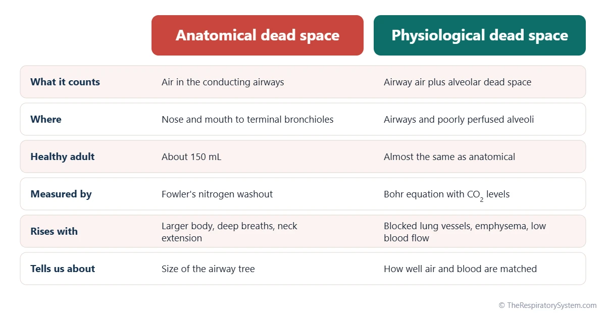
Anatomical dead space is fixed by the size of the conducting airways, while physiological dead space also counts alveoli that are ventilated but not perfused, so physiological dead space is always equal to or larger than anatomical dead space.
The two are measured in different ways and answer different questions. Anatomical dead space describes the structure of the airway tree, whereas physiological dead space describes how efficiently the whole lung is working. In a healthy adult at rest both are around 150 mL; in a patient with a blood clot in a lung artery, anatomical dead space stays about the same while physiological dead space rises.
Apparatus dead space
Equipment adds its own dead space. Any tube, mask or mouthpiece that air passes through in both directions, such as a snorkel, a face mask or the part of a ventilator circuit beyond the point where the breaths in and out separate, adds its volume to the airway. This apparatus dead space is why snorkels are kept short and why breathing circuits are designed to keep the shared section small.
| Term | What it includes | Typical healthy adult value | Larger when |
|---|---|---|---|
| Anatomical dead space | Nose or mouth, pharynx, larynx, trachea, bronchi and bronchioles to the terminal bronchioles | About 150 mL (about 2 mL per kg of body weight) | Breaths are very deep, the neck is extended or the airways widen |
| Alveolar dead space | Alveoli that receive air but little or no blood | Close to zero | A lung artery is blocked or blood pressure is low |
| Physiological dead space | Anatomical plus alveolar dead space | About 150 mL; VD/VT about 0.2 to 0.35 | Lung disease mismatches air and blood flow |
| Apparatus dead space | Masks, mouthpieces, snorkels and shared breathing tubes | Equal to the volume of the shared tubing | The shared tube is longer or wider |
| Tidal volume | Air moved in one quiet breath | About 500 mL | Breathing is deeper, as during exercise |
| Minute ventilation | Total air breathed per minute | About 6 L per minute | Breathing is faster or deeper |
| Alveolar ventilation | Fresh air reaching the alveoli per minute | About 4.2 L per minute | Breaths are deeper, so less of each is dead space |
| Source: Typical adult values, our editors | |||
How Is Dead Space Measured? The Anatomical Dead Space Formula
Anatomical dead space is measured with Fowler's single-breath nitrogen method or estimated from body weight, and physiological dead space is calculated with the Bohr equation from the carbon dioxide levels in arterial blood and exhaled air.
Anatomical dead space formula and Fowler's method
A quick estimate of anatomical dead space is about 2 mL per kilogram of body weight, close to 1 mL per pound, which gives about 150 mL for a 70 kg adult. In Fowler's method, the person takes one breath of pure oxygen and then breathes out steadily while the nitrogen level of the exhaled air is recorded. The first air out comes from the airways and contains only oxygen; as alveolar air follows, nitrogen rises. The volume breathed out up to the midpoint of that rise equals the anatomical dead space.
The Bohr equation for physiological dead space
The Bohr equation uses the fact that dead-space air contains almost no carbon dioxide, so it dilutes the carbon dioxide coming from the alveoli. In its common clinical form it reads:
VD / VT = (PaCO2 − PECO2) / PaCO2
Here VD is dead space, VT is tidal volume, PaCO2 is the arterial carbon dioxide pressure and PECO2 is the carbon dioxide pressure of mixed exhaled air. For example, with a PaCO2 of 40 mmHg and a PECO2 of 28 mmHg, VD/VT is 12 divided by 40, or 0.3, so 150 mL of a 500 mL breath is dead space. In healthy adults the ratio is usually about 0.2 to 0.35 at rest.
Why Does Dead Space Matter?
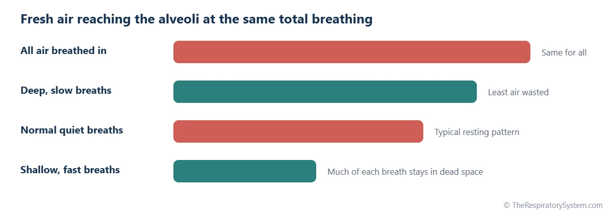
Dead space matters because it sets how much of each breath is actually useful: only the air beyond the dead space refreshes the alveoli, so the same amount of breathing can deliver very different amounts of fresh air depending on how deep the breaths are.
The air that reaches the alveoli each minute is the alveolar ventilation, calculated as (tidal volume minus dead space) times breaths per minute. At rest, a typical adult breathes about 500 mL per breath at 12 breaths per minute, for a minute ventilation of 6 liters. Subtracting 150 mL of dead space from each breath leaves 350 mL, so alveolar ventilation is about 4.2 liters per minute. Our lung volumes and capacities page places these numbers among the other lung volumes.
- Shallow, fast breathing: 250 mL breaths at 24 per minute still move 6 liters, but only 100 mL of each breath passes the dead space, so alveolar ventilation falls to about 2.4 liters per minute.
- Deep, slow breathing: 1,000 mL breaths at 6 per minute also move 6 liters, but 850 mL of each breath reaches the alveoli, giving about 5.1 liters per minute.
This is why the respiratory rate alone says little about ventilation. The conducting airways that create the dead space are not wasted, though: they warm, humidify and filter the air before it reaches the delicate alveoli.
Common Conditions That Increase Dead Space
Dead space increases mainly in conditions that cut off blood flow to ventilated alveoli, destroy the capillary bed, or force a pattern of rapid, shallow breathing. Our respiratory diseases section explains these conditions, and anyone with breathing symptoms should see a doctor.
- Pulmonary embolism: a clot blocks a pulmonary artery, so the alveoli it supplied are ventilated but not perfused, a classic cause of alveolar dead space.
- Emphysema and COPD: destruction of alveolar walls removes their capillaries and creates large air spaces with poor blood supply.
- ARDS: injury to small lung vessels raises the dead space fraction, which is used as a marker of severity.
- Low blood pressure or low cardiac output: less blood reaches the upper parts of the lungs, so alveolar dead space grows.
- Rapid shallow breathing: weakness of the breathing muscles or stiff lungs make each breath small, so a larger share of it is dead space.
Key Facts
- Dead space is the part of each breath that takes no part in gas exchange, also called wasted ventilation.
- Anatomical dead space is the volume of the conducting airways, about 150 mL in an adult, or roughly 2 mL per kilogram of body weight.
- Alveolar dead space is air in alveoli that receive too little blood; in healthy lungs it is close to zero.
- Physiological dead space equals anatomical plus alveolar dead space, and in health it is about 20 to 35 percent of a resting breath.
- Alveolar ventilation equals (tidal volume minus dead space) times breaths per minute, about 4.2 liters per minute in a resting adult.
- Shallow, fast breathing wastes a larger share of each breath in dead space than deep, slow breathing.
- A pulmonary embolism raises alveolar dead space by leaving ventilated alveoli without blood flow.
Quick Quiz: Test What You Know About the Dead Space
6 questions. Tap an answer to check it.
What is anatomical dead space?
Anatomical dead space is the volume of the conducting airways, from the nose and mouth to the terminal bronchioles.
What is the typical anatomical dead space of an adult?
A typical adult has about 150 mL of anatomical dead space, roughly 2 mL per kilogram of body weight.
Physiological dead space equals anatomical dead space plus what?
Physiological (total) dead space is anatomical dead space plus alveolar dead space, the alveoli that are ventilated but poorly perfused.
Where does anatomical dead space end?
The terminal bronchioles are the last purely conducting airways; gas exchange begins at the respiratory bronchioles.
Which equation is used to calculate physiological dead space?
The Bohr equation compares carbon dioxide in arterial blood and mixed exhaled air to find the dead space fraction of each breath.
Which breathing pattern wastes the largest share of each breath in dead space?
With small breaths, the fixed dead space takes up a larger share of each breath, so less fresh air reaches the alveoli.
Frequently Asked Questions
How much of each breath is dead space?
In a healthy adult breathing quietly, about 150 mL of a 500 mL breath stays in the conducting airways, so roughly 30 percent of each breath is dead space. The remaining 350 mL or so reaches the alveoli. The share falls when breaths are deeper, as during exercise, and rises when breaths become small and fast.
Is dead space the same as residual volume?
No. Residual volume is the air that stays in the lungs after the hardest possible breath out, about 1.2 liters in an adult, and most of it sits in the alveoli, where it keeps exchanging gases with the blood. Dead space is the part of each breath that takes no part in gas exchange, mainly the air filling the airways.
Does dead space change during exercise?
The volume of anatomical dead space rises slightly during exercise because deeper breaths stretch the airways open. Tidal volume rises much more, however, so the fraction of each breath lost to dead space falls. That makes breathing more efficient during exertion, letting more of the extra air reach the alveoli where oxygen enters the blood.
Why does exhaled air still contain oxygen?
Exhaled air is a mix of alveolar air and dead-space air. The first part of each breath out comes from the airways and is almost unchanged inhaled air, rich in oxygen. Alveolar air follows, with less oxygen and more carbon dioxide. Mixed together, exhaled air still holds roughly 16 percent oxygen, compared with about 21 percent in fresh air.
How does a snorkel affect dead space?
A snorkel adds its own volume to the anatomical dead space, because air moves in and out through the same tube. Every breath must first clear the stale air left in the snorkel before fresh air reaches the lungs. A longer or wider tube adds more dead space, which is one reason snorkels are made short and narrow.
Does a tracheostomy reduce dead space?
Yes. A tracheostomy is an opening in the front of the neck into the trachea, so air no longer passes through the nose, mouth, pharynx and larynx. Those passages make up a sizable part of anatomical dead space, so bypassing them reduces it. The trade-off is that the air also misses much of the warming, humidifying and filtering that the upper airway normally provides.
Why is dead space important for patients on a ventilator?
On a mechanical ventilator, the shared part of the breathing circuit adds apparatus dead space, and many lung conditions add alveolar dead space. Clinicians track carbon dioxide in exhaled air and blood to estimate the dead space fraction, because when it is high, more ventilation is needed to clear the same amount of carbon dioxide. The dead space fraction is also used to judge how sick the lungs are.
Keep Exploring


