At a Glance
Alveolar ducts are the airways that follow the respiratory bronchioles, at about generations 20 to 22 of the bronchial tree; their walls are lined with alveoli and rimmed by small knobs of smooth muscle and elastic fibers. Each duct ends in alveolar sacs, at about generation 23, which are clusters of alveoli opening into a common space. Both belong to the respiratory zone, where oxygen and carbon dioxide are exchanged.
- Location
- Respiratory zone of both lungs, after the respiratory bronchioles
- Airway generation
- Alveolar ducts about 20 to 22; alveolar sacs about 23, the last
- Tissue
- Alveolar epithelium of type I and type II pneumocytes; small rings of smooth muscle and elastic fibers at duct openings
- Function
- Carry air to the alveoli and exchange gases across the alveoli in their walls
- Connects to
- Respiratory bronchioles above; alveoli open directly off them
- Part of
- The pulmonary acinus, the gas-exchange unit fed by one terminal bronchiole
On this page
- What Are the Alveolar Ducts and Alveolar Sacs?
- Where Are the Alveolar Ducts Located?
- Alveolar Ducts Anatomy: What Are Their Walls Made Of?
- What Is the Function of the Alveolar Ducts in the Respiratory System?
- Alveolar Ducts vs Sacs vs Alveoli: What Is the Difference?
- How Do Alveolar Ducts and Sacs Develop and Change With Age?
- Common Conditions That Affect the Alveolar Ducts and Sacs
- Key Facts
- Quiz
- FAQ
What Are the Alveolar Ducts and Alveolar Sacs?
Alveolar ducts and alveolar sacs are the final branches of the airway tree, where the passages stop being tubes with solid walls and become chains and clusters of tiny air pockets called alveoli. They sit at the very end of the bronchial tree, beyond the bronchioles, and hold a large share of all the alveoli in the lungs.
Alveolar ducts definition in anatomy
In anatomy, an alveolar duct is an elongated airway that arises from a respiratory bronchiole and whose wall consists of a continuous series of alveolar openings. Between neighboring openings, the only true wall left is a small knob of tissue containing smooth muscle, elastic fibers and collagen. Under a microscope these knobs look like a row of beads lining both sides of the duct.
What is the alveolar sac?
An alveolar sac is the dead-end space at the tip of an alveolar duct, into which two or more alveoli open. It is often compared to a bunch of grapes, with each grape an alveolus and the stem the duct. Some textbooks name the small shared chamber where several sacs open off the duct an atrium.
Explained simply
Picture the airways as a tree. The trunk and branches carry air, but the leaves are where the work is done. The alveolar ducts are the thinnest twigs, already covered in leaves along their whole length, and the alveolar sacs are the leaf clusters at the very tips.
Where Are the Alveolar Ducts Located?
Alveolar ducts are located deep inside each lung, in the respiratory zone, where they branch off the respiratory bronchioles and end in alveolar sacs. They are found throughout every lobe, from the region near the hilum out to the tissue just beneath the pleura.
Place in the airway generations
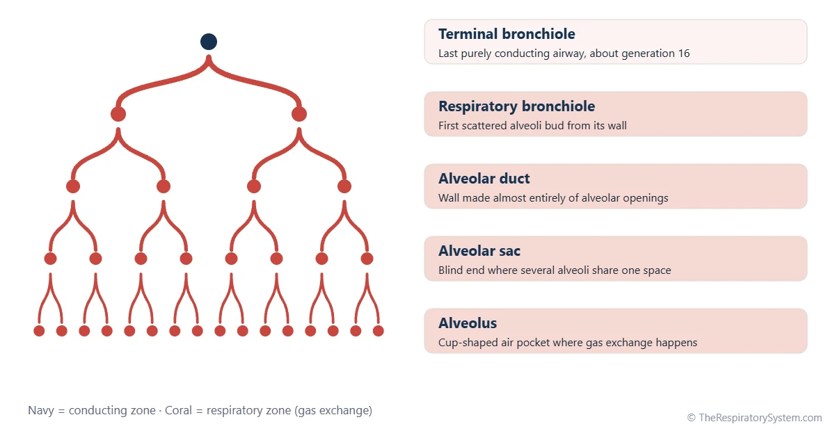
A common model of the human airway counts 23 generations of branching from the trachea, which is generation 0. Generations 0 to 16 form the conducting zone and only move air. The respiratory zone starts with the respiratory bronchioles at about generations 17 to 19, continues with the alveolar ducts at about generations 20 to 22, and ends with the alveolar sacs at about generation 23. The conducting zone vs respiratory zone guide explains this division.
Part of the pulmonary acinus
Each terminal bronchiole supplies one small unit called a pulmonary acinus: its respiratory bronchioles, alveolar ducts, alveolar sacs and alveoli. Several acini together form a secondary pulmonary lobule, the smallest lung unit wrapped in its own connective tissue septa. Each respiratory bronchiole gives rise to several alveolar ducts, which may branch again before they end in sacs.
| Part | Approximate generation | Wall structure | Role |
|---|---|---|---|
| Trachea | 0 | C-shaped hyaline cartilage, smooth muscle, ciliated epithelium | Conducts air only |
| Bronchi | About 1 to 10 | Cartilage plates, smooth muscle, ciliated epithelium | Conducts air only |
| Terminal bronchiole | About 16 | Smooth muscle, cuboidal epithelium with club cells, no cartilage | Last purely conducting airway |
| Respiratory bronchiole | About 17 to 19 | Cuboidal epithelium with scattered alveoli in the wall | Conducts air and begins gas exchange |
| Alveolar duct | About 20 to 22 | Alveolar openings separated by knobs of smooth muscle and elastic fibers | Carries air to alveoli and exchanges gas |
| Alveolar sac | About 23 | Cluster of alveoli around a shared space, no smooth muscle | Final dead end; gas exchange |
| Alveolus | Opens off ducts and sacs | Type I and type II pneumocytes on a thin basement membrane | Main site of gas exchange |
| Source: Typical adult values (Weibel airway model), our editors | |||
Alveolar Ducts Anatomy: What Are Their Walls Made Of?
The walls of alveolar ducts and sacs are made of alveoli themselves, lined by flat type I and cuboidal type II pneumocytes, with only small rings of smooth muscle, elastic fibers and collagen around the duct openings. There is no cartilage, no glands and no ciliated lining at this level.
The duct wall
- Alveolar openings: nearly the whole surface of a duct is taken up by the mouths of alveoli, so the duct is more of a corridor than a tube.
- Knobs of smooth muscle: between the openings, small bundles of smooth muscle encircle each alveolar mouth. They are the last smooth muscle of the airway and disappear at the sacs.
- Elastic and collagen fibers: a network of fibers runs along the duct and around the sacs, stretching during a breath in and springing back during a breath out.
- Thin lining: the knobs are covered by a simple squamous to low cuboidal epithelium that blends into the lining of the alveoli.
The sac and its alveoli
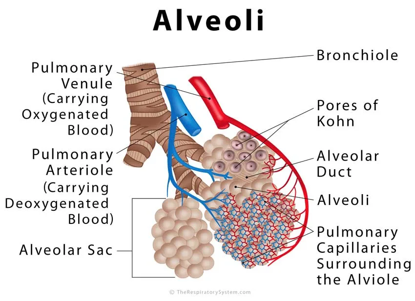
Each alveolar sac is a rounded cluster of alveoli opening into a shared central space. Neighboring alveoli share thin walls called interalveolar septa, which carry a dense web of pulmonary capillaries. Small holes in these walls, the pores of Kohn, connect neighboring alveoli and let air pass sideways. Alveolar macrophages move over the inner surface and clear dust and microbes, and a thin film of pulmonary surfactant made by type II cells lowers surface tension.
Blood supply
Branches of the pulmonary arteries follow the airways down to the acinus and break into capillaries in the walls of the ducts and sacs. Oxygen-rich blood is collected into venules that run in the connective tissue at the edges of the lobules, as described on our pulmonary circulation page.
What Is the Function of the Alveolar Ducts in the Respiratory System?
The function of the alveolar ducts is to deliver air to the alveoli that line them and end in the sacs, and to take part in gas exchange across those same alveoli. Alveolar sacs add the largest number of alveoli at the very end of each pathway, where gas exchange is at its peak.
Where airflow turns into diffusion
With each branching the airways get narrower, but there are so many more of them that their combined cross-section grows enormously. By the level of the alveolar ducts, the incoming air slows almost to a stop. From here on, oxygen moves forward mostly by diffusion through the gas already in the ducts and sacs, rather than by bulk airflow, and the short distances make this fast enough.
Other jobs
- Elastic recoil: the fibers around the ducts and sacs stretch during inhalation and recoil during exhalation, helping push air out without muscle effort.
- Keeping the unit open: because the alveoli share walls, each one helps hold its neighbors open, a support called interdependence.
- Fine control: the smooth muscle rings at the duct openings can narrow the mouths of the alveoli slightly.
- Side channels: pores of Kohn let air reach an alveolus even if its own duct is partly blocked, a process called collateral ventilation.
Alveolar Ducts vs Sacs vs Alveoli: What Is the Difference?
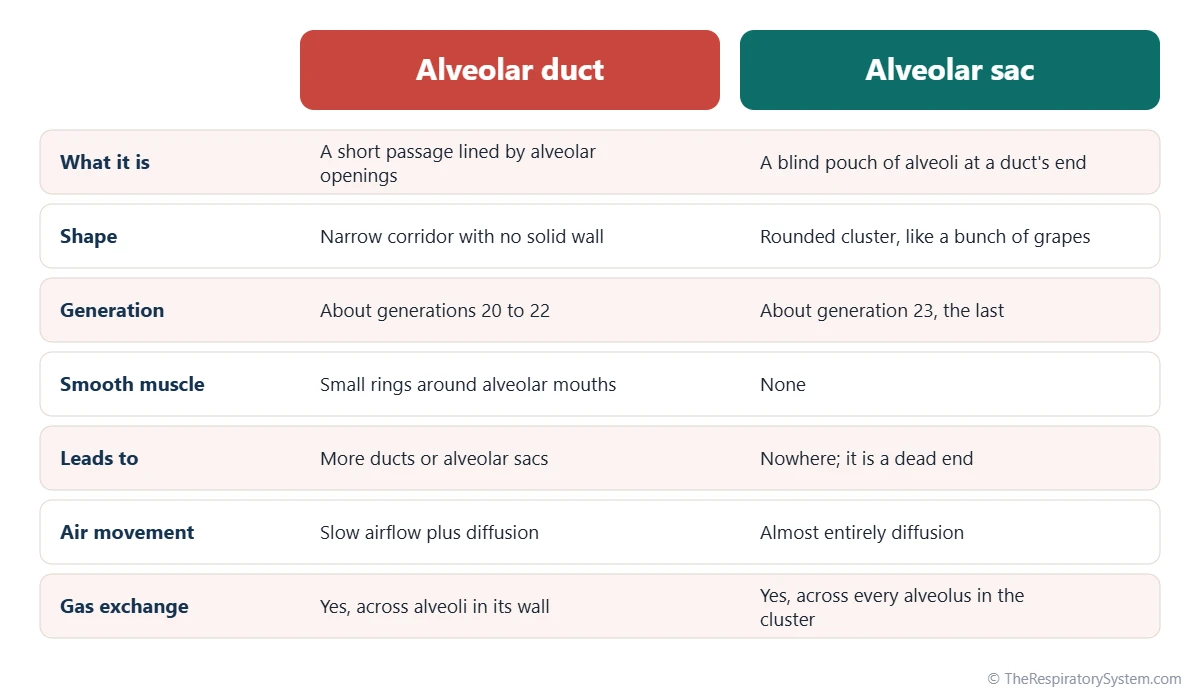
An alveolar duct is a passage lined with alveoli, an alveolar sac is the blind cluster of alveoli at the end of that passage, and an alveolus is a single cup-shaped air pocket, the basic unit of gas exchange. A duct and a sac are both made of alveoli, but only the duct leads on somewhere.
A simple way to tell them apart is to follow the air. It leaves a respiratory bronchiole and enters an alveolar duct, which it may follow through one or two more branchings, with alveoli opening on every side. At the end of the duct, the air reaches a sac with no way forward, only alveoli all around. The full structure of a single alveolus, including its cells and the respiratory membrane, is covered on our alveoli page. The chart below compares the duct and the sac directly.
How Do Alveolar Ducts and Sacs Develop and Change With Age?
Alveolar ducts and sacs begin forming before birth, from the ends of the branching airway buds, and continue to mature and multiply after birth through childhood. In old age they slowly widen and lose some of their surface.
During the saccular stage of fetal life, roughly from the end of the second trimester to birth, the tips of the airway tree widen into thin-walled terminal sacs, and type II cells begin making surfactant. True alveoli then form by dividing these sacs with new walls, a process that starts late in pregnancy and continues for years after birth, so most alveoli are formed after a baby is born. Our respiratory system development page follows these stages in order.
In older adults, the elastic fibers lose some of their spring. The alveolar ducts become wider, the alveoli around them become shallower, and the total gas-exchange surface shrinks slightly. These changes are a normal part of aging and are much milder than the destruction seen in disease.
Common Conditions That Affect the Alveolar Ducts and Sacs
Diseases that affect the alveolar ducts and sacs usually damage their thin walls, fill them with fluid, or collapse them, which reduces the surface available for gas exchange. Our respiratory diseases section covers each condition in depth.
- Emphysema: the walls of the acinus break down into fewer, larger air spaces. In the centriacinar type, linked mainly to smoking, the damage starts at the respiratory bronchioles; in the panacinar type, linked to alpha-1 antitrypsin deficiency, the ducts, sacs and alveoli are enlarged evenly. Both are forms of COPD.
- Neonatal respiratory distress syndrome: in very premature babies, too little surfactant lets the sacs and alveoli collapse, and protein-rich hyaline membranes line the alveolar ducts.
- ARDS: widespread lung injury in adults also floods the alveoli and lays down hyaline membranes along the ducts.
- Pneumonia: infection fills alveoli and sacs with fluid and inflammatory cells.
- Dust diseases: very fine particles that reach the acinus can settle around the respiratory bronchioles and alveolar ducts, where they may cause scarring over years.
Key Facts
- Alveolar ducts arise from respiratory bronchioles and have walls made almost entirely of alveolar openings.
- In the common 23-generation airway model, alveolar ducts are about generations 20 to 22 and alveolar sacs are generation 23, the last.
- An alveolar sac is a blind-ended space into which two or more alveoli open, often compared to a bunch of grapes.
- Small knobs of smooth muscle around the alveolar mouths of the ducts are the last smooth muscle in the airway tree.
- Alveolar ducts and sacs have no cartilage, glands or cilia; dust that reaches them is cleared by alveolar macrophages.
- At this level, air moves forward mainly by diffusion rather than bulk flow, because the combined airway cross-section is so large.
- Respiratory bronchioles, alveolar ducts, sacs and alveoli fed by one terminal bronchiole form a pulmonary acinus, part of a pulmonary lobule.
Quick Quiz: Test What You Know About the Alveolar Ducts and Alveolar Sacs
6 questions. Tap an answer to check it.
Which airway directly gives rise to the alveolar ducts?
Alveolar ducts branch from respiratory bronchioles, the first airways with alveoli in their walls.
What is an alveolar sac?
An alveolar sac is the dead end of an alveolar duct, with two or more alveoli opening into a shared space.
What forms the small knobs between alveolar openings in an alveolar duct?
The knobs contain smooth muscle, elastic fibers and collagen, the last smooth muscle of the airway tree.
In the 23-generation airway model, which generation are the alveolar sacs?
Alveolar sacs are the last generation, about 23; the trachea is generation 0.
Which zone of the respiratory system do the alveolar ducts belong to?
Alveolar ducts and sacs are lined with alveoli, so they are part of the respiratory zone, where gas exchange occurs.
What lets air pass sideways between neighboring alveoli in a sac?
Pores of Kohn are small holes in the alveolar walls that allow collateral ventilation.
Frequently Asked Questions
Are alveolar sacs the same as alveoli?
No. An alveolus is a single tiny air pocket, while an alveolar sac is a group of two or more alveoli that open into one shared space at the end of an alveolar duct. The words are often mixed up in everyday use, but in anatomy the sac is the cluster and each alveolus is one unit within it, much like a bunch of grapes and a single grape.
Do alveolar ducts have cilia?
No. Cilia become fewer along the bronchioles and disappear around the respiratory bronchioles, so alveolar ducts and sacs have no ciliated lining and no mucus-producing goblet cells. Instead of being swept upward by the mucus escalator, particles that reach this deep level are engulfed by alveolar macrophages, which can carry them up to the ciliated airways or into the lymphatic vessels.
Do alveolar ducts have cartilage?
No. Cartilage supports the trachea and the bronchi but becomes smaller and patchier as the airways branch, and it is gone by the level of the bronchioles. Alveolar ducts and sacs are held open instead by the elastic fiber network of the lung, by the shared walls of neighboring alveoli, and by surfactant, which lowers the surface tension that would otherwise pull them closed.
Is the alveolar duct part of the conducting or the respiratory zone?
The alveolar duct is part of the respiratory zone. The conducting zone runs from the nose down to the terminal bronchioles and only moves air. The respiratory zone begins with the respiratory bronchioles, which have scattered alveoli, and continues through the alveolar ducts and sacs, which are lined with alveoli along their whole length and take part directly in gas exchange.
What is the atrium of an alveolar duct?
Some anatomy texts use the word atrium for the small common chamber at the end of an alveolar duct, into which two or more alveolar sacs open. Other texts skip this term and describe the sacs as opening straight off the duct. In either description, the space is lined with alveoli and works the same way, as a final mixing space before the dead-end sacs.
How does fresh air reach a dead-end alveolar sac?
Each breath in pushes fresh air down to the alveolar ducts, where it mixes with the air already inside. From there, oxygen spreads into the alveolar sacs by diffusion, which is fast over such tiny distances, while carbon dioxide spreads outward the same way. Pores of Kohn between neighboring alveoli add extra routes, so air can still arrive if a small airway is partly blocked.
What cells line the alveolar sacs?
Alveolar sacs are lined by two kinds of epithelial cells. Type I pneumocytes are very thin, flat cells that cover most of the surface and form part of the barrier that gases cross. Type II pneumocytes are rounder cells that make surfactant and can divide to replace damaged type I cells. Alveolar macrophages move freely over the surface to clear dust and microbes.
Can damaged alveolar sacs grow back?
In children, the lungs keep adding new alveoli for years, so some growth is possible. In adults, type II pneumocytes can repair the lining of alveoli after injury, but the walls lost in conditions such as emphysema do not grow back in a meaningful way. This is why damage to the acinus from smoking or disease is usually described as permanent.
Keep Exploring


