At a Glance
The respiratory membrane, also called the alveolar-capillary membrane or blood-air barrier, is the wall that separates alveolar air from pulmonary capillary blood. It is made of a fluid film with surfactant, the alveolar epithelium, a largely fused basement membrane and the capillary endothelium, and it is typically only about 0.5 micrometer thick across a surface of roughly 50 to 75 square meters.
- Location
- Walls of the alveoli, wherever they touch the pulmonary capillaries
- Also called
- Alveolar-capillary membrane, blood-air barrier, blood-gas barrier
- Layers
- Surfactant fluid film, type I pneumocytes, basement membrane(s), thin interstitium, capillary endothelium
- Thickness
- About 0.5 micrometer on average, as thin as about 0.2 micrometer in places
- Surface area
- About 50 to 75 square meters in an adult
- Function
- Lets oxygen and carbon dioxide diffuse while keeping blood and fluid out of the air sacs
On this page
- What Is the Respiratory Membrane?
- Where Is the Respiratory Membrane Located?
- Respiratory Membrane Structure: What Are Its Layers?
- What Are the Thin and Thick Sides of the Blood-Air Barrier?
- What Is the Function of the Respiratory Membrane?
- Which Structures Are Related to the Respiratory Membrane?
- Common Conditions That Affect the Respiratory Membrane
- Key Facts
- Quiz
- FAQ
What Is the Respiratory Membrane?
The respiratory membrane is the combined wall of an alveolus and the capillary pressed against it, forming the only thing that separates inhaled air from blood. It is not a single cell membrane but a sandwich of thin layers, and every molecule of oxygen you absorb crosses it on the way into the bloodstream.
Textbooks use several names for the same structure. Alveolar capillary membrane describes its two halves, blood-air barrier (or blood-gas barrier) describes its job, and respiratory membrane is the name most physiology courses use. It is where the conducting airways finally give way to the working surface of the lungs.
Explained simply
Picture a balloon of air touching a garden hose full of blood. The rubber of the balloon and the wall of the hose are pressed so tightly together that they act like one sheet, thinner than a soap bubble in most places. Oxygen slips through that sheet into the blood, and carbon dioxide slips the other way into the air.
Where Is the Respiratory Membrane Located?
The respiratory membrane is located in the walls of the alveoli throughout the respiratory zone of both lungs, wherever an alveolus touches a pulmonary capillary. It does not exist in the trachea, bronchi or terminal bronchioles, whose walls are far too thick for gas exchange.
The respiratory zone starts at the respiratory bronchioles, which have a few alveoli budding from their walls, and continues into the alveolar ducts and sacs. The conducting zone vs respiratory zone guide marks the exact boundary.
Between neighboring alveoli lies a thin wall called the interalveolar septum, filled with a dense web of capillaries. Each capillary is usually exposed to air on more than one side, so a single red blood cell can load oxygen from two alveoli at once. The blood reaches this web through the pulmonary arteries and leaves through the pulmonary veins, as explained on our pulmonary circulation page.
Respiratory Membrane Structure: What Are Its Layers?
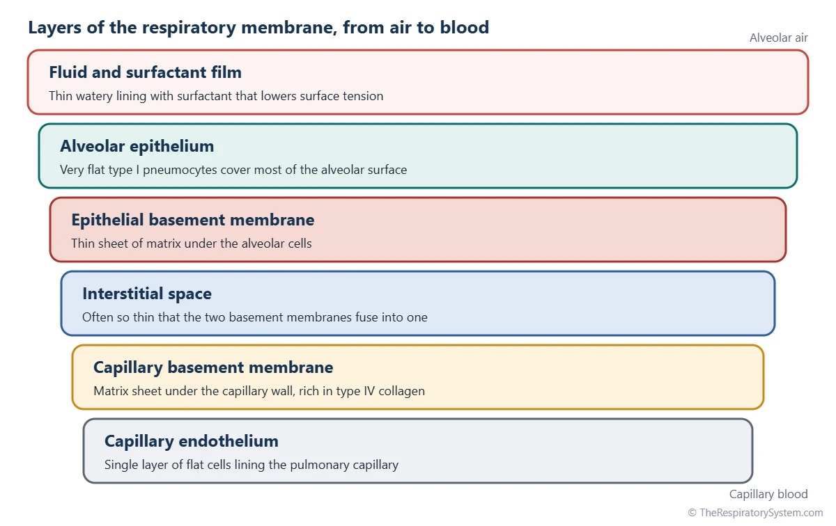
The respiratory membrane has three essential layers, the alveolar epithelium, a basement membrane and the capillary endothelium, and most textbooks describe six layers when the fluid lining and the interstitium are counted separately. From the air side to the blood side they are:
- Fluid and surfactant film, a very thin watery layer topped by pulmonary surfactant.
- Alveolar epithelium, mostly flat type I pneumocytes.
- Epithelial basement membrane, a thin sheet of protein matrix under those cells.
- Interstitial space, which in many places is so narrow that the two basement membranes fuse into one.
- Capillary basement membrane.
- Capillary endothelium, a single layer of flat cells lining the capillary.
The cells of the barrier
Type I pneumocytes are so thin and broad that, although they are fewer in number than type II cells, they cover about 95% of the alveolar surface. Type II pneumocytes, tucked into corners, make surfactant and can divide to replace damaged type I cells. The capillary endothelium is continuous, with no open pores, so blood cells and most proteins stay inside the vessel. Alveolar macrophages crawl over the air side and clear dust and germs, and our respiratory system histology page shows these cells under the microscope.
| Layer | What it is made of | What it does |
|---|---|---|
| Fluid and surfactant film | Thin watery lining topped by surfactant lipids and proteins | Lets gases dissolve; lowers surface tension so alveoli stay open |
| Alveolar epithelium | Type I pneumocytes, with scattered type II cells | Forms the air side of the barrier; type II cells make surfactant |
| Epithelial basement membrane | Protein matrix rich in type IV collagen and laminin | Anchors the alveolar cells and adds strength |
| Interstitial space | Very little on the thin side; fibers and fibroblasts on the thick side | Supports the wall and collects leaked fluid for lymph drainage |
| Capillary basement membrane | Protein matrix, fused with the epithelial one on the thin side | Anchors the endothelium and resists capillary pressure |
| Capillary endothelium | Single layer of flat endothelial cells with no open pores | Forms the blood side; keeps blood cells and proteins in the vessel |
| Beyond the membrane: plasma | Liquid part of the blood in the capillary | Oxygen dissolves here on its way to the red blood cells |
| Beyond the membrane: red cell wall | Cell membrane of the red blood cell | Last step before oxygen binds hemoglobin |
| Source: Standard histology of the adult lung, our editors | ||
What Are the Thin and Thick Sides of the Blood-Air Barrier?
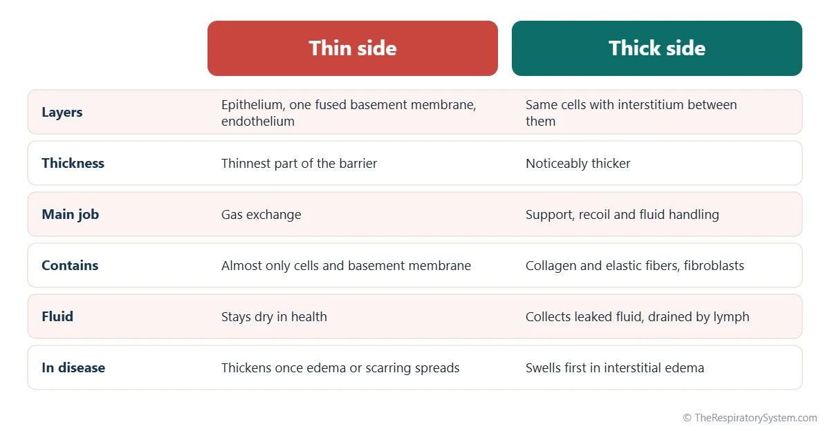
Around each capillary, the blood-air barrier has a thin side, where the basement membranes are fused and gas exchange is fastest, and a thick side, where a little connective tissue lies between them to give strength and handle fluid. The two sides alternate around the capillary like the two faces of a coin.
Thin side
On the thin side, nothing lies between the alveolar cell and the endothelial cell except one shared basement membrane. This is where most oxygen and carbon dioxide cross. In health it stays almost completely dry.
Thick side
On the thick side, the interstitium holds collagen and elastic fibers and a few fibroblasts. These fibers form a skeleton that keeps the alveolar walls in shape and helps the lungs recoil during exhalation. Fluid that leaks out of the capillaries collects here first and is carried away by lymph vessels before it can reach the air sacs.
Thin but strong
The barrier faces a tricky trade-off: it must be thin for diffusion yet strong enough to resist the pressure of the blood inside the capillaries. Much of that strength comes from type IV collagen in the basement membrane. When capillary pressure becomes very high, the barrier can tear, a process physiologists call stress failure.
What Is the Function of the Respiratory Membrane?
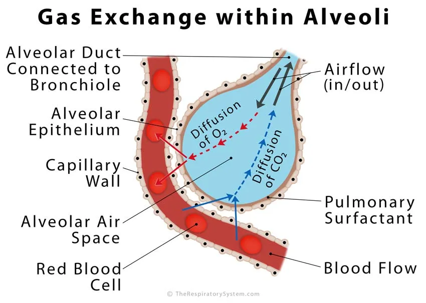
The main function of the respiratory membrane is to let oxygen diffuse from alveolar air into the blood and carbon dioxide diffuse from the blood into the air, while keeping blood, plasma and fluid out of the air spaces. It does this passively, with no pumps, by being very large, very thin and always moist.
Why its shape speeds diffusion
The rate at which a gas crosses a membrane follows Fick's law of diffusion: it rises with surface area and with the difference in partial pressure, and it falls as the membrane gets thicker. The respiratory membrane pushes every factor in the right direction:
- Huge area: about 50 to 75 square meters, spread over hundreds of millions of alveoli.
- Tiny thickness: about 0.5 micrometer on average.
- Steep gradients: kept in place by breathing on one side and constant blood flow on the other.
- Moist surface: gases dissolve in the lining fluid before diffusing, and carbon dioxide dissolves about 20 times more readily than oxygen.
Fast enough with time to spare
At rest, a red blood cell spends about three-quarters of a second in a lung capillary, but its oxygen level normally matches the alveolar air within the first third of that time. This reserve lets the blood stay fully loaded during exercise, when it moves through faster. The full process of loading and unloading is described on our gas exchange in the lungs page.
Other jobs
The capillary side of the membrane also handles chemicals in the blood. Its endothelium carries angiotensin-converting enzyme, which turns angiotensin I into angiotensin II, a hormone that helps control blood pressure.
Common Conditions That Affect the Respiratory Membrane
Diseases harm the respiratory membrane by thickening it, flooding it with fluid, damaging it so it leaks, or destroying its surface area, and each of these slows the diffusion of oxygen into the blood. Our respiratory diseases section covers them in more depth, and anyone with breathing symptoms should see a doctor.
- Pulmonary fibrosis: scar tissue fills the interstitium and thickens the barrier.
- Pulmonary edema: fluid builds up first in the thick side, then spills into the alveoli, lengthening the path for gases.
- ARDS: widespread injury makes the barrier leaky, and protein-rich fluid and debris line the alveoli.
- Pneumonia: infection fills alveoli with fluid and inflammatory cells so air cannot reach the membrane.
- Emphysema: alveolar walls break down, so less membrane is left for gas exchange.
- High-altitude pulmonary edema: very high pressure in the lung capillaries pushes fluid across the barrier; see breathing at high altitude.
When the membrane cannot keep up, the oxygen level of the blood falls, a state called hypoxemia. Oxygen usually suffers first, because it is far less soluble than carbon dioxide.
Key Facts
- The respiratory membrane separates the air in the alveoli from the blood in the pulmonary capillaries.
- It is also called the alveolar-capillary membrane or the blood-air barrier.
- Its three essential layers are the alveolar epithelium, a fused basement membrane and the capillary endothelium.
- It is about 0.5 micrometer thick on average, with a total surface of about 50 to 75 square meters.
- Type I pneumocytes cover about 95% of the alveolar surface and form the air side of the barrier.
- A red blood cell normally finishes loading oxygen within the first third of its time in a lung capillary.
- Fibrosis, edema, ARDS and emphysema all slow gas exchange by thickening, flooding, damaging or destroying the membrane.
Quick Quiz: Test What You Know About the Respiratory Membrane
6 questions. Tap an answer to check it.
What does the respiratory membrane separate?
The respiratory membrane is the barrier between the air in the alveoli and the blood in the pulmonary capillaries.
Which cells form most of the air side of the respiratory membrane?
Very flat type I pneumocytes cover about 95% of the alveolar surface.
About how thick is the respiratory membrane on average?
The barrier averages about 0.5 micrometer, roughly a hundredth of the width of a hair.
By what process do gases cross the respiratory membrane?
Oxygen and carbon dioxide diffuse passively from higher to lower partial pressure.
What happens on the thick side of the blood-air barrier?
The thick side holds collagen and elastic fibers and collects fluid for lymph drainage, while the thin side handles most gas exchange.
Which condition thickens the respiratory membrane with scar tissue?
In pulmonary fibrosis, scar tissue builds up in the interstitium and lengthens the path for gases.
Frequently Asked Questions
How many layers does the respiratory membrane have?
It depends on how they are counted. The three essential layers are the alveolar epithelium, a fused basement membrane and the capillary endothelium. Most physiology textbooks list six: the fluid and surfactant lining, the alveolar epithelium, its basement membrane, the interstitial space, the capillary basement membrane and the capillary endothelium. Both answers describe the same structure.
Is the respiratory membrane the same as the pleura?
No. The pleura is a pair of membranes that wraps the outside of each lung and lines the chest wall, so the lungs can slide smoothly during breathing. The respiratory membrane lies deep inside the lung, in the walls of the alveoli, and separates air from blood. The pleura takes no part in gas exchange.
Why is the respiratory membrane so thin?
Diffusion slows down sharply as the distance a gas must travel increases, so a thin barrier lets oxygen and carbon dioxide cross in a fraction of a second. Keeping the barrier thin also means it can be spread over a huge surface without filling the chest with tissue. Its basement membrane gives it enough strength to resist the pressure of the blood.
How is the function of the respiratory membrane measured?
Doctors use a test called the diffusing capacity of the lungs for carbon monoxide, often written DLCO. The person breathes in a tiny, harmless amount of carbon monoxide and holds the breath for a few seconds. Measuring how much was taken up shows how easily gas passes from the alveoli into the blood across the respiratory membrane.
Why does carbon dioxide cross the respiratory membrane more easily than oxygen?
Carbon dioxide dissolves in water about 20 times more readily than oxygen. Because the membrane is wet and gases must dissolve before they diffuse, carbon dioxide crosses very quickly even though its pressure difference is small. This is why lung diseases that thicken the membrane usually lower blood oxygen long before carbon dioxide starts to build up.
Can the respiratory membrane repair itself?
Yes, to a degree. When type I pneumocytes are damaged, type II pneumocytes can divide and change into new type I cells, rebuilding the alveolar lining. Endothelial cells on the capillary side can also regrow. Repair works best after mild injury. Severe or repeated damage can instead lead to scarring, which thickens the barrier permanently.
How does the respiratory membrane change with age?
With age, some alveolar walls are slowly lost and the remaining air spaces become larger, so the total area of the respiratory membrane gradually shrinks. The lungs also lose some elastic recoil. Diffusing capacity therefore declines slowly over adult life, but in healthy older people the remaining membrane still provides plenty of reserve for everyday breathing.
What is the difference between the respiratory membrane and the alveoli?
The alveoli are the tiny air sacs at the end of the airways, each a hollow space filled with air. The respiratory membrane is the wall where an alveolus meets a capillary. In other words, the alveolus is the room, and the respiratory membrane is the thin shared wall between that room and the blood flowing past it.
Keep Exploring


