At a Glance
Pulmonary circulation runs from the right ventricle through the pulmonary trunk and pulmonary arteries to the capillaries around the alveoli, then back through the pulmonary veins (usually four) to the left atrium. It carries the whole output of the right heart, about 5 liters per minute at rest, at a pulmonary artery pressure of only about 25/8 mmHg, compared with about 120/80 mmHg in the systemic circulation.
- Starts
- Right ventricle, through the pulmonary valve into the pulmonary trunk
- Ends
- Left atrium, through the pulmonary veins (usually two from each lung)
- Main job
- Gas exchange: blood picks up oxygen and gives off carbon dioxide in the alveolar capillaries
- Blood flow
- About 5 liters per minute at rest, the entire output of the right ventricle
- Pressure
- Low: about 25/8 mmHg in the pulmonary artery, mean about 15 mmHg
- Part of
- The double circulation of the heart; works hand in hand with the respiratory system
On this page
- What Is Pulmonary Circulation?
- What Is the Pathway of Pulmonary Circulation?
- What Does the Pulmonary Artery Do?
- What Do the Pulmonary Capillaries Do?
- What Do the Pulmonary Veins Do?
- What Are the Functions of Pulmonary Circulation?
- Pulmonary Circulation vs Systemic Circulation: What Is the Difference?
- What Is the Blood Supply of the Lungs?
- How Is Blood Flow Through the Lungs Controlled?
- Common Conditions That Affect Pulmonary Circulation
- Key Facts
- Quiz
- FAQ
What Is Pulmonary Circulation?
Pulmonary circulation is the loop of blood vessels that carries blood from the heart to the lungs and back, so that the blood can unload carbon dioxide and take up oxygen. It begins at the right ventricle and ends at the left atrium, and every drop of blood that returns from the body must pass through it before it can be pumped out again.
Humans have a double circulation, two circuits joined in series by the heart. The systemic circulation, driven by the left ventricle, supplies the whole body; the pulmonary circulation, driven by the right ventricle, supplies only the lungs. Because the two sides pump the same amount of blood each minute, the lungs receive as much blood as the rest of the body combined.
Pulmonary circulation definition
In one line, pulmonary circulation is the low-pressure circuit of the cardiovascular system that carries deoxygenated blood from the right ventricle through the pulmonary arteries to the pulmonary capillaries for gas exchange, and returns oxygenated blood through the pulmonary veins to the left atrium. It is sometimes called the lesser circulation, because its path is much shorter than the systemic one.
Explained simply
Think of the heart as two pumps glued side by side. The right pump sends tired, used blood on a short trip to the lungs, where it drops off carbon dioxide and fills up with fresh oxygen, like a delivery truck stopping at a depot to swap its load. The refreshed blood then returns to the left pump, which sends it on the long trip around the body.
What Is the Pathway of Pulmonary Circulation?
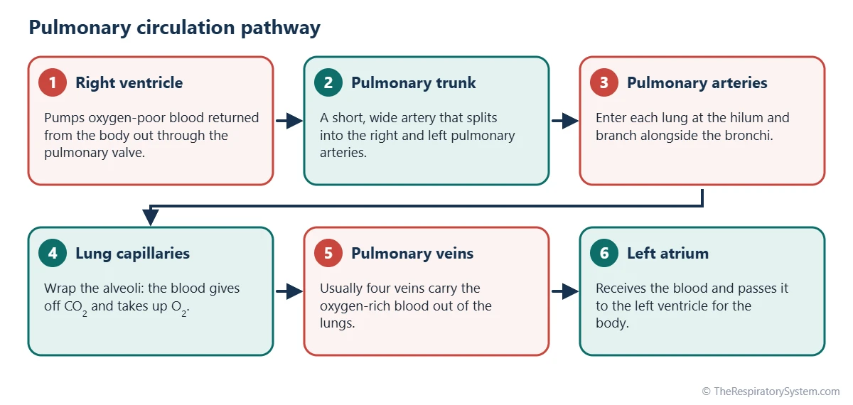
The pathway of pulmonary circulation is right ventricle, pulmonary valve, pulmonary trunk, right and left pulmonary arteries, smaller arteries and arterioles, pulmonary capillaries, venules, pulmonary veins and left atrium. The steps below follow one red blood cell around the circuit, starting where it arrives back at the heart from the body.
- Return to the right atrium. Oxygen-poor blood from the body enters the right atrium through the superior and inferior venae cavae.
- Into the right ventricle. It passes through the tricuspid valve into the right ventricle, the pump of the pulmonary circuit.
- Out through the pulmonary valve. The right ventricle contracts and pushes the blood through the pulmonary valve into the pulmonary trunk.
- Into each lung. The pulmonary trunk divides into the right and left pulmonary arteries, which enter the lungs at the hilum.
- Branching with the airways. The arteries divide again and again alongside the bronchi and bronchioles, ending as arterioles.
- Gas exchange. In the capillaries around the alveoli, the blood gives off carbon dioxide and takes up oxygen.
- Back to the heart. Oxygen-rich blood drains into venules and then into the pulmonary veins, which empty into the left atrium.
From the left atrium, the blood passes through the mitral valve into the left ventricle, which pumps it into the aorta and the systemic circulation. The way the two circuits share this work is explained on our respiratory and circulatory system page, and the matching route of the air is traced on pathway of air.
| Step | Structure | Blood | What happens |
|---|---|---|---|
| 1 | Right atrium | Oxygen-poor | Receives blood from the body through the superior and inferior venae cavae |
| 2 | Tricuspid valve and right ventricle | Oxygen-poor | The right ventricle fills, then contracts to start the pulmonary circuit |
| 3 | Pulmonary valve and pulmonary trunk | Oxygen-poor | Blood is pushed into a short, wide artery about 5 cm long |
| 4 | Right and left pulmonary arteries | Oxygen-poor | One artery enters each lung at the hilum |
| 5 | Lobar and segmental arteries, arterioles | Oxygen-poor | Branches follow the bronchi and bronchioles into every part of the lung |
| 6 | Pulmonary capillaries | Oxygen-poor to oxygen-rich | Oxygen enters and carbon dioxide leaves across the alveolar walls |
| 7 | Venules and pulmonary veins | Oxygen-rich | Small veins between the lobules join into usually four pulmonary veins |
| 8 | Left atrium | Oxygen-rich | Receives the blood and passes it through the mitral valve to the left ventricle |
| Source: Typical adult anatomy, our editors | |||
What Does the Pulmonary Artery Do?
The pulmonary artery carries oxygen-poor blood from the right ventricle to the lungs, making it the only artery in the adult body that carries deoxygenated blood. The name artery describes the direction of flow, away from the heart, not the oxygen content of the blood.
Pulmonary trunk
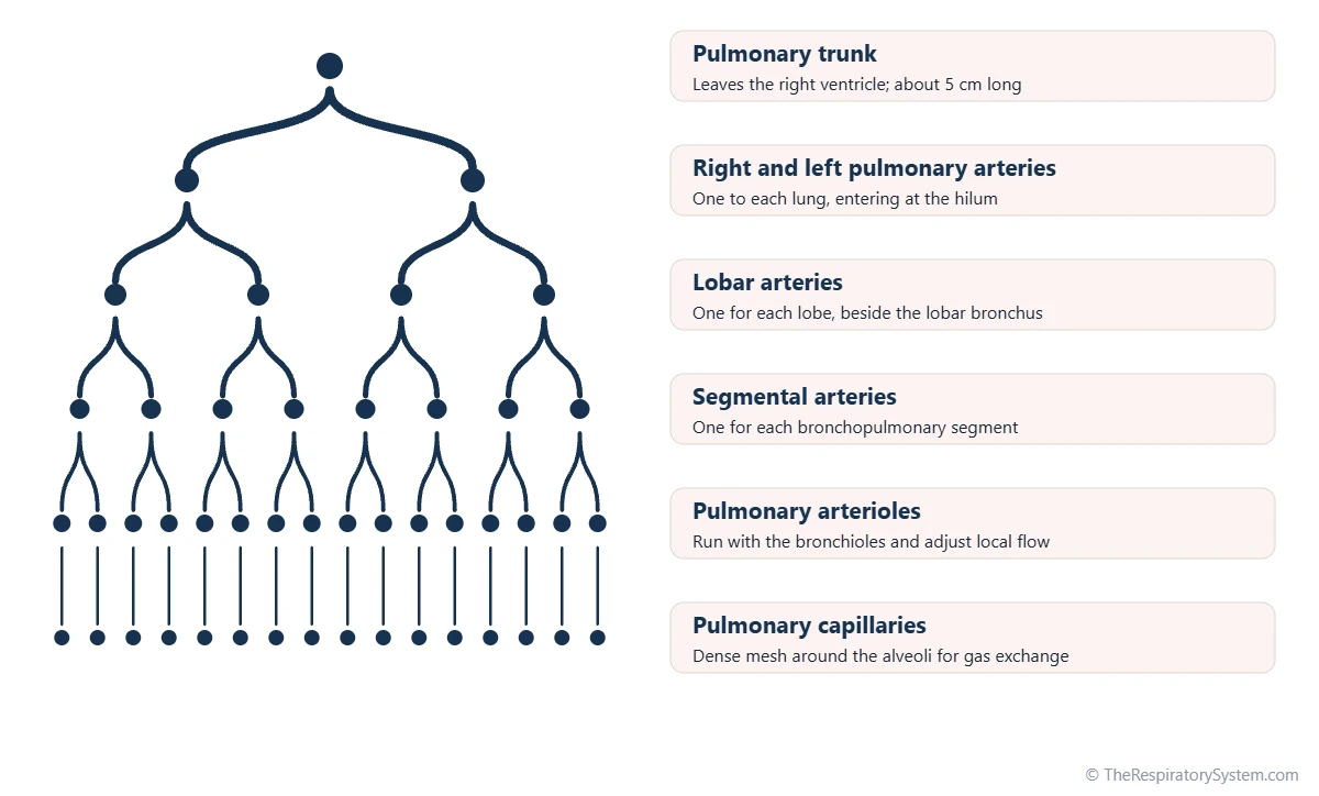
The pulmonary trunk is a short, wide vessel, about 5 cm long and about 3 cm across in an adult, that leaves the top of the right ventricle in front of the aorta. It climbs upward and backward and divides under the arch of the aorta, at about the level of the fourth to fifth thoracic vertebrae, into the right and left pulmonary arteries. A fibrous cord called the ligamentum arteriosum joins the start of the left pulmonary artery to the aortic arch; it is the remnant of a fetal blood vessel.
Right and left pulmonary arteries
The right pulmonary artery is longer and passes behind the ascending aorta and the superior vena cava to reach the right lung. The left pulmonary artery is shorter and runs in front of the descending aorta to the left lung. Inside each lung, the arteries branch into lobar arteries for each of the lung lobes and segmental arteries for each of the bronchopulmonary segments, always running beside the airway they supply.
Pulmonary artery function and wall
Because the pressure in the pulmonary circuit is low, the pulmonary arteries have thin, stretchy walls, about one-third as thick as the wall of the aorta. This stretchiness lets them absorb each beat of the right ventricle and hold extra blood without a large rise in pressure. Their small branches, the pulmonary arterioles, can narrow or widen to steer blood toward the best-ventilated parts of the lung.
What Do the Pulmonary Capillaries Do?
The pulmonary capillaries are where the actual work of the pulmonary circulation happens: their blood exchanges oxygen and carbon dioxide with the air in the alveoli by diffusion. They form such a dense mesh over the alveolar walls that the blood flows almost as a thin sheet rather than through separate tubes.
Each capillary is so narrow that red blood cells pass through it in single file, pressed against the wall. Only the very thin respiratory membrane separates this blood from the alveolar air, and at rest a red blood cell spends about 0.75 second in a capillary, usually becoming fully loaded with oxygen in the first third of that time. The details of the exchange are on our gas exchange page.
Why capillary pressure stays low
The pressure inside the pulmonary capillaries is only about 7 to 10 mmHg, far lower than in the capillaries of the body. This low pressure matters, because it keeps fluid from being pushed out of the blood into the air spaces. When capillary pressure rises, for example when the left side of the heart fails, fluid can leak into the lung tissue and alveoli, a condition called pulmonary edema.
Pulmonary capillaries during exercise
At rest, some pulmonary capillaries carry little or no blood. During exercise, the rising flow opens these closed capillaries, a process called recruitment, and widens those already open, called distension. Together they lower the resistance of the lungs, so the blood flow can rise several-fold while pulmonary artery pressure rises only a little.
What Do the Pulmonary Veins Do?
The pulmonary veins carry oxygen-rich blood from the lungs to the left atrium of the heart, which makes them the only veins in the adult body that carry oxygenated blood. Most people have four pulmonary veins, a superior and an inferior vein from each lung.
The veins begin as venules collecting blood from the alveolar capillaries. Unlike the arteries, which follow the airways, the small veins run in the thin walls of connective tissue between neighboring pulmonary lobules, and only join the bronchi near the hilum. They have no valves, and they leave each lung through its root to open into the back wall of the left atrium.
Pulmonary veins function
Besides returning blood to the heart, the pulmonary veins and the lung's blood vessels as a whole act as a blood reservoir for the left ventricle. Strips of heart muscle extend a short way into the openings of the pulmonary veins, which is why these openings are an important site in the study of heart rhythm. A small amount of oxygen-poor blood from the bronchial veins also drains into the pulmonary veins, one reason arterial blood carries slightly less oxygen than blood leaving the alveoli.
What Are the Functions of Pulmonary Circulation?
The main function of pulmonary circulation is gas exchange, but the lungs' vessels also filter the blood, store a reserve of blood and chemically change some substances that pass through them. These extra jobs are possible because the entire output of the heart flows through the lungs every minute.
- Gas exchange: loading oxygen into the blood and removing carbon dioxide, linking breathing with oxygen transport and carbon dioxide transport.
- Filtering: the narrow capillaries trap small blood clots, clumps of cells and tiny air bubbles from the veins before they can reach the brain or heart arteries.
- Blood reservoir: the lungs hold about 450 mL of blood, about 9% of the body's total, and can shift some of it into the systemic circulation when needed.
- Metabolic work: the lining cells of the lung vessels carry angiotensin-converting enzyme (ACE), which turns angiotensin I into the blood-pressure hormone angiotensin II, and they also break down or remove substances such as bradykinin and serotonin.
- Fluid balance: the low pressure in the capillaries keeps the alveoli dry while the lung lymphatics drain the small amount of fluid that does leak out.
Gas exchange also depends on the flow of blood matching the flow of air in each region of the lung, which is measured by the ventilation-perfusion ratio.
Pulmonary Circulation vs Systemic Circulation: What Is the Difference?
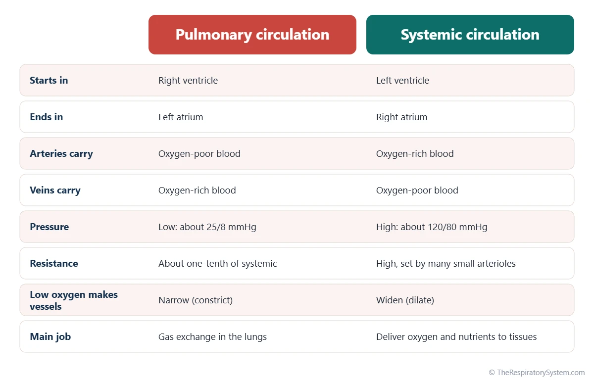
Pulmonary circulation carries blood between the right side of the heart and the lungs at low pressure, while systemic circulation carries blood between the left side of the heart and the rest of the body at high pressure. Both carry the same volume of blood each minute, but almost everything else about them differs.
- Oxygen in the vessels: pulmonary arteries carry oxygen-poor blood and pulmonary veins carry oxygen-rich blood, the reverse of systemic arteries and veins.
- Pressure: the pulmonary artery pressure is about 25/8 mmHg, against about 120/80 mmHg in the aorta.
- Resistance: the pulmonary vessels offer only about one-tenth of the resistance of the systemic vessels, so a much smaller push moves the same flow.
- The pump: the right ventricle has a thinner wall than the left ventricle because it works against this low pressure.
- Response to low oxygen: pulmonary vessels narrow where oxygen is low, while systemic vessels widen.
The last difference is the most surprising. In the body, a tissue short of oxygen needs more blood, so its vessels open up. In the lungs, a region short of oxygen is one with poor airflow, so sending blood there would be wasted, and the vessels close down instead.
| Measure | Pulmonary circulation | Systemic circulation |
|---|---|---|
| Pumping chamber | Right ventricle, thinner wall | Left ventricle, thick muscular wall |
| Main artery pressure | About 25/8 mmHg | About 120/80 mmHg |
| Mean arterial pressure | About 15 mmHg | About 93 mmHg |
| Capillary pressure | About 7 to 10 mmHg | About 30 to 40 mmHg at the arterial end, 10 to 15 at the venous end |
| Vascular resistance | About one-tenth of systemic | About ten times higher |
| Blood flow at rest | About 5 liters per minute | About 5 liters per minute |
| Blood held at one time | About 450 mL, about 9% of the total | Most of the remaining blood, much of it in the veins |
| Main artery wall | Thin, about one-third as thick as the aorta | Thick and elastic (aorta) |
| Response to low oxygen | Vessels constrict | Vessels dilate |
| Source: Typical adult values at rest, our editors | ||
What Is the Blood Supply of the Lungs?
The lungs have a dual blood supply: the pulmonary circulation brings the blood that is to be oxygenated, while the much smaller bronchial circulation brings oxygen-rich blood that feeds the lung tissue itself. The bronchial circulation carries only about 1 to 2% of the heart's output.
Bronchial arteries and veins
The bronchial arteries are small branches of the systemic circulation, usually two on the left from the descending thoracic aorta and one on the right, often from a posterior intercostal artery. They run along the back of the bronchi and supply the walls of the airways down to the terminal bronchioles, the connective tissue of the lung, the lymph nodes at the root of the lung and much of the visceral pleura. The alveolar walls themselves are fed by the pulmonary circulation.
Bronchial veins drain the larger airways into the azygos and hemiazygos veins, which return to the right side of the heart. Blood from the smaller airways drains into the pulmonary veins instead, so it reaches the left atrium without passing through the alveolar capillaries. Because of this double supply, lung tissue often survives when a branch of the pulmonary artery is blocked.
How Is Blood Flow Through the Lungs Controlled?
Blood flow through the lungs is set mainly by the output of the right ventricle and distributed by local factors: the oxygen level in each region, gravity and the opening of reserve capillaries. Nerves and hormones have only a small effect on the pulmonary vessels compared with the body's arteries.
Hypoxic pulmonary vasoconstriction
When an area of lung receives too little air, its alveolar oxygen falls and the small arteries that supply it constrict. This response, called hypoxic pulmonary vasoconstriction, diverts blood to better-ventilated areas and keeps air and blood matched. If the whole lung is short of oxygen, as at high altitude, the same response narrows vessels everywhere and raises pulmonary artery pressure.
The effect of gravity
Because pulmonary pressures are so low, gravity has a large effect on them. In an upright person, the lower parts of the lungs receive much more blood than the tops, where the capillaries may be almost collapsed between heartbeats. Lying down evens out the flow between top and bottom.
How pulmonary circulation changes at birth
Before birth the lungs are filled with fluid, their vessels are tightly constricted and most blood bypasses them through two shortcuts: the foramen ovale between the atria and the ductus arteriosus between the pulmonary trunk and the aorta. With the first breaths, the alveoli fill with air, pulmonary resistance falls sharply and blood floods into the lungs. The foramen ovale closes as left atrial pressure rises, and the ductus arteriosus closes over the following days, leaving the ligamentum arteriosum, as described on our development of the respiratory system page.
Common Conditions That Affect Pulmonary Circulation
Conditions of the pulmonary circulation block its vessels, raise its pressure or let fluid leak from its capillaries, and each of them reduces how well the lungs can load the blood with oxygen. Our respiratory diseases section covers lung conditions in more depth, and sudden breathlessness or chest pain is a reason to see a doctor.
- Pulmonary embolism: a blood clot, usually from the deep veins of the legs, lodges in a pulmonary artery and stops blood from reaching part of the lung.
- Pulmonary hypertension: abnormally high pressure in the pulmonary arteries, which makes the right ventricle work harder and can strain it over time.
- Cor pulmonale: enlargement and failure of the right ventricle caused by long-standing lung disease or pulmonary hypertension.
- Pulmonary edema: fluid leaking from the capillaries into the lung tissue and alveoli, most often because of left-sided heart failure.
- Congenital heart defects: openings between the heart's chambers or vessels, such as a ductus arteriosus that stays open, can send too much or too little blood through the lungs.
Long-term lung diseases such as COPD and pulmonary fibrosis can also raise pulmonary pressure, because low oxygen and damaged tissue narrow or destroy the small vessels. When gas exchange suffers, blood oxygen falls, a state described on our hypoxia and hypoxemia page.
Key Facts
- Pulmonary circulation carries blood from the right ventricle to the lungs and back to the left atrium.
- The pulmonary arteries are the only adult arteries that carry oxygen-poor blood, and the pulmonary veins the only adult veins that carry oxygen-rich blood.
- The pulmonary trunk is about 5 cm long and divides under the aortic arch into the right and left pulmonary arteries.
- Pulmonary artery pressure is about 25/8 mmHg, roughly one-fifth of the pressure in the aorta, and pulmonary resistance is about one-tenth of systemic resistance.
- At rest a red blood cell spends about 0.75 second in a pulmonary capillary, where gas exchange takes place.
- The lungs hold about 450 mL of blood, about 9% of the body's total, and also filter small clots from the venous blood.
- Low oxygen makes pulmonary vessels constrict, the opposite of systemic vessels, which steers blood toward well-ventilated alveoli.
Quick Quiz: Test What You Know About the Pulmonary Circulation
6 questions. Tap an answer to check it.
Which chamber of the heart pumps blood into the pulmonary circulation?
The right ventricle pumps oxygen-poor blood through the pulmonary valve into the pulmonary trunk.
What kind of blood do the pulmonary veins carry?
The pulmonary veins return oxygen-rich blood from the lungs to the left atrium.
What is the typical pressure in the pulmonary artery of a healthy adult at rest?
Pulmonary artery pressure is about 25/8 mmHg, far lower than the systemic pressure of about 120/80 mmHg.
Where does gas exchange take place in the pulmonary circulation?
Oxygen and carbon dioxide are exchanged in the capillaries that wrap around the alveoli.
How do pulmonary arterioles respond to low oxygen in an area of lung?
Hypoxic pulmonary vasoconstriction narrows the vessels so blood is diverted to better-ventilated areas.
Which vessels supply oxygen-rich blood to the walls of the bronchi?
The bronchial arteries, small branches of the systemic circulation, feed the airway walls and supporting lung tissue.
Frequently Asked Questions
Does the pulmonary artery carry oxygenated or deoxygenated blood?
The pulmonary artery carries deoxygenated, oxygen-poor blood. It takes blood that has already delivered oxygen to the body from the right ventricle to the lungs. Arteries are defined by carrying blood away from the heart, not by their oxygen content, so the pulmonary artery is an exception to the usual rule that arteries carry oxygen-rich blood, along with the umbilical arteries before birth.
Why is pulmonary circulation called the lesser circulation?
It is called the lesser or minor circulation because its route is much shorter than the systemic circulation and its pressure is much lower. Blood travels only from the heart to the lungs and straight back, a path of a few tens of centimeters. The systemic circulation, the greater circulation, reaches every organ from the brain to the toes, so it needs a stronger pump.
How long does it take blood to pass through the pulmonary circulation?
At rest, blood takes roughly 4 to 6 seconds to travel from the right ventricle through the lungs to the left atrium, estimated from the lungs holding about 450 mL of blood with a flow of about 5 liters per minute. Of that time, a red blood cell spends only about 0.75 second in the capillaries where gas exchange happens, and less during hard exercise.
Who discovered pulmonary circulation?
The first known accurate description of pulmonary circulation came from Ibn al-Nafis, a physician in Cairo, in the 13th century. He argued that blood travels from the right side of the heart to the lungs and back rather than passing through the wall between the ventricles. In Europe, Michael Servetus and Realdo Colombo described it in the 16th century, and William Harvey explained the full circulation in 1628.
Is pulmonary circulation part of the respiratory or circulatory system?
Pulmonary circulation is part of the circulatory system, because it consists of the heart's right ventricle and a set of blood vessels. It is also the point where the circulatory and respiratory systems meet: the capillaries of the pulmonary circuit lie against the alveoli of the lungs, so neither system could exchange gases without the other.
How many pulmonary veins are there?
Most people have four pulmonary veins: a superior and an inferior vein from the right lung and the same pair from the left lung. Variation is common, though. Some people have a single shared vein on one side, and others have an extra vein, most often a separate right middle lobe vein, giving five. These variations usually cause no problems.
What is pulmonary capillary wedge pressure?
Pulmonary capillary wedge pressure is a measurement taken by passing a thin catheter with a small balloon through the right side of the heart into a branch of the pulmonary artery. When the balloon wedges in the vessel and blocks the flow, the pressure measured beyond it reflects the pressure in the left atrium. In healthy adults it is typically about 6 to 12 mmHg.
Do all animals have a pulmonary circulation?
No. Fish have a single circulation: the heart pumps blood through the gills and then straight on to the body. Amphibians and most reptiles have a partly divided double circulation, so some mixing of oxygen-rich and oxygen-poor blood occurs. Birds and mammals, including humans, have a fully separated pulmonary circulation driven by its own ventricle, which supports their high energy use.
Keep Exploring


