At a Glance
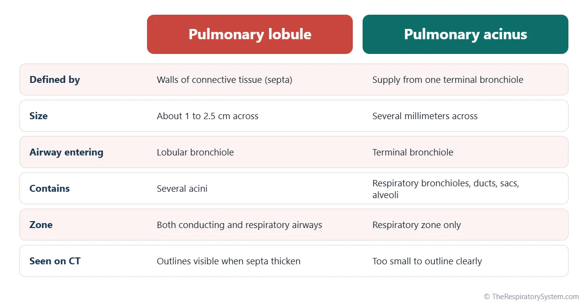
The pulmonary lobule, usually meaning the secondary pulmonary lobule, is the smallest unit of the lung surrounded by its own connective tissue septa, about 1 to 2.5 cm across. A lobular bronchiole and a pulmonary artery branch enter its center, while veins and lymphatics run in its walls. Inside, each terminal bronchiole supplies one pulmonary acinus, the gas-exchange unit of respiratory bronchioles, alveolar ducts, sacs and alveoli.
- Location
- Throughout both lungs, inside each bronchopulmonary segment; most clearly outlined near the lung surface
- Size
- Secondary lobule about 1 to 2.5 cm across; acinus only several millimeters
- Bounded by
- Interlobular septa, thin walls of connective tissue carrying veins and lymphatics
- Function
- Packages the gas-exchange tissue into small units, each with its own airway and blood supply
- Contains
- Several acini, each beyond one terminal bronchiole
- Part of
- Lung lobes and bronchopulmonary segments, in the lower respiratory tract
On this page
What Is a Pulmonary Lobule?
A pulmonary lobule is a small, many-sided compartment of lung tissue that is outlined by thin walls of connective tissue and fed by one small airway, the lobular bronchiole, together with a branch of the pulmonary artery. It is the smallest part of the lung that can be seen as a separate unit with the naked eye, and it is often called a lung lobule.
Pulmonary lobule definition
In anatomy and radiology, the term usually means the secondary pulmonary lobule, defined as the smallest unit of lung bounded by connective tissue septa. Each one is roughly polyhedral, about 1 to 2.5 cm across, with its narrower end often pointing toward the center of the lung. An older term, the primary lobule, describes the tiny unit beyond the last respiratory bronchiole, but it is rarely used today.
Where it fits in the lung
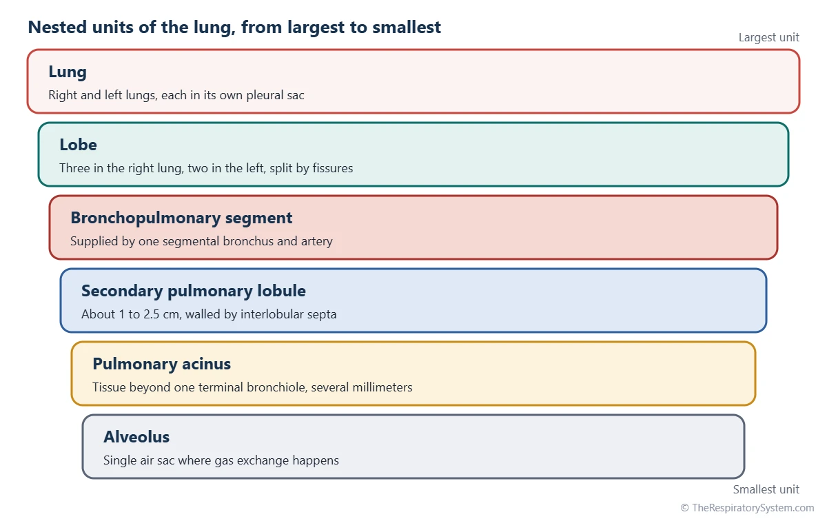
The lungs are built as nested units. Each lung is divided into lobes, each lobe into bronchopulmonary segments, each segment into many lobules, and each lobule into several acini, which finally open into the alveoli. The diagram above shows this ladder from largest to smallest.
Explained simply
Think of the lung as a huge bunch of grapes. The lobes are the big branches of the bunch, the lobules are the small clusters on each branch, wrapped in thin skin, and the acini are the tiny grape groups inside each cluster where the real work of exchanging gases happens.
What Is the Anatomy of a Pulmonary Lobule?
A pulmonary lobule has a central core that holds the airway and artery, a mass of acini filling its interior, and an outer wall of interlobular septa that holds the veins and lymphatic vessels. This split between a central supply and a peripheral drainage is the key to its anatomy.
The center: lobular core
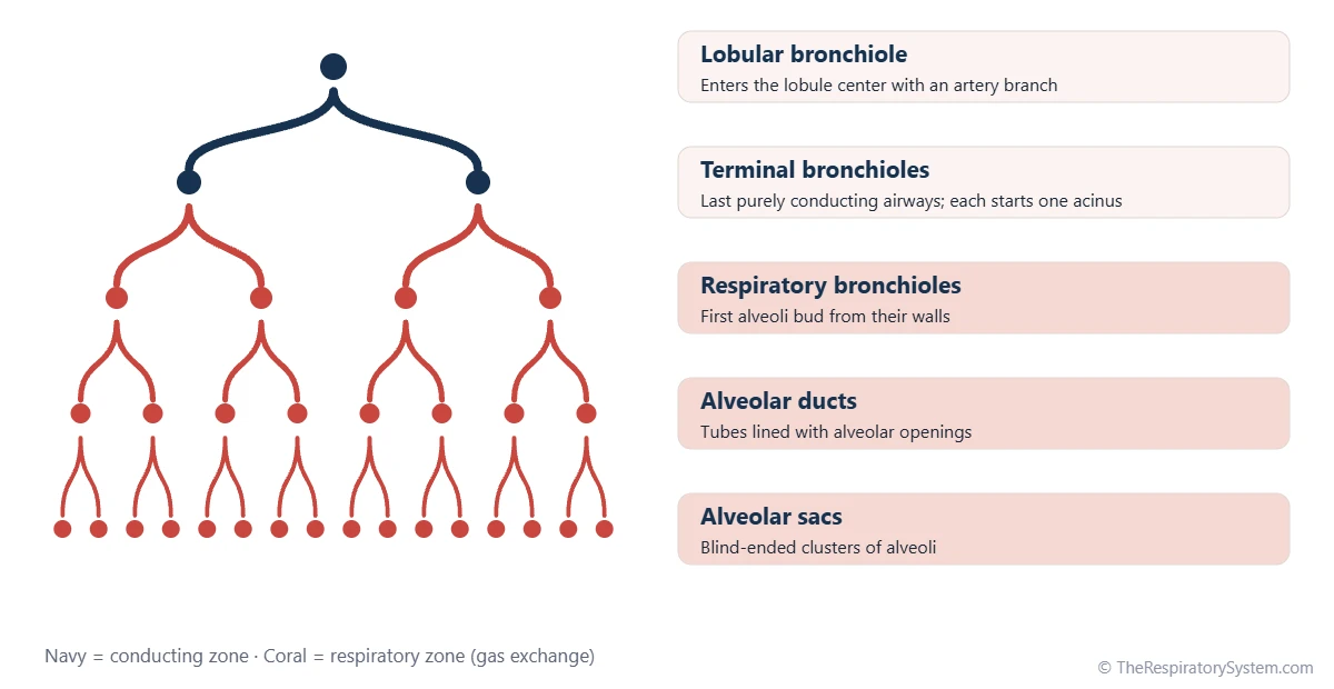
A lobular bronchiole, about a millimeter wide, enters the middle of the lobule side by side with a branch of the pulmonary artery. Inside the lobule the bronchiole divides into several terminal bronchioles, and the artery branches with it, so every airway has a matching blood vessel. Small lymphatic vessels also run along this central bundle.
The middle: the acini
Most of the lobule is filled with acini and their alveoli, the working gas-exchange tissue. Each terminal bronchiole leads to one acinus, so a lobule holds as many acini as it has terminal bronchioles, usually around a dozen or fewer, though the number varies widely from one lobule to the next.
The edge: interlobular septa
The thin walls between lobules are called interlobular septa. They carry the small pulmonary veins, which collect oxygen-rich blood from the edges of the lobule, along with lymphatic vessels that drain fluid. Near the surface of the lung the septa join the visceral pleura, which is why faint many-sided outlines of lobules, often marked by dark carbon pigment in adults, can be seen on the lung surface.
| Part | Where in the lobule | What it does |
|---|---|---|
| Lobular bronchiole | Enters at the center | Brings air into the lobule |
| Pulmonary artery branch | Center, beside the bronchiole | Brings oxygen-poor blood to the acini |
| Terminal bronchioles | Branch from the lobular bronchiole | Last purely conducting airways; each starts one acinus |
| Respiratory bronchioles | Beginning of each acinus | First airways with alveoli in their walls |
| Alveolar ducts and sacs | Filling the acini | Lead air to the alveoli |
| Alveoli | Throughout the acini | Site of gas exchange |
| Interlobular septa | Outer walls of the lobule | Separate lobules and carry veins and lymphatics |
| Pulmonary veins | Within the septa | Carry oxygen-rich blood out of the lobule |
| Lymphatic vessels | Septa and central bundle | Drain excess fluid to the lymph nodes |
| Visceral pleura | Covers lobules at the lung surface | Joins the septa; shows lobule outlines |
| Source: Standard adult anatomy, our editors | ||
What Is the Pulmonary Acinus?
The pulmonary acinus is all the lung tissue supplied by one terminal bronchiole: its respiratory bronchioles, alveolar ducts, alveolar sacs and alveoli. It is the largest unit of the lung in which every part takes part in gas exchange, and it is often called the functional unit of the lung.
An acinus is tiny, only several millimeters across, yet it holds thousands of alveoli. Its airways begin with the respiratory bronchioles, whose walls carry the first scattered alveoli, and continue into the alveolar ducts and sacs, which are lined almost entirely by alveoli. Everything before the acinus belongs to the conducting zone and everything inside it to the respiratory zone, a boundary explained on our conducting zone vs respiratory zone page.
Pulmonary acinus vs alveoli
An alveolus is a single, cup-shaped air sac with a wall only a cell thick. An acinus is a whole cluster of airways and thousands of alveoli grouped around one terminal bronchiole. In other words, alveoli are the building blocks, and the acinus is the small neighborhood they form together.
What Is the Function of the Pulmonary Lobule?
The function of the pulmonary lobule is to organize the lung's gas-exchange tissue into small, self-contained units, each with its own airway and artery in the center and its own venous and lymphatic drainage around the edge. This layout keeps the paths for air, blood and fluid short and orderly.
- Delivering air: the lobular bronchiole spreads incoming air to all the acini of the lobule.
- Matching air and blood: because each airway branch travels with an artery branch, the blood reaching each acinus is lined up with the air reaching it, supporting efficient gas exchange.
- Draining blood: oxygen-rich blood leaves through veins in the septa and joins the pulmonary veins of the pulmonary circulation.
- Draining fluid: lymphatics in the septa and the central bundle remove excess fluid, keeping the alveoli dry.
- Limiting spread: the septa partly separate neighboring lobules, so some disease processes stay confined to one lobule while others spread through the septa.
Inside each acinus, small openings between alveoli and between neighboring airways allow some collateral air flow, which helps keep alveoli open if a small airway becomes blocked.
Why Are Pulmonary Lobules Important on CT Scans?
Pulmonary lobules are important on high-resolution CT scans because many lung diseases affect a predictable part of the lobule, so the pattern of change inside the lobules helps radiologists narrow down the cause. In healthy lungs the lobules are hard to see; they become visible when their walls or centers are altered.
Radiologists describe findings by their position in the lobule. Changes near the center point to processes in the small airways or arteries, thickening of the septa points to the veins or lymphatics, and changes spread evenly through the whole lobule point to the alveoli themselves. A thickened septum seen at the lung base on a plain chest X-ray is called a Kerley B line. Studying the bronchial tree and the lobule together is a basis of chest imaging, and the tissue itself is described on our respiratory system histology page.
Common Conditions That Affect the Pulmonary Lobule
Conditions that affect the pulmonary lobule are often classified by which part of the lobule or acinus they damage, such as the center, the whole lobule or the septa at its edge. Our respiratory diseases section covers these conditions in more depth.
- Centrilobular emphysema: damage starts around the respiratory bronchioles in the center of the lobule and is strongly linked to smoking; it is the most common form of emphysema in COPD and usually affects the upper lobes most.
- Panlobular emphysema: the whole acinus and lobule are destroyed evenly, typically in the lower lobes; it is the classic pattern in alpha-1 antitrypsin deficiency.
- Paraseptal emphysema: damage lies along the septa and the pleura at the edge of lobules and can form air blisters that may burst, causing a pneumothorax.
- Septal thickening: fluid in the septa, as in pulmonary edema from heart failure, or cancer spreading through the lymphatics thickens the walls between lobules.
- Small airway infection: infections of the bronchioles can fill the center of lobules, producing small branching shadows on CT described as a tree-in-bud pattern.
Anyone with ongoing breathlessness or cough should see a doctor.
Key Facts
- A secondary pulmonary lobule is the smallest unit of the lung surrounded by its own connective tissue walls, about 1 to 2.5 cm across.
- A lobular bronchiole and a pulmonary artery branch enter the center of each lobule, while veins and lymphatics run in its walls.
- Each lobule holds several acini, one for each terminal bronchiole inside it.
- The pulmonary acinus includes the respiratory bronchioles, alveolar ducts, alveolar sacs and alveoli beyond one terminal bronchiole.
- The acinus is the largest lung unit in which every part takes part in gas exchange.
- Faint many-sided outlines on the lung surface are the edges of lobules, often marked by carbon pigment in adults.
- Types of emphysema are named by where they strike the lobule: centrilobular, panlobular or paraseptal.
Quick Quiz: Test What You Know About the Pulmonary Lobule and Acinus
6 questions. Tap an answer to check it.
What forms the walls of a secondary pulmonary lobule?
Each secondary lobule is outlined by interlobular septa, thin walls of connective tissue.
What enters the center of a pulmonary lobule?
The lobular bronchiole and its matching pulmonary artery branch run together into the lobule's center.
A pulmonary acinus is the lung tissue supplied by one:
Everything beyond one terminal bronchiole forms one acinus.
Where do the small pulmonary veins of a lobule run?
Veins run in the septa at the edge of the lobule, away from the central bronchiole and artery.
About how wide is a secondary pulmonary lobule?
Secondary lobules are typically about 1 to 2.5 cm across.
Which form of emphysema starts in the center of the lobule and is linked to smoking?
Centrilobular emphysema begins around the respiratory bronchioles in the lobule's center.
Frequently Asked Questions
What is the difference between a lung lobe and a lung lobule?
A lobe is one of the five large divisions of the lungs, three on the right and two on the left, separated by deep fissures and supplied by a lobar bronchus. A lobule is a much smaller unit, about 1 to 2.5 cm across, outlined by thin connective tissue walls and supplied by a lobular bronchiole. Each lobe contains a very large number of lobules.
What is the difference between a primary and a secondary pulmonary lobule?
The secondary pulmonary lobule is the unit outlined by connective tissue septa, about 1 to 2.5 cm across, and is the meaning used in most textbooks and radiology reports. The primary lobule is an older term for the small unit beyond the last respiratory bronchiole, made of alveolar ducts, sacs and alveoli. Because it is hard to identify, the primary lobule is rarely mentioned today.
Is the acinus the functional unit of the lung?
The pulmonary acinus is widely described as the functional unit of the lung because it is the largest unit in which every airway and air space takes part in gas exchange. Some books call the alveolus the functional unit instead, since it is the site of exchange itself. Both views are correct at different scales: the alveolus is the basic structure, and the acinus is the working group.
How many acini are in a pulmonary lobule?
The number of acini in a secondary lobule varies widely, because lobules differ in size and in how many terminal bronchioles they contain. A typical lobule holds roughly a dozen or fewer, but small lobules may have only a few and large ones many more. Since each terminal bronchiole supplies one acinus, counting terminal bronchioles gives the number of acini.
Can you see pulmonary lobules on an X-ray?
Normal pulmonary lobules are generally not visible on a plain chest X-ray because their walls are too thin. When the septa between lobules thicken, for example with fluid in heart failure, they can appear as short horizontal lines near the lung base called Kerley B lines. High-resolution CT shows lobules in much more detail and is the main imaging tool for studying them.
Are lobules the same as bronchopulmonary segments?
No. A bronchopulmonary segment is a much larger unit, supplied by a segmental bronchus and an artery branch, and each lung has about ten of them. Segments can be removed surgically as separate units. A pulmonary lobule is a far smaller subdivision within a segment, and many lobules pack together to build each segment.
Why are pulmonary lobules shaped like polygons?
Lobules are packed tightly together inside the lung, and like soap bubbles or cells in a honeycomb, units pressed against their neighbors take on flat-sided, many-sided shapes. Near the lung surface, the lobules that touch the pleura often look like irregular pyramids or cones, with their base at the surface and their narrower end pointing toward the center of the lung.
Keep Exploring


