At a Glance
Terminal bronchioles are the narrowest purely air-conducting airways, about 0.5 mm wide, lined by ciliated cuboidal cells and club cells with no cartilage or goblet cells. Respiratory bronchioles branch from them and carry scattered alveoli in their walls, so they are the first part of the respiratory zone and the start of each pulmonary acinus.
- Location
- Deep inside each lung, at the end of the bronchial tree, inside the pulmonary lobules
- Function
- Terminal: conduct air only. Respiratory: conduct air and begin gas exchange
- Tissue
- Simple cuboidal epithelium with club cells, smooth muscle, elastic fibers; no cartilage
- Size
- Terminal bronchioles about 0.5 mm wide; respiratory bronchioles smaller
- Connects to
- Lobular bronchioles above, alveolar ducts and alveolar sacs below
- Part of
- Bronchial tree, lower respiratory tract
On this page
- What Are Terminal and Respiratory Bronchioles?
- Where Are the Terminal Bronchioles Located?
- Terminal and Respiratory Bronchioles Histology
- What Is the Function of the Terminal Bronchioles?
- What Do the Respiratory Bronchioles Do?
- Related Structures: Lobular Bronchioles, Alveolar Ducts and the Acinus
- Common Conditions That Affect the Terminal and Respiratory Bronchioles
- Key Facts
- Quiz
- FAQ
What Are Terminal and Respiratory Bronchioles?
Terminal and respiratory bronchioles are the two smallest kinds of airway in the lungs: terminal bronchioles end the air-conducting system, and respiratory bronchioles begin the gas-exchange system. They are the final branches of the bronchioles, which in turn are the smallest tubes of the bronchial tree. This entry belongs to our respiratory system glossary.
The terminal bronchiole is defined in anatomy as the last airway without alveoli. Everything above it, from the nose to the terminal bronchiole, only moves air. The respiratory bronchiole is the first airway with alveoli opening from its wall, which is why it marks the line between the conducting zone and the respiratory zone.
Explained simply
Picture the airways as a tree turned upside down. The terminal bronchioles are the thinnest twigs that still have plain bark. The respiratory bronchioles are the next twigs, and they already carry a few small grapes, the alveoli, along their sides. From here on, the twigs end in full bunches of grapes where oxygen enters the blood.
Where Are the Terminal Bronchioles Located?
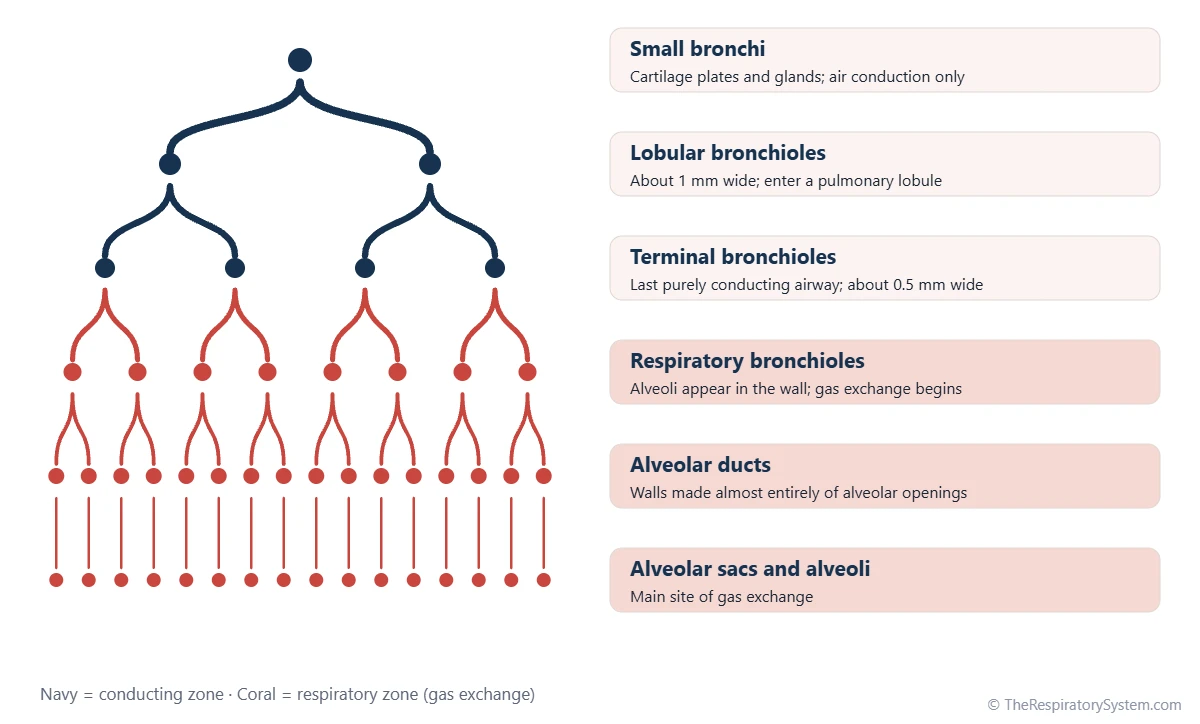
The terminal bronchioles are located deep inside the lungs, inside the small units of lung tissue called pulmonary lobules, after many rounds of branching from the trachea. In the classic Weibel model of the airways, the trachea is generation 0 and the terminal bronchioles sit at about generation 16.
Each lobule receives a lobular bronchiole that splits into several terminal bronchioles. Each terminal bronchiole then supplies one pulmonary acinus, the cluster of respiratory bronchioles, alveolar ducts and alveolar sacs that hangs from it. The pulmonary lobule and acinus entry explains how these units fit together.
- Terminal bronchioles: about generation 16 in the Weibel model, the end of the conducting zone.
- Respiratory bronchioles: about generations 17 to 19, usually three successive orders, each with more alveoli than the last.
- Alveolar ducts and sacs: about generations 20 to 23, the end of the tree.
Terminal and Respiratory Bronchioles Histology
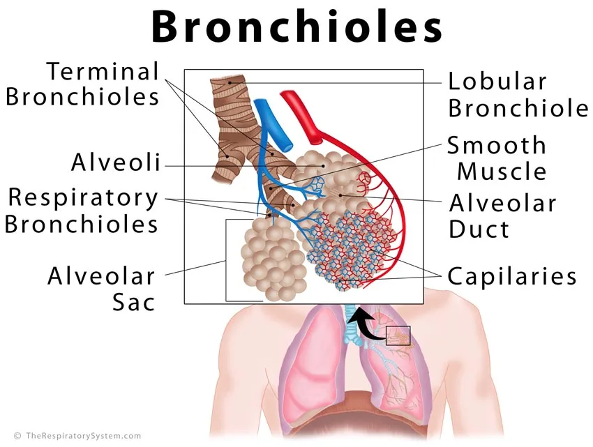
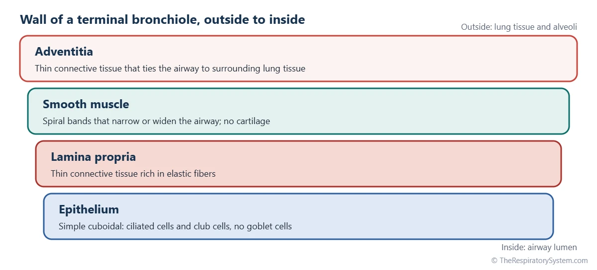
Under the microscope, terminal bronchioles have a thin wall lined by simple cuboidal epithelium made of ciliated cells and club cells, a narrow layer of connective tissue, and a ring of smooth muscle, with no cartilage, no glands and no goblet cells. Respiratory bronchioles have a similar but thinner wall that is broken up by alveolar openings.
Terminal bronchiole histology
The lining of a terminal bronchiole is a single layer of cube-shaped cells. Many still carry cilia, which keep the mucociliary escalator running up to this level, while dome-shaped club cells bulge into the lumen between them. Beneath the lining lies a thin lamina propria rich in elastic fibers, then spiral bands of smooth muscle, then a thin adventitia that ties the airway to the surrounding lung tissue.
Respiratory bronchiole histology
The respiratory bronchiole starts with the same cuboidal lining, but the cells become lower, cilia become fewer and club cells more common. At intervals the wall opens into single alveoli lined by type I and type II pneumocytes. Between the openings, thin bands of smooth muscle and elastic fibers remain. The overall shift from tall ciliated cells in larger airways to these flat cells is traced in our respiratory epithelium and respiratory system histology pages.
What club cells do
Club cells, formerly called Clara cells, are the signature cell of the terminal bronchiole. They secrete a protective protein known as club cell secretory protein, help break down inhaled toxins with enzymes of the cytochrome P450 family, and can divide to replace damaged lining cells.
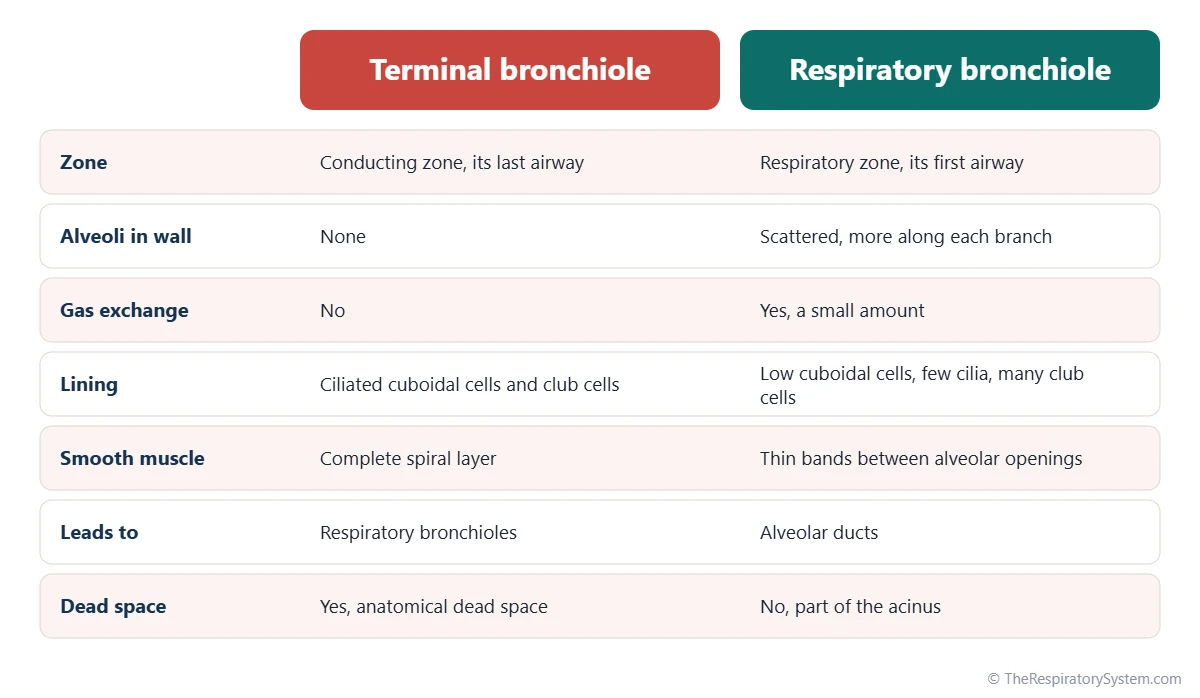
| Feature | Terminal bronchioles | Respiratory bronchioles |
|---|---|---|
| Zone | Conducting zone (last airway of it) | Respiratory zone (first airway of it) |
| Weibel generation | About 16 | About 17 to 19 |
| Width | About 0.5 mm | Smaller than terminal bronchioles |
| Epithelium | Ciliated simple cuboidal with club cells | Low cuboidal, few cilia, many club cells |
| Goblet cells | None | None |
| Cartilage and glands | None | None |
| Smooth muscle | Complete layer of spiral bands | Thin bands between alveolar openings |
| Alveoli in the wall | None | Scattered, more numerous along each branch |
| Gas exchange | None, part of anatomical dead space | Some, through the alveoli in the wall |
| Branches into | Respiratory bronchioles | Alveolar ducts |
| Source: Typical adult anatomy, our editors | ||
What Is the Function of the Terminal Bronchioles?
The function of the terminal bronchioles is to deliver air to the gas-exchange region and spread it evenly among the acini, without taking part in gas exchange themselves. Because they contain no alveoli, the air inside them belongs to the anatomical dead space.
- Air distribution: they hand inhaled air to the respiratory bronchioles of each acinus.
- Airflow control: their smooth muscle narrows the airway when the vagus nerve releases acetylcholine and relaxes in response to adrenaline acting on beta-2 receptors.
- Clearance: ciliated cells move mucus and trapped particles upward, although mucus is scarce at this level.
- Protection and repair: club cells secrete protective proteins, neutralize some inhaled toxins and renew the lining after injury.
No cartilage holds these tiny tubes open. Instead, the elastic fibers of the surrounding lung tissue pull outward on their walls, which keeps them open during breathing in and lets them narrow slightly during breathing out.
What Do the Respiratory Bronchioles Do?
Respiratory bronchioles do two jobs at once: they conduct air onward to the alveolar ducts, and the alveoli along their walls carry out a small share of gas exchange. They are the transition airway between pure conduction and full exchange.
The number of airways at each level doubles roughly with every branching, so by the time air reaches the respiratory bronchioles, the total cross-sectional area of all the tubes together is enormous. Air slows almost to a stop here. Beyond this point, oxygen and carbon dioxide move mainly by diffusion rather than by bulk flow, which is why the change from tube to exchange surface happens at this level.
The alveoli on respiratory bronchioles are lined with the same cells and covered by the same pulmonary surfactant as all other alveoli, and they share the same capillary network of the pulmonary circulation. Wandering alveolar macrophages patrol them and remove fine dust that has reached this far.
Common Conditions That Affect the Terminal and Respiratory Bronchioles
Conditions of the smallest airways usually narrow, inflame or destroy the terminal and respiratory bronchioles, and they are grouped under the name small airway disease. The wider picture is covered in our respiratory diseases section.
- Centriacinar emphysema: the form of emphysema most linked to cigarette smoke, which destroys the respiratory bronchioles in the center of each acinus.
- Respiratory bronchiolitis: a mild inflammation seen in smokers, with pigmented macrophages collecting in the respiratory bronchioles.
- Small airway narrowing in COPD: thickened, inflamed bronchiole walls are an early part of COPD.
- Asthma: in asthma, smooth muscle spasm and inflammation narrow the small airways along with the larger ones.
- Bronchiolitis: a viral infection of the bronchioles that is common in infants, often caused by respiratory syncytial virus (RSV).
- Bronchiolitis obliterans: scarring that blocks the small airways after certain injuries, infections or transplants.
Anyone with ongoing cough or shortness of breath should see a doctor for a proper assessment.
Key Facts
- Terminal bronchioles are the last airways of the conducting zone and have no alveoli in their walls.
- Respiratory bronchioles are the first airways of the respiratory zone, with alveoli opening from their walls.
- A terminal bronchiole is about 0.5 mm wide and sits at about generation 16 of the bronchial tree in the Weibel model.
- Both are lined by simple cuboidal epithelium with club cells and contain no cartilage, glands or goblet cells.
- Each terminal bronchiole supplies one pulmonary acinus, the basic gas-exchange unit of the lung.
- Centriacinar emphysema, linked to smoking, destroys the respiratory bronchioles first.
Quick Quiz: Test What You Know About the Terminal and Respiratory Bronchioles
6 questions. Tap an answer to check it.
What is the terminal bronchiole?
The terminal bronchiole is the last airway of the conducting zone; it has no alveoli in its wall.
Which airway is the first part of the respiratory zone?
Respiratory bronchioles have alveoli in their walls, so gas exchange starts there.
Which cell type is typical of the terminal bronchiole lining?
Dome-shaped club cells sit between ciliated cuboidal cells; goblet cells are absent at this level.
What supports the terminal bronchioles instead of cartilage?
With no cartilage, the outward pull of elastic lung tissue keeps these small airways open.
What do respiratory bronchioles lead into?
Each respiratory bronchiole ends in alveolar ducts, which open into alveolar sacs.
Which form of emphysema mainly damages the respiratory bronchioles?
Centriacinar emphysema, linked to smoking, destroys the respiratory bronchioles in the center of the acinus.
Frequently Asked Questions
What is the difference between terminal and respiratory bronchioles?
Terminal bronchioles only carry air and have no alveoli, so they belong to the conducting zone and the anatomical dead space. Respiratory bronchioles branch from them and have alveoli scattered along their walls, so they take part in gas exchange and form the start of the respiratory zone. Their linings are similar, but the respiratory bronchiole wall is thinner, with fewer cilia and more club cells.
Do terminal bronchioles have cartilage?
No. Cartilage plates disappear once the airways become bronchioles, at a width of about 1 mm, so terminal and respiratory bronchioles have none. Glands and goblet cells are also gone. Their walls are held open by the elastic tissue of the surrounding lung, which pulls outward on them, and their diameter is adjusted by spiral bands of smooth muscle.
Is gas exchange possible in terminal bronchioles?
No. The terminal bronchiole is defined as the last airway without alveoli, and its wall is too thick for oxygen and carbon dioxide to cross into the blood. Gas exchange begins one step farther down, in the respiratory bronchioles, and most of it happens in the alveolar ducts, alveolar sacs and alveoli beyond them.
How many orders of respiratory bronchioles are there?
Histology textbooks usually describe three successive orders of respiratory bronchioles, often called first, second and third order. Each order carries more alveoli in its wall than the one before, so the airway gradually turns from a tube into an exchange surface. The last order branches into alveolar ducts, which lead to the alveolar sacs.
Why are terminal bronchioles part of the dead space?
Dead space is the volume of air in the airways that never reaches an exchange surface. Because terminal bronchioles have no alveoli, the air that stays in them at the end of a breath is breathed out again without giving up oxygen. All conducting airways, from the nose down to the terminal bronchioles, make up the anatomical dead space.
What are small airways?
Small airways is a term for airways narrower than about 2 mm, which includes the smallest bronchi, the bronchioles and the terminal and respiratory bronchioles. Because there are so many of them, their combined cross-section is huge and they normally add little to airway resistance. Disease can therefore narrow them for some time before ordinary breathing tests pick up a change.
What is a pulmonary acinus?
A pulmonary acinus is all the lung tissue supplied by a single terminal bronchiole: its respiratory bronchioles, alveolar ducts, alveolar sacs and alveoli. It is regarded as the basic functional unit of gas exchange. Several acini together make up a pulmonary lobule, the small unit of lung tissue that is outlined by thin connective tissue walls.
Where are club cells found?
Club cells are found mainly in the terminal and respiratory bronchioles, where they become the most common lining cell as goblet cells disappear. They have a dome-shaped top that bulges into the airway. Club cells secrete protective proteins, help break down some inhaled toxins and can divide to rebuild the bronchiolar lining after injury.
Keep Exploring


