At a Glance
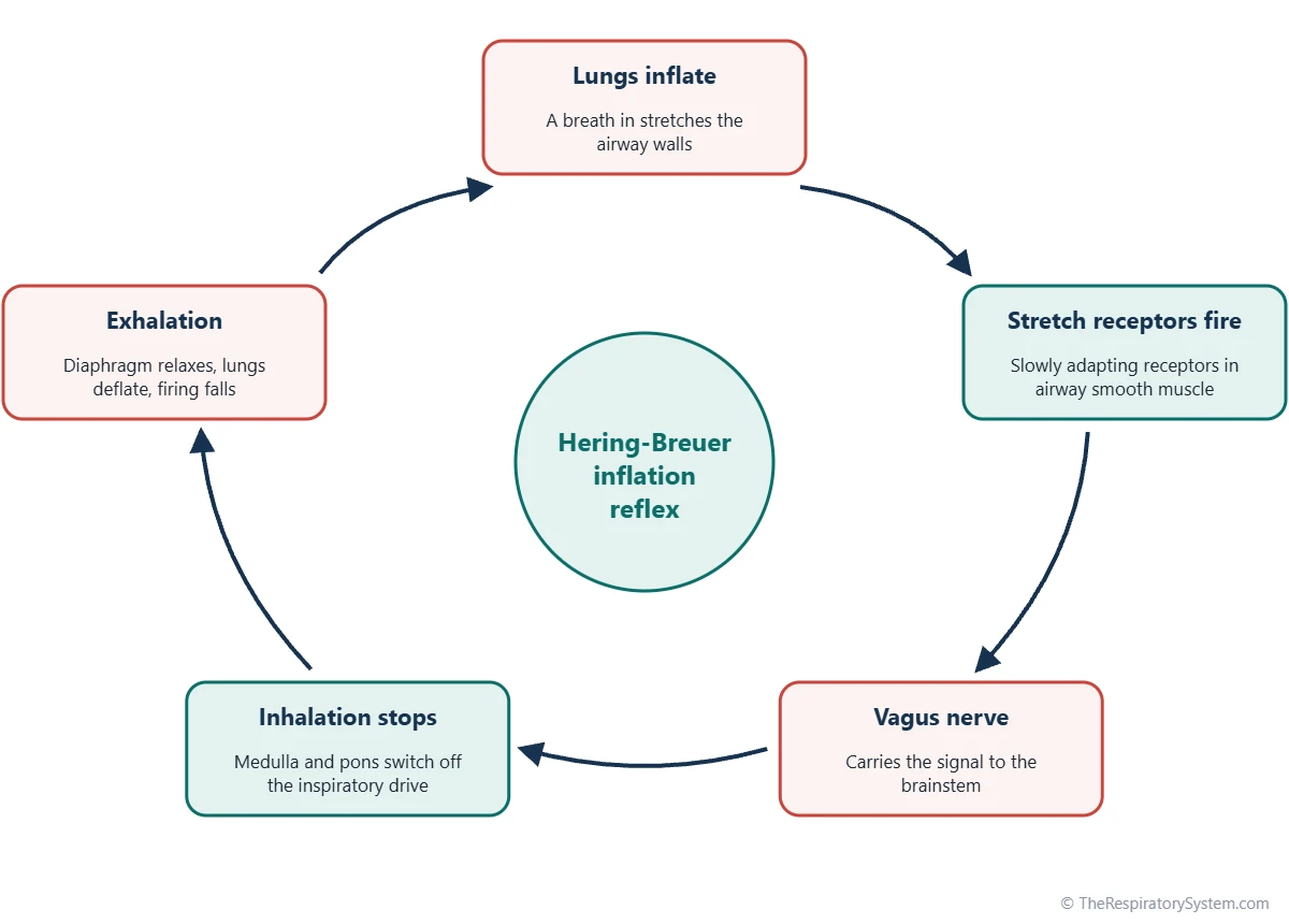
The Hering-Breuer inflation reflex is a negative feedback reflex: slowly adapting stretch receptors in the smooth muscle of the airways sense lung inflation and send signals along the vagus nerve to the medulla, which switches off inspiration. Described in 1868 by Ewald Hering and Josef Breuer, it is strong in infants but in adults acts mainly during large breaths.
- Type
- Protective reflex of breathing; negative feedback
- Function
- Ends inhalation when the lungs are stretched; prevents overinflation
- Receptors
- Slowly adapting pulmonary stretch receptors in airway smooth muscle
- Nerves
- Afferent: vagus nerve. Efferent: phrenic and intercostal nerves
- Center
- Respiratory centers of the medulla and pons
- Part of
- Control of breathing
On this page
- What Is the Hering-Breuer Reflex?
- Where Are the Receptors of the Hering-Breuer Reflex Located?
- Hering-Breuer Reflex Arc: Afferent and Efferent Pathways
- What Is the Function of the Hering-Breuer Reflex?
- Hering-Breuer Inflation vs Deflation Reflex
- How Does the Hering-Breuer Reflex Differ in Infants and Adults?
- Common Conditions That Affect the Hering-Breuer Reflex
- Key Facts
- Quiz
- FAQ
What Is the Hering-Breuer Reflex?
The Hering-Breuer reflex is an automatic reflex that shortens a breath in once the lungs have stretched enough, so the lungs do not overinflate. It is one of the lung reflexes that feed information into the wider system described in control of breathing, and it is part of our respiratory system glossary.
The reflex is named after two physiologists working in Vienna, Ewald Hering and his assistant Josef Breuer, who described it in 1868. They showed that inflating an animal's lungs stopped its breathing movements, and that the effect disappeared when the vagus nerves were cut. This was one of the first examples of a feedback reflex found in physiology.
Explained simply
Think of filling a balloon with a sensor in its skin. When the balloon stretches enough, the sensor sends a message to stop blowing. In the lungs, the sensors are stretch receptors in the airway walls, the message travels up the vagus nerve, and the brain answers by ending the breath in.
Where Are the Receptors of the Hering-Breuer Reflex Located?
The receptors of the Hering-Breuer reflex are located in the smooth muscle of the airway walls, from the trachea through the bronchi to the bronchioles. They are called slowly adapting pulmonary stretch receptors.
Slowly adapting means they keep firing for as long as the airway stays stretched instead of falling silent after a moment. Their firing rate rises as the lungs fill, so the brainstem receives a running measure of lung volume during each breath in. They are different from the rapidly adapting, or irritant, receptors in the airway lining, which respond to dust, smoke and sudden changes and help drive the cough reflex.
Hering-Breuer Reflex Arc: Afferent and Efferent Pathways
The Hering-Breuer reflex arc runs from stretch receptors in the airways, along vagal afferent fibers to the medulla, and back out along the phrenic and intercostal efferent nerves to the breathing muscles. Each link of the arc has a fixed job.
- Stimulus: the lungs and airways stretch during a breath in.
- Receptor: slowly adapting stretch receptors in airway smooth muscle start firing faster.
- Afferent pathway: large myelinated sensory fibers of the vagus nerve, cranial nerve X, carry the signal to the brainstem.
- Integration center: the signal reaches the nucleus of the solitary tract in the medulla, home of the dorsal respiratory group, which works with the pontine respiratory group in the pons.
- Efferent pathway: the inspiratory neurons are inhibited, so firing falls in the phrenic nerve and the intercostal nerves.
- Effector and response: the diaphragm and the external intercostal muscles stop contracting, the breath in ends and passive exhalation begins.
Because a bigger stretch produces a stronger stop signal, the reflex is a classic negative feedback loop.
| Part of the arc | Hering-Breuer inflation reflex | Detail |
|---|---|---|
| Stimulus | Stretching of the lungs during a breath in | Response grows as lung volume rises |
| Receptor | Slowly adapting pulmonary stretch receptors | In the smooth muscle of the trachea, bronchi and bronchioles |
| Afferent pathway | Vagus nerve (cranial nerve X) | Large myelinated sensory fibers |
| Integration center | Nucleus of the solitary tract, medulla | Dorsal respiratory group, working with the pontine respiratory group |
| Efferent pathway | Phrenic nerve and intercostal nerves | Their firing is reduced |
| Effectors | Diaphragm and external intercostal muscles | Stop contracting |
| Response | Inhalation ends early, exhalation is prolonged | Tidal volume is limited |
| Type of feedback | Negative feedback | More stretch gives a stronger stop signal |
| First described | 1868, Ewald Hering and Josef Breuer | Experiments on animal lungs in Vienna |
| Source: Standard physiology, our editors | ||
What Is the Function of the Hering-Breuer Reflex?
The function of the Hering-Breuer reflex is to protect the lungs from overinflation and to help set the depth and timing of each breath. It acts as an off-switch for inspiration that is triggered by lung volume rather than by blood gases.
- Prevents overinflation: it stops a breath in before the lungs are stretched too far.
- Limits tidal volume: by cutting inhalation short, it keeps each breath, the tidal volume, within a safe range.
- Sets the rhythm: shorter breaths in allow breaths to follow each other faster, so the reflex tends to raise the respiratory rate.
- Prolongs exhalation: continued stretch at the end of a breath in delays the start of the next one.
How does it affect ventilation?
The Hering-Breuer reflex mainly changes the pattern of ventilation rather than its total amount. It trades depth for rate, giving shallower and more frequent breaths, while the overall level of breathing is set by the respiratory chemoreceptors that sense carbon dioxide, oxygen and acidity in the blood. In experiments, animals whose vagus nerves were cut breathed more slowly and much more deeply, showing what breathing looks like without the reflex.
Hering-Breuer Inflation vs Deflation Reflex
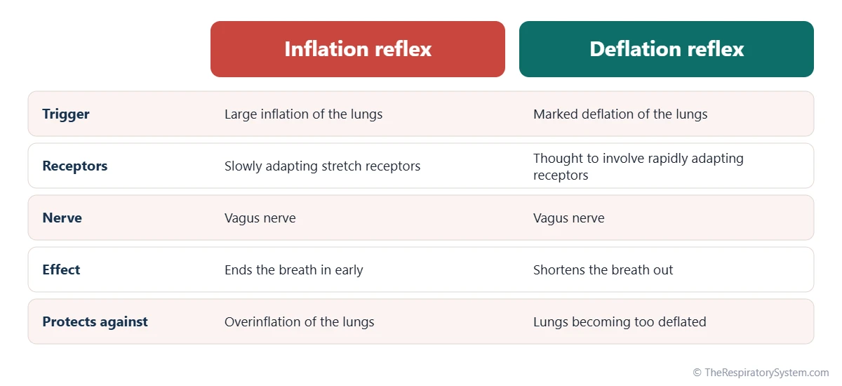
The Hering-Breuer inflation reflex ends a breath in when the lungs are well inflated, while the Hering-Breuer deflation reflex shortens a breath out when the lungs become unusually small. Most textbooks mean the inflation reflex when they say Hering-Breuer reflex.
The deflation reflex works in the opposite direction: strong deflation of the lungs speeds up the start of the next breath in. It is thought to involve rapidly adapting receptors as well as the drop in firing of stretch receptors, and it may help keep the lungs from sinking to too low a volume at the end of each exhalation.
Head's paradoxical reflex
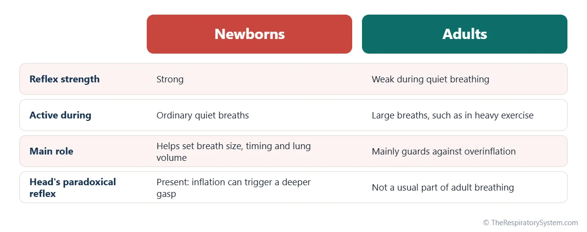
A third, related reflex was described by the British physiologist Henry Head. In Head's paradoxical reflex, a quick inflation of the lungs makes the subject take an even deeper breath instead of stopping. It is seen mainly in newborns, and it is thought to help with the first deep breaths that open the lungs after birth and with the occasional deep sighs that reopen small airspaces.
How Does the Hering-Breuer Reflex Differ in Infants and Adults?
The Hering-Breuer reflex is strong in newborns and young infants, where it helps regulate every breath, but in adults it is weak during quiet breathing and becomes active mainly when breaths are much larger than usual. This change with age is one of its best-known features.
In newborns, the reflex helps control the size and timing of breaths and is thought to help hold the lungs at a stable volume between breaths, which matters because a baby's chest wall is soft. In adults, a normal resting breath does not stretch the lungs enough to trigger much of a response. During heavy exercise, when breaths become deep, the reflex is thought to help limit tidal volume, as covered in lung volumes and capacities.
Evidence from people who have received a lung transplant supports this view. Their new lungs have no vagal connection to the brainstem, yet their resting breathing pattern is close to normal, which suggests that adults depend little on the reflex at rest.
Common Conditions That Affect the Hering-Breuer Reflex
Conditions that damage the vagus nerve, change how stiff the lungs are or remove their nerve supply can alter the Hering-Breuer reflex and the other vagal lung reflexes. These changes affect the pattern of breathing more than its overall control.
- Lung transplantation: the transplanted lung is cut off from its vagal nerve supply, so stretch signals from it no longer reach the brainstem.
- Stiff lungs: in conditions such as pulmonary fibrosis, rapid, shallow breathing is common, and stretch receptors together with other vagal receptors are thought to contribute.
- Overinflated lungs: in emphysema and COPD, air trapping leaves the lungs larger than normal, which changes the signals that stretch receptors send.
- Newborn breathing: because the reflex is strong early in life, it is studied in the breathing patterns of premature and newborn babies.
Different breathing patterns and wider respiratory diseases are covered on their own pages.
Key Facts
- The Hering-Breuer reflex ends a breath in when stretch receptors in the airways sense that the lungs are well inflated.
- Its receptors are slowly adapting pulmonary stretch receptors in the smooth muscle of the airways.
- The afferent signal travels in the vagus nerve; the efferent response is reduced firing of the phrenic and intercostal nerves.
- Ewald Hering and Josef Breuer described the reflex in Vienna in 1868.
- The reflex is strong in newborns but weak in adults during quiet breathing.
- It changes the pattern of breathing, while the chemoreceptors set its overall level.
Quick Quiz: Test What You Know About the Hering-Breuer Reflex
6 questions. Tap an answer to check it.
What triggers the Hering-Breuer inflation reflex?
Stretch receptors in the airway walls fire as the lungs inflate and trigger the reflex.
Which nerve carries the afferent signal of the reflex?
Sensory fibers of the vagus nerve carry the stretch signals from the lungs to the brainstem.
Where are the receptors of the Hering-Breuer reflex?
Slowly adapting pulmonary stretch receptors lie in the smooth muscle of the trachea, bronchi and bronchioles.
What is the main effect of the inflation reflex?
The reflex switches off inspiration so the lungs do not overinflate, and exhalation follows.
In whom is the Hering-Breuer reflex strongest?
The reflex helps control every breath in newborns but is weak in adults during quiet breathing.
What type of control does the Hering-Breuer reflex provide?
More stretch produces a stronger signal to stop inhaling, which is negative feedback.
Frequently Asked Questions
What is the purpose of the Hering-Breuer reflex?
The purpose of the Hering-Breuer reflex is to protect the lungs from being stretched too far and to help time each breath. By switching off the breath in once the lungs are well filled, it keeps tidal volume within a safe range. In newborns it also helps set breathing rhythm and lung volume, while in adults it acts mainly as a safety limit during very deep breaths.
What are the afferent and efferent limbs of the Hering-Breuer reflex?
The afferent limb is made of sensory fibers in the vagus nerve, which carry signals from stretch receptors in the airways to the nucleus of the solitary tract in the medulla. The efferent limb is the reduced activity of the motor nerves to the breathing muscles, chiefly the phrenic nerve to the diaphragm and the intercostal nerves to the external intercostal muscles.
Is the Hering-Breuer reflex important in adults?
Only to a limited degree. During quiet breathing, an adult's breaths are too small to trigger much of the reflex, and people with transplanted lungs, which lack a vagal connection, breathe almost normally at rest. The reflex becomes more active when breaths are much deeper than usual, as in heavy exercise, where it is thought to help limit the size of each breath.
What happens if the vagus nerves are cut?
In classic animal experiments, cutting both vagus nerves removed the stretch signals from the lungs. The animals then breathed more slowly and much more deeply, because nothing ended each breath in early. This finding was part of the original evidence for the Hering-Breuer reflex and showed that the vagus nerve carries its sensory signals.
Is the Hering-Breuer reflex the same as the pneumotaxic center?
No, but they work toward a similar result. The pneumotaxic center, now usually called the pontine respiratory group, is a group of neurons in the pons that helps end each breath in and set breathing rate. The Hering-Breuer reflex is a sensory reflex from the lungs. Both can shorten inhalation, and the brainstem combines their signals.
What is the difference between the Hering-Breuer reflex and chemoreceptor control?
The Hering-Breuer reflex responds to a mechanical signal, the stretch of the lungs, and mainly shapes the depth and timing of each breath. Chemoreceptor control responds to chemical signals, the levels of carbon dioxide, oxygen and acid in the blood, and sets how much air the body breathes overall. Chemoreceptors are the main drive of breathing in adults.
Who discovered the Hering-Breuer reflex?
The reflex was described in 1868 by Ewald Hering, a German physiologist, and Josef Breuer, an Austrian physician who was working with him in Vienna. Breuer carried out much of the experimental work. Hering is also known for research on vision and color, and Breuer later became known for his role in the early history of psychoanalysis.
Does the Hering-Breuer reflex work during sleep?
Yes. The reflex is automatic and does not depend on being awake, so it continues to act during sleep, as do the chemoreceptor reflexes. In infants, who sleep for much of the day, the stretch reflexes are part of the normal control of breathing during sleep. In adults the reflex stays weak during quiet sleeping breaths, as it is when awake.
Keep Exploring


