At a Glance
Respiratory chemoreceptors are chemical sensors that adjust breathing to keep blood carbon dioxide near about 40 mmHg and pH near 7.4. Central chemoreceptors, just beneath the ventral surface of the medulla oblongata, respond to hydrogen ions formed from carbon dioxide in the cerebrospinal fluid. Peripheral chemoreceptors, in the carotid bodies and aortic bodies, respond quickly to low arterial oxygen (especially below about 60 mmHg), high carbon dioxide and acid.
- Location
- Central: ventral surface of the medulla oblongata. Peripheral: carotid bodies in the neck and aortic bodies on the aortic arch
- Function
- Sense carbon dioxide, pH and oxygen and adjust the rate and depth of breathing
- Type
- Sensory receptors: carbon dioxide-sensitive brainstem neurons and type I glomus cells
- Connects to
- Respiratory centers of the medulla, via the glossopharyngeal and vagus nerves for the peripheral receptors
- Part of
- The chemoreflex, the negative feedback loop of the control of breathing
On this page
- What Are Respiratory Chemoreceptors?
- Where Are Central and Peripheral Chemoreceptors Located?
- What Do Central Chemoreceptors Do?
- What Is the Function of the Carotid Bodies?
- Central vs Peripheral Chemoreceptors: How Do They Work Together?
- Which Other Sensors Help Control Breathing?
- Common Conditions That Affect the Respiratory Chemoreceptors
- Key Facts
- Quiz
- FAQ
What Are Respiratory Chemoreceptors?
Respiratory chemoreceptors are specialized sensors that monitor the chemistry of the blood and the fluid around the brain, chiefly carbon dioxide, acidity (pH) and oxygen, and signal the brainstem to change how fast and how deeply a person breathes. They are the sensing arm of a feedback loop that keeps arterial carbon dioxide close to about 40 mmHg and blood pH close to 7.4.
There are two groups. Central chemoreceptors sit in the brainstem and respond mainly to carbon dioxide through the acid it forms in brain fluid. Peripheral chemoreceptors sit in the carotid bodies and aortic bodies, small clusters of cells beside major arteries, and they are the only sensors that react quickly to a fall in arterial oxygen.
What are central chemoreceptors?
Central chemoreceptors are groups of nerve cells near the front (ventral) surface of the medulla oblongata that detect changes in the hydrogen ion concentration of the cerebrospinal fluid and the fluid between brain cells. Because carbon dioxide in the blood quickly sets the acidity of that fluid, they act in practice as the body's main carbon dioxide sensor.
Explained simply
Think of the chemoreceptors as alarms for breathing. When carbon dioxide, the waste gas of your cells, starts to pile up, or when oxygen runs low, these alarms go off. The breathing centers in the brainstem answer by making you breathe faster and deeper until the levels are back to normal.
Where Are Central and Peripheral Chemoreceptors Located?
Central chemoreceptors are located in the brainstem, just beneath the ventral surface of the medulla oblongata, while peripheral chemoreceptors are located in the carotid bodies at the fork of each common carotid artery in the neck and in the aortic bodies along the arch of the aorta.
Central chemoreceptors in the medulla
The central chemoreceptor areas lie a fraction of a millimeter under the surface of the medulla, near the points where the glossopharyngeal (IX) and vagus (X) nerves leave the brainstem. Here they are bathed by cerebrospinal fluid and sit close to, but separate from, the respiratory centers that generate the breathing rhythm. Carbon dioxide-sensitive cells have also been found in other brainstem sites, such as the retrotrapezoid nucleus, so the central system is best thought of as a network rather than a single spot.
Carotid bodies
There is one carotid body on each side of the neck, tucked into the angle where the common carotid artery divides into the internal and external carotid arteries. Each is about the size of a grain of rice, yet it receives one of the highest blood flows per gram of any tissue in the body, so it samples fresh arterial blood almost continuously. The carotid body should not be confused with the carotid sinus, a nearby widening of the artery wall that senses blood pressure rather than blood chemistry.
Aortic bodies
The aortic bodies are several small clusters of similar tissue scattered around the arch of the aorta, near the origins of its large branches, just above the heart in the mediastinum. In humans they contribute less to breathing control than the carotid bodies and have a larger part in adjusting the circulation.
What Do Central Chemoreceptors Do?
Central chemoreceptors increase breathing when the carbon dioxide level of arterial blood climbs and decrease it when carbon dioxide falls, by sensing the acidity that carbon dioxide produces in the fluid around the brainstem. They supply most of the everyday drive to breathe and most of the response to a rise in carbon dioxide.
How carbon dioxide reaches them
The blood-brain barrier lets carbon dioxide pass freely but keeps hydrogen ions and bicarbonate in the blood from crossing easily. Once carbon dioxide diffuses into the cerebrospinal fluid, it reacts with water to form carbonic acid, which splits into hydrogen ions and bicarbonate. Cerebrospinal fluid contains very little protein to absorb those hydrogen ions, so a small rise in blood carbon dioxide produces a clear drop in local pH that the chemoreceptors detect.
What they do not sense
Central chemoreceptors do not respond to low oxygen; severe lack of oxygen actually depresses brainstem neurons rather than stimulating them. They also react only slowly and weakly to acids made elsewhere in the body, because hydrogen ions in the blood cross into the brain fluid poorly. Those two jobs fall to the peripheral chemoreceptors.
Resetting over time
If carbon dioxide stays high or low for a day or more, the bicarbonate level of the cerebrospinal fluid is adjusted, which brings its pH back toward normal. The central chemoreceptors then fire less strongly than the carbon dioxide level alone would suggest. This is one reason the breathing response to long-standing carbon dioxide retention is weaker than the response to a sudden rise.
| Change in the blood | Central chemoreceptors | Peripheral chemoreceptors | Effect on breathing |
|---|---|---|---|
| Rise in arterial PCO2 | Strong response (main sensor) | Smaller but faster response | Breathing becomes deeper and faster |
| Fall in arterial PCO2 after overbreathing | Firing falls | Firing falls | Breathing slows, sometimes with brief pauses |
| Fall in arterial PO2 below about 60 mmHg | No direct response | Strong, fast response | Breathing increases |
| Mild fall in arterial PO2, still above about 60 mmHg | No response | Small response | Little change in breathing |
| Acid added to the blood (metabolic acidosis) | Slow, weak response | Main response, mostly from the carotid bodies | Deep, rapid breathing |
| Anemia (less hemoglobin, normal PO2) | No response | Little response | Breathing changes little |
| Carbon monoxide in the blood (normal PO2) | No response | Little response | Breathing changes little |
| Carbon dioxide high for days | Response blunted as fluid bicarbonate adjusts | Response kept | Low oxygen becomes a larger share of the drive |
| Source: Typical adult physiology, our editors | |||
What Is the Function of the Carotid Bodies?
The carotid bodies detect a fall in arterial oxygen, a rise in carbon dioxide and a rise in blood acidity, and they send fast nerve signals to the medulla that increase breathing within seconds. They are the body's main oxygen sensor for breathing, and the aortic bodies play a smaller, similar role.
Inside a carotid body
Each carotid body is made of clusters of type I glomus cells, the true sensing cells, wrapped by supporting type II (sustentacular) cells and threaded with wide capillaries. Endings of the carotid sinus nerve, a branch of the glossopharyngeal nerve, surround the glomus cells. Signals from the aortic bodies travel instead in the vagus nerve, and both pathways end in the nucleus of the solitary tract in the medulla.
How a glomus cell senses low oxygen
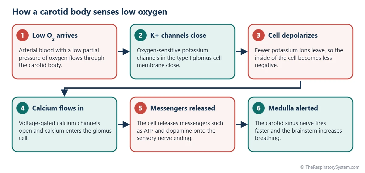
When oxygen falls, oxygen-sensitive potassium channels in the glomus cell membrane close, the cell depolarizes, and calcium flows in through voltage-gated channels. The cell then releases chemical messengers such as ATP and dopamine onto the nerve ending, which fires faster and alerts the brainstem. The diagram below follows these steps in order.
The oxygen threshold
The carotid bodies respond to the partial pressure of oxygen in arterial plasma, not to the total amount of oxygen carried by hemoglobin. Their firing rises only gently until arterial PO2 falls below about 60 mmHg and then increases steeply, close to the point where hemoglobin starts to lose saturation quickly on the oxygen-hemoglobin dissociation curve. For that reason, anemia or carbon monoxide poisoning, which lower oxygen content while leaving PO2 near normal, stimulate them only weakly.
Carbon dioxide and acid
The carotid bodies also respond to carbon dioxide, faster than the central chemoreceptors but with a smaller share of the total effect. They are the main sensors of acids that build up in the blood from metabolism, such as lactic acid during very hard exercise, and they drive the deep, rapid breathing that blows off carbon dioxide in those states.
Central vs Peripheral Chemoreceptors: How Do They Work Together?
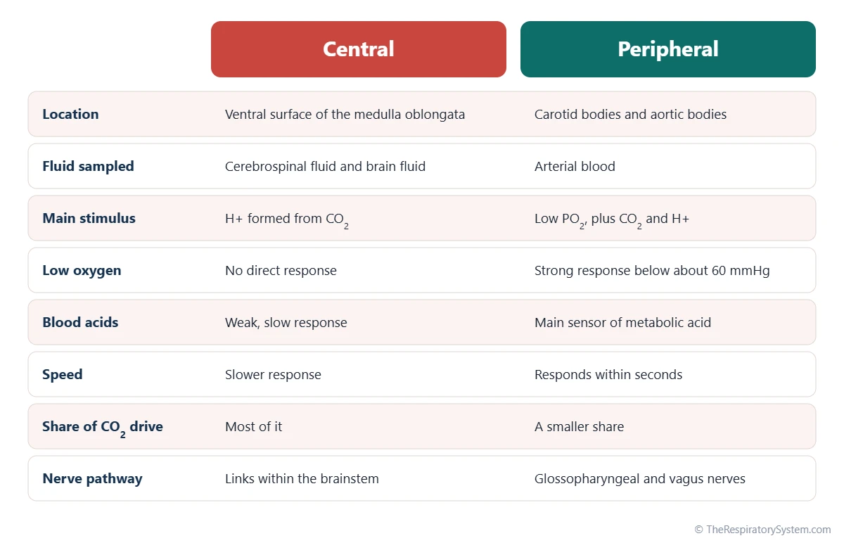
Central and peripheral chemoreceptors work together as two inputs to the same breathing centers: the central ones give a strong but slower response to carbon dioxide, and the peripheral ones give a fast response to low oxygen, carbon dioxide and acid. The medulla adds up their signals and adjusts the output to the breathing muscles.
The chemoreflex loop
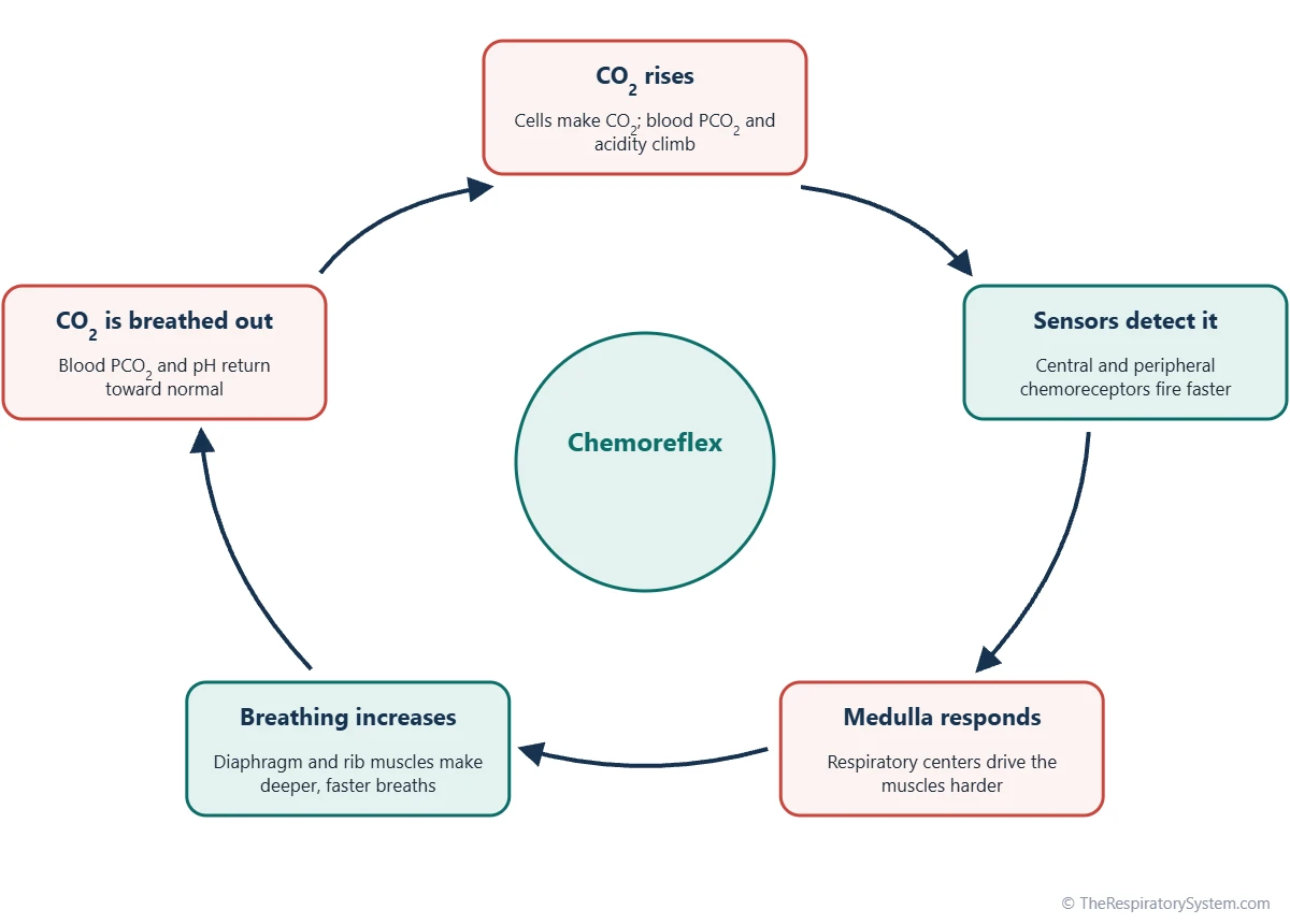
The control system works as a negative feedback loop. When carbon dioxide rises, the chemoreceptors fire faster, and the respiratory centers send stronger bursts down the phrenic nerve and the intercostal nerves, so the diaphragm and rib muscles produce larger, more frequent breaths. The extra airflow washes carbon dioxide out of the alveoli, the blood level falls, and the signal fades. Our page on the control of breathing describes the respiratory centers that receive these signals.
Why carbon dioxide, not oxygen, drives breathing
In a healthy person at sea level, carbon dioxide is the main moment-to-moment regulator of breathing. Arterial oxygen normally sits well above the range where the carotid bodies respond strongly, so the oxygen drive matters most when oxygen falls, as at high altitude or in lung disease. The two drives also strengthen each other, because low oxygen makes the response to carbon dioxide steeper.
Part of acid-base balance
Carbon dioxide and hydrogen ions are linked through the bicarbonate buffer, so the chemoreceptors let the lungs help regulate blood pH within minutes. This is the respiratory part of acid-base balance described on our respiratory system homeostasis page. How the blood carries that carbon dioxide to the lungs is covered on the carbon dioxide transport page.
Which Other Sensors Help Control Breathing?
Besides chemoreceptors, breathing is adjusted by mechanical and irritant receptors in the lungs, airways, muscles and joints, which report stretch, irritation and movement rather than blood chemistry.
- Pulmonary stretch receptors: in the smooth muscle of the airways, they sense lung inflation and help limit very deep breaths through the Hering-Breuer reflex.
- Irritant receptors: nerve endings in the airway lining react to dust, smoke and cold air and can trigger the cough reflex, airway narrowing or rapid shallow breathing.
- J receptors (juxtacapillary receptors): in the alveolar walls next to the capillaries, they respond to fluid or congestion in the lung tissue.
- Proprioceptors: in muscles and joints, they help raise breathing at the very start of exercise, before blood gases have changed.
- Baroreceptors: in the carotid sinus and aortic arch, they sense blood pressure and have only a small effect on breathing.
Higher brain areas can override the chemoreceptors for a while, which is what makes speech, singing and voluntary breath-holding possible. During a breath-hold, the rising carbon dioxide level detected by the chemoreceptors is the main source of the growing urge to breathe, as explained on our breath-holding page.
Common Conditions That Affect the Respiratory Chemoreceptors
Chemoreceptor function can be changed by long-standing lung disease, by life at high altitude, by sleep-related breathing disorders and, rarely, by inherited conditions or tumors. Our respiratory diseases section covers the lung conditions themselves, and anyone with breathing symptoms should see a doctor.
- Chronic carbon dioxide retention: in some people with severe COPD, blood carbon dioxide stays high, the central chemoreceptors reset, and low oxygen sensed by the carotid bodies becomes a larger part of the drive to breathe.
- High altitude: thinner air lowers arterial PO2, the carotid bodies raise breathing, and over several days their sensitivity increases, a change called ventilatory acclimatization.
- Sleep apnea: in central sleep apnea, an unstable chemoreflex loop can cause cycles of breathing pauses and overbreathing during sleep.
- Congenital central hypoventilation syndrome: a rare inherited disorder, usually linked to the PHOX2B gene, in which automatic breathing is weak, especially during sleep, because the response to carbon dioxide is greatly reduced.
- Carotid body tumors: rare, usually slow-growing growths of glomus tissue, a type of paraganglioma, that appear as a lump high in the side of the neck.
- Hyperventilation and hypoventilation: breathing too much or too little shifts carbon dioxide and pH, and the chemoreceptors work to pull them back.
A low oxygen level in the arterial blood, called hypoxemia, is the main signal that activates the carotid bodies.
Key Facts
- Central chemoreceptors lie just beneath the ventral surface of the medulla oblongata and respond to hydrogen ions formed from carbon dioxide in the cerebrospinal fluid.
- Peripheral chemoreceptors are the carotid bodies, at the fork of each common carotid artery, and the aortic bodies on the arch of the aorta.
- Only the peripheral chemoreceptors raise breathing in response to low oxygen, and their response becomes strong once arterial PO2 falls below about 60 mmHg.
- The carotid bodies signal the brainstem through the glossopharyngeal nerve, and the aortic bodies through the vagus nerve.
- Carbon dioxide, not oxygen, is the main moment-to-moment regulator of breathing in healthy people at sea level.
- Type I glomus cells are the sensing cells of the carotid body; low oxygen makes them release messengers that excite the sensory nerve.
- By holding carbon dioxide steady, chemoreceptors protect blood pH and keep gas exchange matched to the body's needs.
Quick Quiz: Test What You Know About the Respiratory Chemoreceptors
6 questions. Tap an answer to check it.
Where are the central chemoreceptors located?
Central chemoreceptors lie just beneath the ventral surface of the medulla, bathed by cerebrospinal fluid.
What do central chemoreceptors directly detect?
Carbon dioxide crosses into the cerebrospinal fluid and forms hydrogen ions, which the central chemoreceptors sense.
Which receptors increase breathing when arterial oxygen falls?
Only the peripheral chemoreceptors in the carotid and aortic bodies respond quickly to low arterial PO2.
Which nerve carries signals from the carotid bodies to the brainstem?
The carotid sinus nerve is a branch of the glossopharyngeal nerve (cranial nerve IX); the aortic bodies use the vagus nerve.
Below roughly what arterial PO2 do the carotid bodies respond strongly?
Carotid body firing rises steeply once arterial PO2 drops below about 60 mmHg.
What are the oxygen-sensing cells of the carotid body called?
Type I glomus cells sense oxygen; type II sustentacular cells support them.
Frequently Asked Questions
What is the main stimulus for central chemoreceptors?
The main stimulus is a rise in hydrogen ions in the cerebrospinal fluid, caused by carbon dioxide diffusing in from the blood. Carbon dioxide crosses the blood-brain barrier easily and reacts with water to form carbonic acid, which releases hydrogen ions. So although the receptors sense acidity, in everyday life they respond mainly to the carbon dioxide level of arterial blood.
Do central chemoreceptors respond to low oxygen?
No. Central chemoreceptors are not stimulated by low oxygen, and a severe lack of oxygen tends to depress the brainstem rather than excite it. The breathing response to low oxygen comes almost entirely from the peripheral chemoreceptors in the carotid bodies, with a smaller contribution from the aortic bodies.
What is the difference between the carotid body and the carotid sinus?
Both sit where the common carotid artery divides in the neck, but they sense different things. The carotid body is a small cluster of glomus cells that detects oxygen, carbon dioxide and acid in the blood. The carotid sinus is a slightly widened part of the artery wall containing baroreceptors, which sense blood pressure and help regulate heart rate and vessel tone.
How quickly do chemoreceptors change breathing?
Peripheral chemoreceptors respond within seconds, because the carotid bodies receive fresh arterial blood almost directly from the heart. Central chemoreceptors respond more slowly, since carbon dioxide must first diffuse into the brain fluid and change its pH. Together they produce a rapid first adjustment followed by a larger, sustained one.
What is the hypoxic drive?
The hypoxic drive is the part of the urge to breathe that comes from low oxygen, sensed by the carotid and aortic bodies. In healthy people it adds little at sea level, because arterial oxygen is normally high. It becomes more important at high altitude and in some people with long-standing carbon dioxide retention, whose central response to carbon dioxide has been blunted.
Can you override your chemoreceptors?
Only for a short time. The cerebral cortex can take temporary control of the breathing muscles, which allows talking, singing and holding the breath. As carbon dioxide builds up, the signal from the chemoreceptors grows until the automatic drive takes over again. This is why the urge to breathe becomes impossible to ignore during a long breath-hold.
Are chemoreceptors part of the nervous system?
Yes. Central chemoreceptors are nerve cells within the brainstem itself. The carotid and aortic bodies are small sensory organs whose glomus cells develop from neural crest tissue and pass their signals to sensory nerve fibers. Both feed into the respiratory centers of the medulla, making them part of the body's automatic, or autonomic, control of breathing.
Do newborn babies have working chemoreceptors?
Yes, but they are still maturing. Before birth the fetus lives at a low oxygen level, and the carotid bodies gradually reset their sensitivity over the days and weeks after birth as blood oxygen rises. Central chemoreceptors work from birth, although the overall control of breathing in newborns, especially premature babies, is less stable than in older children.
Keep Exploring


