At a Glance
Oxygen is transported in the blood in two forms: about 98% bound to hemoglobin in red blood cells (oxyhemoglobin) and about 1.5 to 2% dissolved in plasma. Each hemoglobin molecule carries up to four oxygen molecules, and each gram of hemoglobin holds about 1.34 mL of oxygen, so arterial blood carries about 20 mL of oxygen per 100 mL. At rest the tissues take about a quarter of it, and blood returns to the lungs still about 75% saturated.
- Main carrier
- Hemoglobin inside red blood cells, as oxyhemoglobin
- Forms
- About 98% bound to hemoglobin, about 1.5 to 2% dissolved in plasma
- Capacity
- Up to four O2 molecules per hemoglobin; about 20 mL O2 per 100 mL of arterial blood
- Loaded in
- Pulmonary capillaries around the alveoli
- Unloaded in
- Systemic capillaries of the body tissues
- Part of
- Gas transport, the step between external and internal respiration
On this page
- What Is Oxygen Transport?
- What Form Is Oxygen Transported in the Blood?
- What Is Hemoglobin and How Does It Carry Oxygen?
- What Is Oxyhemoglobin?
- Oxygen Transport Process: What Are the Steps?
- How Does Hemoglobin Release Oxygen in the Tissues?
- How Much Oxygen Does the Blood Carry?
- Oxygen Transport and Storage: Myoglobin and Fetal Hemoglobin
- Common Conditions That Affect Oxygen Transport
- Key Facts
- Quiz
- FAQ
What Is Oxygen Transport?
Oxygen transport is the carriage of oxygen by the blood from the lungs, where it is picked up, to the body's tissues, where it is released for cells to use. It links breathing and gas exchange in the lungs to the use of oxygen inside cells, and it depends on both the lungs and the heart.
Oxygen transport definition
In physiology, oxygen transport means the movement of oxygen through the circulation in two forms, bound to hemoglobin and dissolved in plasma, together with the loading and unloading steps at each end. Physiologists also speak of oxygen delivery, the amount of oxygen pumped to the tissues each minute, which equals the oxygen content of arterial blood multiplied by the cardiac output.
Why the body needs a carrier
Oxygen dissolves poorly in water. If blood relied only on dissolved oxygen, the heart would have to pump many times more blood each minute just to supply the body at rest. Hemoglobin solves the problem by raising the oxygen-carrying capacity of blood about seventy-fold, so a normal cardiac output can meet the body's needs, as explained on our why do we need oxygen page.
Explained simply
Think of red blood cells as delivery trucks and hemoglobin as the seats inside them. In the lungs every seat fills with an oxygen passenger. The trucks drive through the arteries to the body, where busy cells call the passengers off, and then return through the veins with empty seats to pick up more.
What Form Is Oxygen Transported in the Blood?
Oxygen is transported in the blood mainly as oxyhemoglobin, oxygen reversibly bound to hemoglobin in red blood cells, which accounts for about 98% of the total; the remaining 1.5 to 2% is dissolved as free oxygen gas in the plasma.
Dissolved oxygen
The amount of oxygen that dissolves in plasma is proportional to its partial pressure, a rule known as Henry's law. At a PO2 of about 100 mmHg, only about 0.3 mL of oxygen dissolves in each 100 mL of blood. Small as it is, this dissolved fraction matters: it is the oxygen that sets the PO2 of the blood, and every oxygen molecule must pass through the dissolved state on its way into and out of a red blood cell.
Oxygen bound to hemoglobin
Each red blood cell contains roughly 250 to 300 million hemoglobin molecules, and each molecule can bind up to four oxygen molecules. In arterial blood, hemoglobin is normally about 97 to 98% saturated, holding about 19.5 mL of oxygen per 100 mL of blood. That is around sixty-five times more than the dissolved fraction, which is why hemoglobin concentration has such a large effect on how much oxygen the blood can carry.
What Is Hemoglobin and How Does It Carry Oxygen?
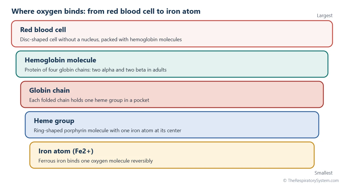
Hemoglobin is an iron-containing protein in red blood cells made of four globin chains, each holding one heme group whose iron atom can bind one oxygen molecule, so one hemoglobin carries up to four oxygen molecules.
Structure of hemoglobin
Adult hemoglobin (hemoglobin A) has two alpha and two beta globin chains folded around each other. Each chain cradles a heme group, a flat ring-shaped porphyrin molecule with a single iron atom at its center. The iron must be in the ferrous (Fe2+) state to bind oxygen, and the binding is reversible, so oxygen can be picked up and released many times during a red blood cell's life of about 120 days.
Cooperative binding
Hemoglobin does not bind its four oxygen molecules independently. When the first oxygen attaches, the molecule shifts from a tense (T) shape to a relaxed (R) shape, which makes the next oxygen molecules bind more easily. The reverse happens on unloading: once one oxygen leaves, the others are released more readily. This cooperative behavior gives the S-shaped oxygen-hemoglobin dissociation curve, which lets hemoglobin fill almost completely in the lungs and release large amounts in the tissues over a narrow range of PO2.
Hemoglobin and oxygen in numbers
Each gram of fully saturated hemoglobin holds about 1.34 mL of oxygen. With a normal hemoglobin concentration of roughly 12 to 17 grams per 100 mL of blood, higher on average in men than in women, the oxygen-carrying capacity of blood is about 20 mL per 100 mL.
What Is Oxyhemoglobin?
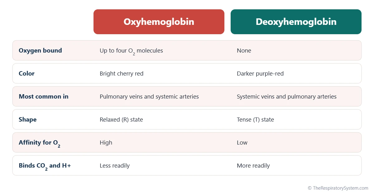
Oxyhemoglobin is hemoglobin with oxygen bound to its iron atoms; it forms in the lungs, gives arterial blood its bright red color and turns back into deoxyhemoglobin when it releases oxygen in the tissues.
The loss and gain of oxygen changes the way hemoglobin absorbs light. Oxyhemoglobin is bright cherry red, while deoxyhemoglobin is a darker purple-red. Veins look bluish through the skin not because the blood inside is blue, but because of the way skin scatters and absorbs different colors of light.
The same color difference is what a pulse oximeter uses. By shining red and infrared light through a fingertip, it estimates the percentage of hemoglobin carrying oxygen, a value called oxygen saturation (SpO2). Oxyhemoglobin should not be confused with carbaminohemoglobin, hemoglobin carrying carbon dioxide on its protein chains, a form described on our carbon dioxide transport page.
Oxygen Transport Process: What Are the Steps?
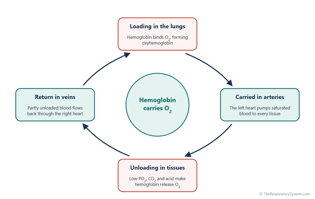
The oxygen transport process has four main stages: oxygen is loaded onto hemoglobin in the lungs, carried in arterial blood, unloaded in the tissue capillaries, and the partly emptied blood returns to the lungs to be reloaded.
- Oxygen reaches the alveoli. Each breath in brings fresh air to the alveoli, where the PO2 is about 100 mmHg.
- Oxygen diffuses into the blood. It crosses the respiratory membrane into the plasma of the pulmonary capillaries, where blood arrives with a PO2 of about 40 mmHg.
- Hemoglobin binds oxygen. Oxygen enters the red blood cells and binds to hemoglobin, forming oxyhemoglobin. This keeps the dissolved oxygen level low, so more oxygen keeps flowing in.
- Blood travels to the heart. Oxygen-rich blood flows through the pulmonary veins to the left atrium and left ventricle, as traced on our pulmonary circulation page.
- Arteries carry it to the body. The left ventricle pumps the blood through the aorta and its branches to every organ.
- Hemoglobin releases oxygen. In the systemic capillaries the PO2 of the tissues is lower, so oxyhemoglobin gives up part of its oxygen, which diffuses into the cells.
- Cells use the oxygen. Mitochondria use it in cellular respiration to make ATP, releasing carbon dioxide.
- Blood returns to the lungs. Venous blood, still about 75% saturated at rest, flows back through the right heart to the lungs.
The whole circuit takes about a minute at rest, and each red blood cell repeats it many thousands of times during its life. The table below lists typical PO2 values along the route.
| Stage | Where | Typical PO2 | What happens |
|---|---|---|---|
| Breathed in | Airways to alveoli | About 100 mmHg in alveolar air | Fresh air keeps alveolar oxygen high |
| Loading | Pulmonary capillaries | Rises from about 40 to about 100 mmHg | Oxygen diffuses in and binds hemoglobin |
| To the heart | Pulmonary veins, left atrium and left ventricle | About 100 mmHg | Oxygen-rich blood is pumped into the aorta |
| Arterial transport | Aorta and systemic arteries | About 95 mmHg | Hemoglobin is about 97% saturated |
| Unloading | Systemic capillaries | Falls to about 40 mmHg at rest | Hemoglobin releases about a quarter of its oxygen |
| Into the cells | Tissue fluid and cells | About 40 mmHg or lower | Oxygen diffuses to the mitochondria |
| Return | Systemic veins and right heart | About 40 mmHg | Blood about 75% saturated returns to the lungs |
| Source: Typical values for a healthy adult at rest at sea level, our editors | |||
How Does Hemoglobin Release Oxygen in the Tissues?
Hemoglobin releases oxygen in the tissues because the PO2 there is lower than in the lungs and because local conditions in active tissues, such as more carbon dioxide, more acid and higher temperature, lower hemoglobin's grip on oxygen.
The pressure difference
Blood reaching the systemic capillaries has a PO2 of about 95 mmHg, while the fluid around working cells is about 40 mmHg or lower. Dissolved oxygen diffuses out toward the cells, the plasma PO2 drops, and hemoglobin releases more oxygen to replace it. At rest, hemoglobin falls from about 97% to about 75% saturated, giving up roughly a quarter of its load.
Factors that increase unloading
- Carbon dioxide and acid: both make hemoglobin release oxygen more easily, a link called the Bohr effect.
- Temperature: warmer, active muscle lowers hemoglobin's affinity for oxygen.
- 2,3-bisphosphoglycerate (2,3-BPG): a by-product of red blood cell metabolism that binds hemoglobin and favors release; its level rises during long stays at high altitude.
These factors shift the dissociation curve to the right, so working tissues automatically receive more oxygen. In hard exercise, active muscle can take well over half of the oxygen from each passing red blood cell. The reverse link, in which oxygen loading helps the blood give up carbon dioxide in the lungs, is the Haldane effect.
How Much Oxygen Does the Blood Carry?
Arterial blood carries about 20 mL of oxygen per 100 mL, so with a resting cardiac output of about 5 liters per minute the heart delivers roughly 1,000 mL of oxygen to the body each minute, while the body uses only about 250 mL.
This fourfold safety margin means blood returning to the lungs at rest is still well loaded with oxygen. During exercise, the heart pumps more blood each minute and the muscles extract more oxygen from it, so delivery and use can both rise many times over without the system running out.
Oxygen delivery depends on three things: the amount of hemoglobin, how saturated that hemoglobin is, and how much blood the heart pumps. A fall in any one of them reduces the oxygen reaching the tissues, which is why both the respiratory and circulatory systems must work together. The table below summarizes typical values for a resting adult.
| Measure | Typical value | What it means |
|---|---|---|
| Hemoglobin concentration | About 12 to 17 g per 100 mL of blood | Higher on average in men than in women |
| Oxygen per gram of hemoglobin | About 1.34 mL when fully saturated | Four oxygen molecules per hemoglobin molecule |
| Oxygen-carrying capacity | About 20 mL per 100 mL of blood | The most the blood can hold at full saturation |
| Arterial oxygen content | About 20 mL per 100 mL of blood | Hemoglobin about 97% saturated |
| Venous oxygen content at rest | About 15 mL per 100 mL of blood | Hemoglobin about 75% saturated |
| Oxygen given to tissues at rest | About 5 mL per 100 mL of blood | Roughly a quarter of the arterial load |
| Dissolved oxygen in arterial blood | About 0.3 mL per 100 mL of blood | Only about 1.5% of the total |
| Oxygen delivered to the body | About 1,000 mL per minute | Arterial content times a cardiac output of about 5 L per minute |
| Oxygen used by the body | About 250 mL per minute | Leaves a large reserve in venous blood |
| Source: Typical adult values, our editors | ||
Oxygen Transport and Storage: Myoglobin and Fetal Hemoglobin
The body stores very little oxygen, but two related proteins help: myoglobin holds a small oxygen reserve inside muscle cells, and fetal hemoglobin lets a developing baby draw oxygen from the mother's blood.
Myoglobin
Myoglobin is a single-chain relative of hemoglobin found in heart and skeletal muscle, with one heme group that binds one oxygen molecule. It has a higher affinity for oxygen than hemoglobin and releases it only when muscle PO2 falls very low, so it acts as a short-term store during bursts of activity and helps move oxygen through the cell to the mitochondria. Muscles rich in myoglobin look darker red, and diving mammals such as whales and seals have especially high amounts, as described on our how do whales breathe page.
Fetal hemoglobin
Fetal hemoglobin (hemoglobin F) has two alpha and two gamma chains instead of two alpha and two beta. It binds 2,3-BPG weakly, so it holds oxygen more tightly than adult hemoglobin, which lets oxygen pass from the mother's blood to the fetus across the placenta. After birth it is gradually replaced by adult hemoglobin over the first months of life.
How much oxygen is stored?
Apart from the oxygen in the blood, the lungs and myoglobin, the body has almost no oxygen reserve. At rest these stores last only a few minutes, which is why the brain, the most oxygen-hungry organ, is so sensitive to any interruption in breathing or circulation.
Common Conditions That Affect Oxygen Transport
Oxygen transport is reduced when the blood has too little working hemoglobin, when hemoglobin cannot bind or release oxygen normally, when too little oxygen enters the blood in the lungs, or when the heart cannot pump enough blood. Our respiratory diseases section covers the lung causes, and anyone with symptoms should see a doctor.
- Anemia: a low hemoglobin level lowers oxygen content even though PO2 and saturation may be normal.
- Carbon monoxide poisoning: carbon monoxide binds hemoglobin more than 200 times more strongly than oxygen, blocking binding sites and making the remaining oxygen harder to release.
- Methemoglobinemia: the heme iron is oxidized to the ferric (Fe3+) state, which cannot carry oxygen.
- Sickle cell disease: an inherited change in the beta chain produces hemoglobin S, which can distort red blood cells when it gives up oxygen.
- Hypoxemia: lung diseases such as pneumonia or COPD, or the thin air of high altitude, lower the PO2 of arterial blood, so hemoglobin leaves the lungs less saturated.
- Heart failure: reduced cardiac output lowers oxygen delivery even when the blood itself is fully loaded.
Key Facts
- About 98% of the oxygen in blood is carried bound to hemoglobin as oxyhemoglobin, and only about 1.5 to 2% is dissolved in plasma.
- Each hemoglobin molecule has four heme groups and can carry up to four oxygen molecules.
- One gram of fully saturated hemoglobin holds about 1.34 mL of oxygen, giving arterial blood about 20 mL of oxygen per 100 mL.
- At rest the tissues take only about a quarter of the oxygen in arterial blood, so venous blood returns about 75% saturated.
- Carbon dioxide, acid, heat and 2,3-BPG help hemoglobin release oxygen in active tissues, shifting the dissociation curve to the right.
- Myoglobin stores a small amount of oxygen in muscle, and fetal hemoglobin binds oxygen more tightly than adult hemoglobin.
- Oxygen delivery depends on hemoglobin level, saturation and cardiac output, so it needs both the lungs and the heart.
Quick Quiz: Test What You Know About the Oxygen Transport in the Blood
6 questions. Tap an answer to check it.
In what form is most oxygen transported in the blood?
About 98% of oxygen travels bound to hemoglobin; only about 1.5 to 2% is dissolved in plasma.
How many oxygen molecules can one hemoglobin molecule carry?
Hemoglobin has four heme groups, each binding one oxygen molecule.
Which atom in hemoglobin binds oxygen?
Oxygen binds to the ferrous (Fe2+) iron atom at the center of each heme group.
About how saturated is hemoglobin in venous blood at rest?
At rest the tissues take about a quarter of the oxygen, so venous blood is still about 75% saturated.
Which change helps hemoglobin release oxygen in active muscle?
Carbon dioxide and acid lower hemoglobin's affinity for oxygen, the Bohr effect.
Which protein stores oxygen inside muscle cells?
Myoglobin, a single-chain relative of hemoglobin, holds a small oxygen reserve in muscle.
Frequently Asked Questions
Why is oxygen not simply dissolved in the blood?
Oxygen dissolves poorly in water, so plasma alone can hold only about 0.3 mL of oxygen per 100 mL of blood at normal arterial PO2. At rest the body needs around 250 mL of oxygen each minute, which would require an impossibly high blood flow. Hemoglobin raises the carrying capacity about seventy-fold, allowing a normal cardiac output to meet the body's needs.
What is the difference between oxygen saturation and oxygen content?
Oxygen saturation is the percentage of hemoglobin binding sites that are carrying oxygen, normally about 95 to 100% in arterial blood. Oxygen content is the actual amount of oxygen in a volume of blood, about 20 mL per 100 mL. A person with anemia can have normal saturation but low content, because there is less hemoglobin to fill.
Is blood ever blue?
No. Human blood is always red. Oxygen-rich blood is bright red, and oxygen-poor blood is a darker red. Veins look blue through the skin because red light reaches deep enough to be absorbed by the blood in the vein, while blue light is scattered back by the skin above it, so the skin over a vein reflects less red light and looks bluish.
What is the oxygen-carrying capacity of blood?
The oxygen-carrying capacity is the maximum amount of oxygen the blood can hold when its hemoglobin is fully saturated. With about 15 grams of hemoglobin per 100 mL of blood and about 1.34 mL of oxygen per gram, it is about 20 mL of oxygen per 100 mL of blood. It rises and falls with the hemoglobin level.
How does carbon monoxide affect oxygen transport?
Carbon monoxide binds to the same iron sites on hemoglobin as oxygen, but more than 200 times more strongly, forming carboxyhemoglobin. This takes binding sites away from oxygen and also makes the remaining oxygen cling more tightly, so less is released to the tissues. Because blood PO2 can stay normal, the body's oxygen sensors may not react strongly.
Why does a fetus need different hemoglobin?
A fetus receives oxygen from its mother's blood across the placenta, where oxygen levels are much lower than in the lungs. Fetal hemoglobin binds oxygen more tightly than adult hemoglobin because it interacts weakly with 2,3-BPG. This difference lets oxygen move from the mother's hemoglobin to the baby's. Fetal hemoglobin is mostly replaced by adult hemoglobin during the first year of life.
How does exercise change oxygen transport?
During exercise, working muscles become warmer, more acidic and richer in carbon dioxide, which makes hemoglobin release more of its oxygen. At the same time the heart pumps much more blood each minute and blood flow is redirected to the active muscles. Together these changes can raise oxygen delivery and use many times above resting levels.
Where does the oxygen go once it leaves the blood?
After leaving the capillaries, oxygen diffuses through the tissue fluid into the cells and on to the mitochondria. There it accepts electrons at the end of the electron transport chain and combines with hydrogen to form water, allowing the cell to make large amounts of ATP. This final use of oxygen is called cellular respiration.
Keep Exploring


