At a Glance
The respiratory system brings oxygen into the lungs and removes carbon dioxide, and the circulatory system (heart, blood vessels and blood) transports both gases between the lungs and the body's tissues. They meet in the alveoli, where about 5 liters of blood per minute at rest flow through capillaries wrapped around the air sacs and exchange gases across a membrane only about 0.5 micrometer thick.
- Respiratory system
- Airways and lungs; moves air and exchanges oxygen and carbon dioxide with the blood
- Circulatory system
- Heart, blood vessels and blood; carries gases, nutrients and wastes around the body
- Where they meet
- Alveoli and the pulmonary capillaries around them, in both lungs
- Shared goal
- Deliver oxygen to every cell and remove carbon dioxide
- Link in the middle
- Pulmonary circulation: right ventricle to lungs, lungs to left atrium
- Controlled by
- Brainstem centers that adjust both breathing and heart rate
On this page
- What Is the Difference Between the Respiratory System and the Circulatory System?
- How Does the Respiratory System Work With the Circulatory System?
- Where Do the Respiratory and Circulatory Systems Meet?
- Respiratory System vs Cardiovascular System: Key Differences
- What Do the Respiratory and Circulatory Systems Have in Common?
- How Does the Body Keep Breathing and Blood Flow Matched?
- Common Conditions That Affect Both Systems
- Key Facts
- Quiz
- FAQ
What Is the Difference Between the Respiratory System and the Circulatory System?
The difference is the job each one does with oxygen: the respiratory system gets oxygen from the air into the blood, and the circulatory system moves that blood to the tissues. One is an air-handling system, the other is a fluid-transport system, and neither can keep a cell alive without the other.
The respiratory system is built around a branching set of air tubes, from the nose and pharynx down through the trachea and bronchi to millions of tiny air sacs called alveoli. It is powered by the diaphragm and the muscles between the ribs, which change the size of the chest so air flows in and out. Its product is fresh alveolar air, rich in oxygen and low in carbon dioxide.
The circulatory system, also called the cardiovascular system, is a closed loop of blood vessels driven by the heart. Arteries carry blood away from the heart, capillaries let substances pass between blood and tissues, and veins bring blood back. Besides oxygen and carbon dioxide, blood carries nutrients, hormones, heat, immune cells and wastes, so the circulatory system serves every organ, not only the lungs.
Explained simply: think of the lungs as a loading dock and the blood as a fleet of delivery trucks. The dock loads oxygen onto the trucks and takes away the carbon dioxide they bring back. The heart is the engine that keeps the trucks moving. A dock with no trucks, or trucks with no dock, delivers nothing.
| Feature | Respiratory system | Circulatory system |
|---|---|---|
| Main job | Bring oxygen into the blood and remove carbon dioxide | Carry oxygen, carbon dioxide, nutrients and wastes around the body |
| Main organs | Nose, pharynx, larynx, trachea, bronchi, lungs | Heart, arteries, capillaries, veins, blood |
| What flows | Air, in and out through the same tubes | Blood, one way around a closed loop |
| Pump | Diaphragm and intercostal muscles (skeletal muscle) | Heart (cardiac muscle) |
| Exchange site | Alveoli | Capillaries in the lungs and in every tissue |
| Typical resting rate | About 12 to 20 breaths per minute | About 60 to 100 beats per minute |
| Volume moved at rest | About 0.5 liter of air per breath | About 5 liters of blood per minute |
| Voluntary control | Partly: breathing can be held or changed for a short time | No: the heartbeat cannot be stopped by choice |
| Control center | Breathing centers in the medulla and pons | Cardiovascular center in the medulla, plus the heart's own pacemaker |
| Role in pH balance | Removes carbon dioxide, an acid-forming gas | Carries carbon dioxide, mostly as bicarbonate, to the lungs |
| Source: Typical adult values, our editors | ||
How Does the Respiratory System Work With the Circulatory System?
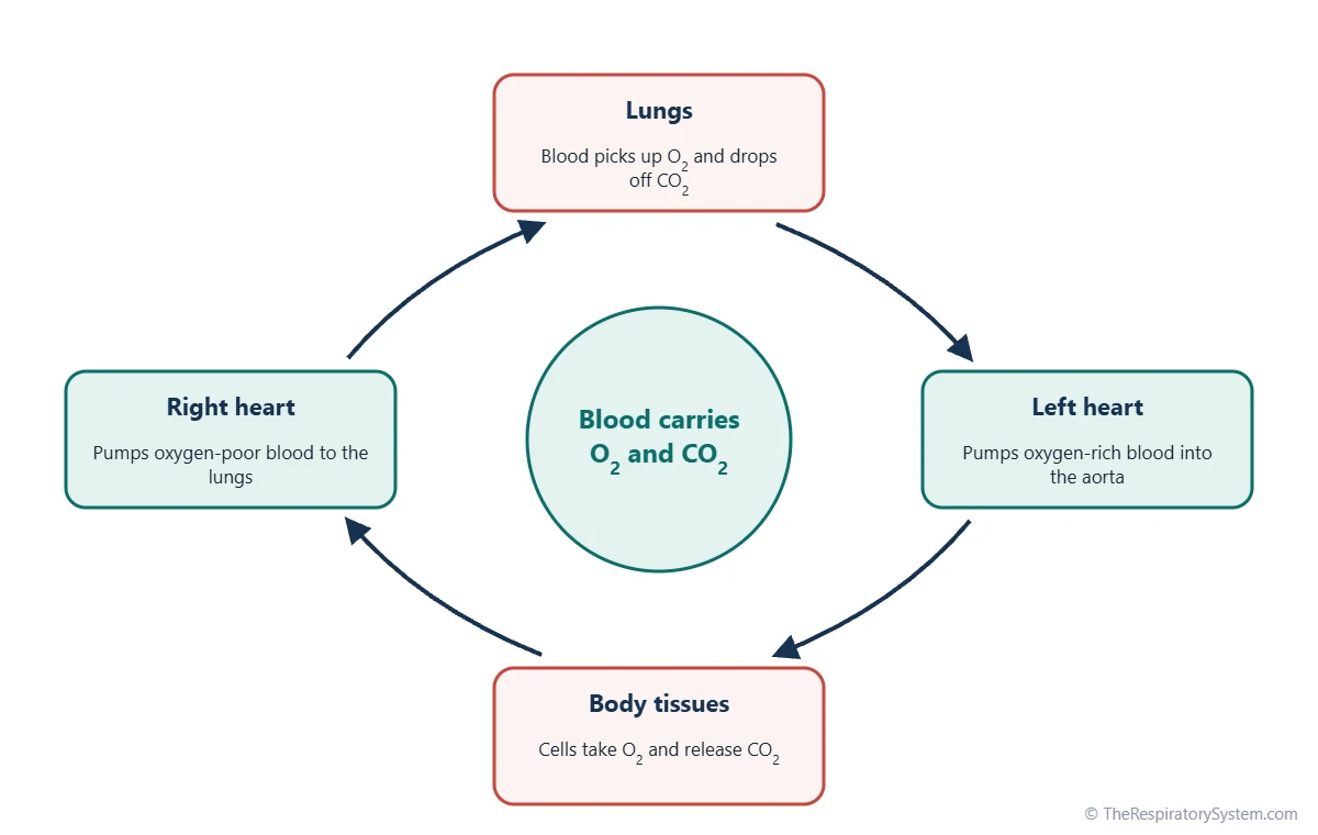
The respiratory system works with the circulatory system in a continuous loop: the lungs add oxygen to the blood, the heart pumps it to the body, the tissues use the oxygen and add carbon dioxide, and the heart sends the blood back to the lungs to unload it. Each trip around the loop follows the same steps.
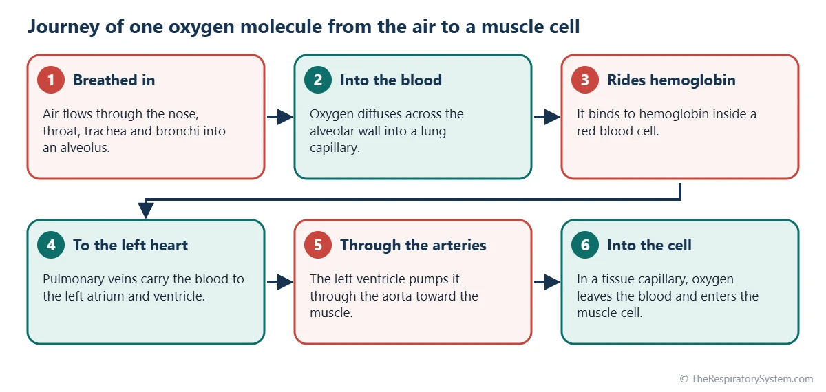
- Ventilation: Breathing in brings fresh air along the pathway of air into the alveoli, keeping their oxygen level high and carbon dioxide level low.
- Gas exchange in the lungs: Oxygen diffuses from the alveoli into the blood of the pulmonary capillaries, and carbon dioxide diffuses the other way, as described on our gas exchange page.
- Return to the heart: Oxygen-rich blood leaves the lungs in the pulmonary veins and enters the left atrium and left ventricle.
- Delivery to the body: The left ventricle pumps the blood into the aorta and out through the systemic arteries to every organ.
- Gas exchange in the tissues: In the body's capillaries, oxygen moves into the cells and carbon dioxide moves into the blood. This step is called internal respiration; see external and internal respiration.
- Back to the lungs: Oxygen-poor blood returns in the veins to the right atrium and right ventricle, which pumps it through the pulmonary arteries into the lungs, and the loop starts again.
The two halves of the heart are therefore two pumps in series. The right side serves the pulmonary circulation, a short, low-pressure loop through the lungs, and the left side serves the systemic circulation, a long, high-pressure loop through the rest of the body. Every drop of blood must pass through the lungs once for each trip around the body.
Where Do the Respiratory and Circulatory Systems Meet?
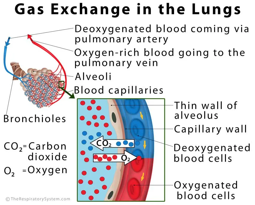
The two systems meet at the walls of the alveoli, where each air sac is wrapped in a dense net of pulmonary capillaries so thin that red blood cells pass through them almost in single file. This meeting point is the only place where air and blood come close enough to exchange gases.
Between the air and the blood lies the respiratory membrane: the flat type I cells of the alveolar wall, a shared basement membrane and the capillary wall. In most places it is only about 0.5 micrometer thick, and in an adult the total surface for exchange is roughly 50 to 75 square meters. Gases cross it by simple diffusion, moving from higher to lower partial pressure, so no energy is spent on the exchange itself.
The lungs are the only organ that receives the entire output of the heart. At rest, about 5 liters of blood flow through the pulmonary capillaries every minute, and each red blood cell spends roughly three-quarters of a second there, which is normally more than enough time to fill with oxygen. The lung tissue itself is fed separately by small bronchial arteries that branch from the aorta, because the blood in the pulmonary arteries is low in oxygen.
Respiratory System vs Cardiovascular System: Key Differences
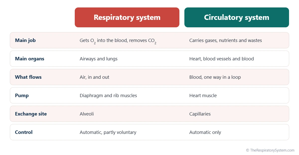
The key differences are in what each system moves, what powers it and how it is controlled: the respiratory system moves air with skeletal muscles that you can partly control, while the cardiovascular system moves blood with heart muscle that works on its own. The table below sets the two side by side.
- What flows: air, a mixture of gases, flows in and out of the same tubes; blood, a liquid, flows one way around a closed circuit.
- The pump: breathing depends on the diaphragm and the muscles of respiration, which are skeletal muscles; the heart is cardiac muscle with its own built-in pacemaker.
- Control: you can hold your breath or speed it up for a short time, but you cannot stop your heart by choice. Both are normally run automatically by the brainstem.
- Pressure: air moves because pressure in the lungs falls slightly below or rises slightly above the air outside, while the heart creates much higher pressures to push blood through the vessels.
- Reach: the respiratory system ends in the lungs; the circulatory system reaches almost every tissue in the body.
Typical resting rates show the difference in rhythm: an adult breathes about 12 to 20 times a minute (see respiratory rate), while the heart beats about 60 to 100 times a minute.
What Do the Respiratory and Circulatory Systems Have in Common?
Both systems are transport networks of branching tubes that end in microscopic, thin-walled spaces where exchange takes place. The airways divide again and again until they reach the alveoli, and the arteries divide again and again until they reach the capillaries, so both pack a huge exchange surface into a small volume.
- Branching design: the bronchial tree and the arterial tree both narrow and multiply at each division.
- Smooth muscle control: smooth muscle in the walls of bronchioles and arterioles adjusts how much air or blood reaches each region.
- Rhythmic pumping: both depend on a repeating cycle, the breathing cycle and the heartbeat.
- Brainstem control: centers in the medulla oblongata and pons set both breathing and heart rate.
- Shared organs in the chest: the heart and lungs sit side by side in the thoracic cavity, separated by the mediastinum.
Both systems also help keep the body's internal conditions stable. Breathing controls how much carbon dioxide stays in the blood, which sets blood pH, and blood flow spreads oxygen, heat and buffers evenly through the body. Our page on respiratory system homeostasis explains this balance in detail.
How Does the Body Keep Breathing and Blood Flow Matched?
The body keeps breathing and blood flow matched through shared sensors and control centers that raise or lower both together. When cells need more oxygen, both the breathing rate and the heart rate climb, so more air and more blood reach the lungs at the same time.
Chemoreceptors and the brainstem
Sensors called respiratory chemoreceptors track carbon dioxide, pH and oxygen. Central chemoreceptors near the medulla respond mainly to carbon dioxide through changes in pH, and peripheral chemoreceptors in the carotid bodies and aortic bodies respond to low oxygen as well. Their signals reach the brainstem, which adjusts the depth and rate of breathing and, through the autonomic nerves, heart rate. The full feedback loop is covered on our control of breathing page.
Matching inside the lungs
Within the lungs, air and blood are also matched region by region. When a group of alveoli receives little air, the small pulmonary arteries supplying it narrow, a response called hypoxic pulmonary vasoconstriction, which sends blood toward better-ventilated areas. The balance between air and blood in each region is described by the ventilation-perfusion ratio.
During exercise
During hard exercise, the amount of air breathed per minute and the heart's output can each rise several times above resting levels. Our page on exercise and the respiratory system shows how both systems respond.
Common Conditions That Affect Both Systems
Because the heart and lungs depend on each other, a problem in one system often shows up in the other. The conditions below are listed for education only; they are explained further in our respiratory diseases section.
- Pulmonary embolism: a blood clot, usually from a leg vein, blocks a pulmonary artery, so part of the lung is ventilated but not supplied with blood.
- Fluid in the lungs from heart disease: when the left side of the heart cannot pump well, pressure backs up into the lung capillaries and fluid can leak into the lung tissue, making gas exchange harder.
- Lung disease straining the heart: long-term low oxygen, as in severe COPD, narrows lung blood vessels and makes the right ventricle work harder.
- Anemia: the lungs may work normally, but blood with too little hemoglobin carries less oxygen, which can lead to hypoxia in the tissues.
Breathlessness and chest pain can come from either system, so they are a reason to see a doctor rather than to guess the cause.
Key Facts
- The respiratory system loads oxygen into the blood and unloads carbon dioxide; the circulatory system carries both gases between the lungs and the tissues.
- The two systems meet at the alveoli, across a respiratory membrane only about 0.5 micrometer thick.
- The lungs are the only organ that receives the whole output of the heart, about 5 liters of blood per minute at rest.
- The pulmonary arteries carry oxygen-poor blood and the pulmonary veins carry oxygen-rich blood, the reverse of the rest of the body.
- The right side of the heart pumps blood through the lungs, and the left side pumps it to the rest of the body.
- Most carbon dioxide travels to the lungs as bicarbonate in the blood, as explained on our carbon dioxide transport page.
- Brainstem centers raise breathing and heart rate together when the body needs more oxygen.
Quick Quiz: Test What You Know About the How the Respiratory and Circulatory Systems Work Together
6 questions. Tap an answer to check it.
Where do the respiratory and circulatory systems exchange gases in the lungs?
Gas exchange happens across the thin walls of the alveoli and the capillaries wrapped around them. The trachea and bronchi only conduct air.
Which chamber of the heart pumps oxygen-poor blood to the lungs?
The right ventricle pumps blood into the pulmonary arteries and through the lungs. The left ventricle pumps oxygen-rich blood to the body.
Which vessels carry oxygen-rich blood from the lungs to the heart?
The pulmonary veins return oxygen-rich blood to the left atrium. They are the only veins in the adult body that carry oxygen-rich blood.
What powers the movement of air into the lungs?
The diaphragm and the muscles between the ribs enlarge the chest, lowering lung pressure so air flows in. The heart moves blood, not air.
In which form is most carbon dioxide carried in the blood to the lungs?
Most carbon dioxide travels as bicarbonate dissolved in the plasma. Smaller amounts ride on hemoglobin or stay dissolved as gas.
What links the respiratory and circulatory systems in the chest?
The pulmonary circulation runs from the right ventricle through the lung capillaries to the left atrium, which is where blood meets air.
Frequently Asked Questions
Is the heart part of the respiratory system?
No. The heart belongs to the circulatory, or cardiovascular, system. The respiratory system includes the airways and lungs. The two are so closely linked that doctors often speak of the cardiopulmonary system, because the right side of the heart exists to pump blood through the lungs, and the lungs exist to refill that blood with oxygen before the left side pumps it to the body.
Why do the pulmonary arteries carry oxygen-poor blood?
Arteries are defined by direction, not by oxygen: they carry blood away from the heart. The pulmonary arteries leave the right ventricle carrying blood that has just returned from the body, low in oxygen and high in carbon dioxide, on its way to the lungs. After gas exchange, the pulmonary veins bring the newly oxygen-rich blood back to the left atrium.
Which is more important, the respiratory system or the circulatory system?
Neither can keep the body alive alone, so they are equally essential. Without breathing, blood keeps circulating but runs out of oxygen within minutes. Without circulation, the lungs can fill with fresh air but no oxygen reaches the cells. This is why basic life support focuses on both airway and circulation together.
What is the cardiopulmonary system?
Cardiopulmonary means heart and lungs. The term describes the heart, the lungs and the pulmonary blood vessels that connect them, treated as one working unit. It is used in physiology, in exercise science and in medicine, for example in cardiopulmonary exercise testing, which measures how well the heart, lungs and muscles work together during effort.
Why do breathing and heart rate both go up during exercise?
Working muscles use more oxygen and make more carbon dioxide. Sensors and brain centers respond by deepening and speeding up breathing to refresh the alveolar air faster, and by raising heart rate and the amount pumped per beat to move blood through the lungs and muscles faster. Both changes are needed, because extra air is useless without extra blood flow to carry it.
Do the lungs get their own blood supply?
Yes. Besides the pulmonary circulation used for gas exchange, the lungs have a small bronchial circulation. Bronchial arteries, which usually branch from the aorta or nearby arteries, deliver oxygen-rich blood to the walls of the bronchi, the connective tissue and the pleura. Much of this blood drains back into the pulmonary veins.
How long does it take blood to pass through the lungs?
At rest, a red blood cell spends about three-quarters of a second in a lung capillary, and it is usually fully loaded with oxygen within the first third of that time. During hard exercise, blood moves faster and the time can drop to well under half a second, yet in healthy lungs it is still normally enough.
How do you draw the respiratory and circulatory system together?
A simple drawing shows the lungs on both sides of the heart. Draw the trachea branching into the bronchi, then a blue pulmonary artery leaving the right ventricle toward each lung and a red pulmonary vein returning to the left atrium. Add an aorta leaving the left ventricle and veins returning to the right atrium. Our worksheet page has printable labeling diagrams.
Keep Exploring


