At a Glance
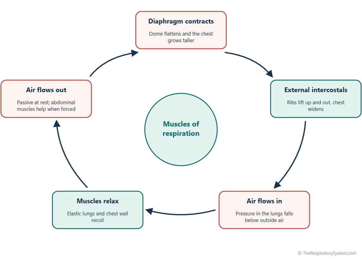
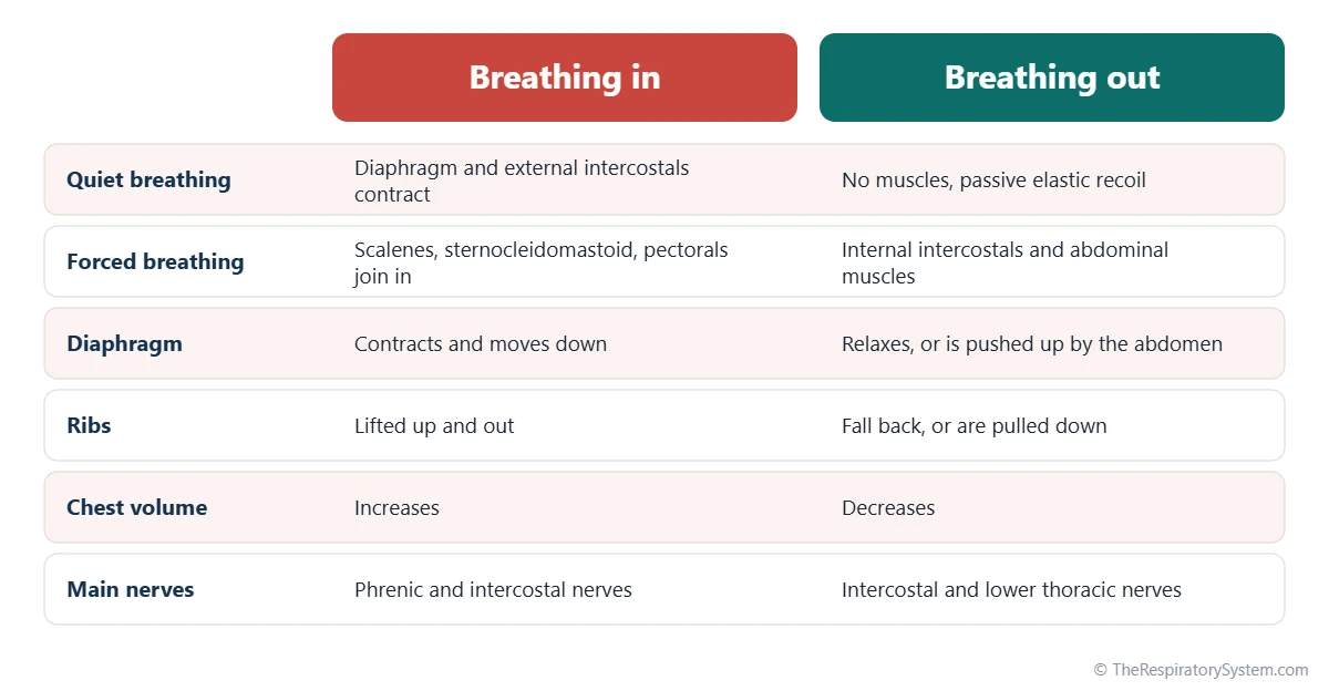
The primary muscles of respiration are the diaphragm, which does about 75 percent of the work of a quiet breath in, and the external intercostal muscles between the ribs. Accessory muscles of inspiration, such as the sternocleidomastoid, scalenes and pectoralis minor, lift the upper ribs during deep or labored breathing. Quiet breathing out is passive, while forced breathing out uses the internal intercostals and the abdominal muscles.
- Primary muscles
- Diaphragm and external intercostal muscles
- Accessory inspiration
- Sternocleidomastoid, scalenes, pectoralis major and minor, serratus anterior and others
- Forced expiration
- Internal and innermost intercostals, rectus abdominis, obliques, transversus abdominis
- Muscle type
- Skeletal muscle, under both automatic and voluntary control
- Main nerves
- Phrenic nerves (C3 to C5) and intercostal nerves (T1 to T11)
- Part of
- The breathing pump that drives pulmonary ventilation
On this page
- What Are the Muscles of Respiration?
- What Muscles Are Involved in Respiration When You Breathe In?
- What Are the Accessory Muscles of Respiration?
- Which Muscles Are Used to Breathe Out?
- How Do the Breathing Muscles Move the Rib Cage?
- How Are the Respiratory Muscles Controlled?
- Which Muscles Keep the Airway Open During Breathing?
- Common Conditions That Affect the Muscles of Respiration
- Key Facts
- FAQ
What Are the Muscles of Respiration?
The muscles of respiration are the muscles that enlarge and shrink the chest cavity to move air in and out of the lungs, and they fall into three groups: primary muscles of inspiration, accessory muscles of inspiration and muscles of expiration. The lungs have no muscle of their own to inflate themselves, so every breath depends on these muscles pulling on the walls of the thoracic cavity.
The three groups
- Primary muscles of inspiration: the diaphragm and the external intercostal muscles, active in every breath in, even during sleep.
- Accessory muscles of inspiration: neck, chest and back muscles that lift the ribs further when the body needs more air, such as during hard exercise.
- Muscles of expiration: the internal and innermost intercostals and the abdominal wall muscles, which push air out only when breathing out must be fast or forceful.
Quiet breathing out needs no muscle at all. When the diaphragm and external intercostals relax, the stretched, elastic lungs and chest wall spring back on their own, as described on our inhalation and exhalation page.
Explained simply
Your chest works like a pair of bellows. The diaphragm is the floor that pulls down and the rib muscles are the sides that lift out, so the bellows get bigger and air rushes in. When those muscles let go, the bellows spring back to their smaller size and push the air out again.
What Muscles Are Involved in Respiration When You Breathe In?
The diaphragm and the external intercostal muscles are the muscles involved in every normal breath in, with the diaphragm doing most of the work.
The diaphragm
The diaphragm is a thin, dome-shaped sheet of skeletal muscle that separates the chest from the abdomen. When it contracts, its central tendon is pulled down and the dome flattens, making the chest taller. It is responsible for about 75 percent of the air that enters the lungs during quiet breathing. Its dome moves down only about 1 cm in a quiet breath, but up to about 10 cm in a very deep one. It is supplied by the right and left phrenic nerves from the C3, C4 and C5 spinal nerve roots.
The external intercostal muscles
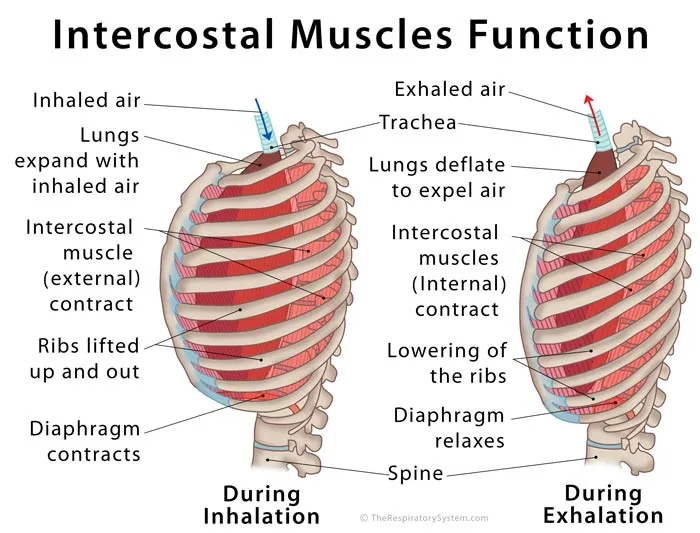
The external intercostals fill the spaces between neighboring ribs, with fibers running downward and forward, like hands in front pockets. When they contract they lift each rib toward the one above, which widens the chest from front to back and from side to side. Each side of the chest has 11 intercostal spaces, and our page on the intercostal muscles describes all three layers in detail.
The parasternal intercostals
Near the breastbone, the front part of the internal intercostal layer between the rib cartilages, called the parasternal or interchondral part, also helps to lift the ribs during inspiration. This is why some textbooks count it among the primary inspiratory muscles, even though the rest of the internal intercostal layer works in expiration.
What Are the Accessory Muscles of Respiration?
The accessory muscles of respiration are muscles of the neck, chest and back whose main job is moving the head, shoulders or arms, but which help lift the ribs and breastbone when the body needs to breathe in more deeply or quickly than the primary muscles can manage alone.
Accessory muscles of respiration location
- Sternocleidomastoid: runs from behind the ear down to the breastbone and collarbone; it lifts the sternum.
- Scalenes (anterior, middle and posterior): run from the neck vertebrae down to the first and second ribs, which they raise and steady.
- Pectoralis minor: lies under the pectoralis major and pulls the third to fifth ribs upward when the shoulder blade is held still.
- Pectoralis major: the large chest muscle; it lifts the ribs and sternum when the arms are fixed in place.
- Serratus anterior: wraps around the side of the chest from the ribs to the shoulder blade and can raise the ribs when the shoulder blade is fixed.
- Serratus posterior superior and levatores costarum: small back muscles that help elevate the ribs.
Accessory muscles of respiration function
Accessory muscles work by reversing their usual pull. Normally the sternocleidomastoid turns the head, but when the head is held steady it pulls up on the breastbone instead. In the same way, a person who is short of breath often leans forward with hands on the knees, fixing the shoulders so the chest muscles can lift the ribs. Accessory muscle use is normal during hard exercise; at rest it is a sign that breathing has become harder work. The scalenes are an exception in being active during quiet breathing in many people, which is why some textbooks list them as primary muscles.
| Muscle | Group | Role in breathing | Nerve supply |
|---|---|---|---|
| Diaphragm | Primary inspiration | Contracts and flattens, making the chest taller | Phrenic nerves (C3 to C5) |
| External intercostals | Primary inspiration | Lift the ribs up and out | Intercostal nerves (T1 to T11) |
| Parasternal (interchondral) intercostals | Primary inspiration | Lift the ribs near the breastbone | Intercostal nerves |
| Scalenes | Accessory inspiration | Raise and steady the first two ribs | Branches of the cervical spinal nerves |
| Sternocleidomastoid | Accessory inspiration | Lifts the sternum when the head is fixed | Accessory nerve (cranial nerve XI) |
| Pectoralis minor | Accessory inspiration | Pulls ribs 3 to 5 upward when the scapula is fixed | Medial pectoral nerve |
| Pectoralis major | Accessory inspiration | Lifts the ribs and sternum when the arms are fixed | Medial and lateral pectoral nerves |
| Serratus anterior | Accessory inspiration | Raises the ribs when the scapula is fixed | Long thoracic nerve |
| Internal intercostals | Forced expiration | Pull the ribs down | Intercostal nerves |
| Innermost intercostals and transversus thoracis | Forced expiration | Help depress the ribs | Intercostal nerves |
| Rectus abdominis | Forced expiration | Pulls the lower ribs and sternum down | Lower intercostal and subcostal nerves (T7 to T12) |
| External and internal obliques | Forced expiration | Compress the abdomen, push the diaphragm up | Lower intercostal, subcostal and first lumbar nerves |
| Transversus abdominis | Forced expiration | Squeezes the abdomen like a belt | Lower intercostal, subcostal and first lumbar nerves |
| Source: Standard anatomy, our editors | |||
Which Muscles Are Used to Breathe Out?
No muscles are needed to breathe out at rest, but during forced expiration the internal and innermost intercostals pull the ribs down and the abdominal muscles push the diaphragm up.
Quiet expiration is passive
During a relaxed breath out, the diaphragm and external intercostals simply relax. The elastic fibers in the lungs, the surface tension of the fluid lining the alveoli and the spring of the rib cage pull the chest back to its resting size, and air flows out without any muscle effort.
Forced expiration muscles
- Internal intercostals: their fibers run at right angles to the external layer, and they pull each rib down toward the one below.
- Innermost intercostals, subcostales and transversus thoracis: thin inner chest wall muscles that help depress the ribs.
- Rectus abdominis: the straight muscle down the front of the abdomen, which pulls the lower ribs and breastbone down.
- External and internal obliques: diagonal abdominal muscles that compress the abdomen and pull the ribs down.
- Transversus abdominis: the deepest abdominal layer, which squeezes the abdomen like a belt.
- Serratus posterior inferior: a back muscle that pulls the lowest ribs down and back.
When the abdominal muscles contract, they raise the pressure inside the abdomen and push the relaxed diaphragm up into the chest, which squeezes air out quickly. This is the force behind a cough, a sneeze, shouting and blowing out candles, and it is how lung function tests measure the vital capacity.
How Do the Breathing Muscles Move the Rib Cage?
The breathing muscles enlarge the chest in three directions: the diaphragm makes it taller, and the intercostal and accessory muscles lift the ribs to make it deeper from front to back and wider from side to side.
Pump handle and bucket handle movements
The ribs are hinged at the spine and slope downward toward the front. When the upper ribs are raised, the breastbone moves forward and up, like the handle of an old water pump, which increases the front-to-back depth of the chest. When the lower ribs are raised, their middle parts swing outward, like the handle of a bucket being lifted, which increases the width. The structure that makes this possible is described on our rib cage and breathing page.
From muscle movement to airflow
As the chest grows, the lungs follow it because the thin layer of fluid in the pleural cavity holds the outer surface of each lung against the inside of the chest wall. A larger lung volume lowers the pressure inside the alveoli a little below the pressure of the outside air, and air flows in until the two pressures are equal. When the muscles relax, the process runs in reverse. Our page on the mechanics of breathing explains these pressure changes and Boyle's law in detail.
How Are the Respiratory Muscles Controlled?
The respiratory muscles are controlled automatically by breathing centers in the brainstem, which send rhythmic signals through the phrenic and intercostal nerves, but they can also be overridden voluntarily, for example when speaking, singing or holding a breath.
Automatic control
Groups of nerve cells in the medulla oblongata and pons set the basic rhythm of breathing, roughly 12 to 20 breaths per minute in a resting adult. They adjust the depth and rate of each breath in response to signals from chemoreceptors that sense carbon dioxide, oxygen and acidity in the blood, and from stretch receptors in the lungs. This is covered on our control of breathing page.
Voluntary control
Because the breathing muscles are skeletal muscles, the motor cortex can also command them directly, which is how you can take a deep breath on purpose or shape airflow for speech. The automatic centers always win in the end: as carbon dioxide builds up, the urge to breathe becomes too strong to resist.
Nerve supply
The diaphragm is supplied only by the phrenic nerves (C3 to C5), the intercostal muscles by the intercostal nerves (T1 to T11), the abdominal muscles by the lower intercostal, subcostal and first lumbar nerves, and the sternocleidomastoid by the accessory nerve, a cranial nerve.
Which Muscles Keep the Airway Open During Breathing?
Besides the pump muscles of the chest, small muscles in the throat and larynx keep the upper airway open with each breath in, so that the falling pressure inside does not suck the soft walls inward.
- Genioglossus: the main muscle of the tongue, which pulls the tongue forward and keeps it from falling back into the pharynx.
- Muscles of the soft palate and pharyngeal walls: stiffen the throat during inspiration.
- Posterior cricoarytenoid: the only muscle that opens the vocal folds, widening the glottis in the larynx as each breath begins.
- Nasal dilator muscles: flare the nostrils slightly during deep or labored breathing.
These muscles fire just before the diaphragm in each breath. During sleep their tone falls, which is one reason the throat can narrow or close in obstructive sleep apnea.
Common Conditions That Affect the Muscles of Respiration
Conditions that weaken the breathing muscles, damage their nerves or make breathing harder can all affect how well air is moved in and out of the lungs. Our respiratory diseases section covers lung conditions in more depth, and anyone with unexplained shortness of breath should see a doctor.
- Diaphragm paralysis: injury to a phrenic nerve can paralyze one half of the diaphragm, which then moves up instead of down during a breath in.
- High spinal cord injury: damage at or above the C3 to C5 level can stop the diaphragm from working, because its nerve roots arise there.
- Neuromuscular diseases: conditions such as amyotrophic lateral sclerosis, muscular dystrophy and myasthenia gravis can weaken the respiratory muscles over time.
- COPD and emphysema: overinflated lungs flatten the diaphragm, leaving it less effective, so neck and chest accessory muscles are used even at rest.
- Hiccups: sudden involuntary contractions of the diaphragm, explained on our why do we hiccup page.
- Muscle strain: overstretched intercostal muscles can make each breath painful.
Key Facts
- The diaphragm is the main muscle of respiration and accounts for about 75 percent of the air drawn in during quiet breathing.
- The external intercostal muscles lift the ribs up and out, widening the chest with every breath in.
- Quiet breathing out is passive and relies on the elastic recoil of the lungs and chest wall.
- Accessory muscles of inspiration, such as the sternocleidomastoid and scalenes, lift the upper ribs during deep or labored breathing.
- Forced breathing out uses the internal intercostals and the abdominal muscles, which push the diaphragm upward.
- The diaphragm is supplied by the phrenic nerves from C3, C4 and C5, and the intercostal muscles by the intercostal nerves.
- Breathing muscles are skeletal muscles under automatic control from the brainstem that can also be overridden voluntarily.
Frequently Asked Questions
What is the main muscle of respiration?
The diaphragm is the main muscle of respiration. This dome-shaped muscle under the lungs contracts and flattens with each breath in, enlarging the chest from top to bottom, and it accounts for about 75 percent of the air that enters the lungs during quiet breathing. The external intercostal muscles between the ribs supply most of the rest by lifting the rib cage.
Are the abdominal muscles muscles of respiration?
Yes, they are the main muscles of forced expiration. The rectus abdominis, external and internal obliques and transversus abdominis squeeze the abdomen and push the relaxed diaphragm up into the chest, driving air out quickly. They are quiet during relaxed breathing but work hard during coughing, sneezing, shouting, singing and heavy exercise.
What muscles are used in forced inspiration?
Forced inspiration uses the diaphragm and external intercostals working harder than usual, plus accessory muscles: the scalenes and sternocleidomastoid in the neck, the pectoralis minor and major in the chest, the serratus anterior on the side of the chest and small back muscles such as the serratus posterior superior. Together they raise the ribs and breastbone higher than in a quiet breath.
Can you breathe without a diaphragm?
Breathing is possible for a time with a paralyzed diaphragm, because the intercostal and accessory muscles can still enlarge the chest, but it is much less effective, especially when lying down. If only one side of the diaphragm is paralyzed, many people have few symptoms at rest. Loss of both sides usually causes marked shortness of breath.
Is breathing controlled by voluntary or involuntary muscles?
Breathing uses skeletal muscles, the type normally under voluntary control, but they are driven automatically by the brainstem most of the time. This dual control lets you breathe without thinking while asleep and still take a deep breath, hold a note or speak on purpose. When carbon dioxide rises, the automatic drive overrides voluntary control.
Why do accessory muscles show during hard breathing?
When the body needs more air than the diaphragm and external intercostals can supply, the brain recruits the neck and chest muscles to lift the rib cage further. Their contraction can be seen as the neck muscles tightening and the spaces above the collarbones sinking with each breath. During intense exercise this is normal; at rest it suggests that breathing has become hard work.
What muscles of respiration are shown on a diagram?
A muscles of respiration diagram usually labels the diaphragm under the lungs, the external and internal intercostals between the ribs, the scalenes and sternocleidomastoid in the neck, the pectoralis minor on the front of the chest and the abdominal muscles below. Many diagrams color the inspiratory muscles and the expiratory muscles differently, matching the two groups described on this page.
Do the respiratory muscles get tired?
Healthy breathing muscles rarely tire during normal life, because the diaphragm is rich in fatigue-resistant muscle fibers and has a generous blood supply. They can tire when breathing becomes very hard work for a long time, for example in severe lung disease, which can lead to breathing failure. Intense exercise can also tire the diaphragm briefly in healthy people.
Keep Exploring


