At a Glance
The rib cage (thoracic cage) is a flexible frame of 24 ribs, the sternum and 12 thoracic vertebrae, joined by costal cartilages and small joints. It protects the heart, lungs and great vessels, and during inhalation its ribs swing up and out in pump-handle and bucket-handle movements that enlarge the chest so air flows into the lungs.
- Location
- Upper trunk, between the neck and the abdomen, around the thoracic cavity
- Made of
- 12 pairs of ribs, costal cartilages, the sternum and 12 thoracic vertebrae (37 bones if the sternum counts as one)
- Rib groups
- True ribs 1 to 7, false ribs 8 to 10, floating ribs 11 and 12
- Function
- Protects chest organs and changes chest volume during breathing
- Connects to
- Spine behind, clavicles above, diaphragm and abdominal muscles below
- Part of
- Axial skeleton; works with the muscles of respiration
On this page
What Is the Rib Cage?
The rib cage is the curved frame of bone and cartilage that forms the walls of the chest and encloses the thoracic cavity. Anatomists call it the thoracic cage, and it belongs to the axial skeleton together with the skull and the spine. Although it is part of the skeletal system, it is built for breathing as much as for protection, which is why it is studied alongside the organs of the lower respiratory tract.
Thoracic cage definition
The thoracic cage is the enclosure of the thorax formed by the 12 thoracic vertebrae behind, the 12 pairs of ribs and their costal cartilages at the sides, and the sternum in front. Counting the sternum as one bone, it contains 37 bones. It is shaped like a cone with its top cut off: narrow where it meets the neck and wide at the bottom, where the diaphragm closes it off.
Explained simply
Picture a birdcage made of curved bars that can bend a little. The bars are the ribs, the spine is the back post and the breastbone is the front post. Every time you breathe in, muscles lift the bars so the cage gets bigger, and the lungs inside get bigger with it.
Where Is the Thoracic Cage?
The thoracic cage sits in the upper trunk, between the neck and the abdomen, and surrounds the chest organs on every side except the floor, which is formed by the diaphragm. Its top opening, the superior thoracic aperture, is a small kidney-shaped ring made by the first thoracic vertebra, the first pair of ribs and the top of the sternum. The trachea, the esophagus and major vessels and nerves pass through it between the neck and the chest.
The bottom opening, the inferior thoracic aperture, is much wider. It is outlined by the 12th thoracic vertebra, ribs 11 and 12 and the costal margin, the sloping edge formed by the cartilages of ribs 7 to 10. The diaphragm attaches all around this rim, and the aorta, esophagus and inferior vena cava reach the abdomen through the openings of the diaphragm.
Landmarks you can feel
- Jugular notch: the dip at the top of the sternum, between the inner ends of the collarbones.
- Sternal angle: a small ridge where the manubrium meets the body of the sternum; the second rib joins here, so clinicians use it as the starting point for counting ribs.
- Costal margin: the lower edge of the cage on each side, curving down and out from the xiphoid process.
Because the diaphragm bulges upward like a dome, the lower ribs also shelter part of the liver, stomach, spleen and kidneys. Inside the cage, the central mediastinum holds the heart, and a lung fills each side.
Rib Cage Anatomy: What Are Its Parts?
The rib cage is made of three groups of parts: the ribs with their costal cartilages, the sternum in front and the thoracic vertebrae behind, held together by a set of small joints. The table below lists each part and where it attaches.
True, false and floating ribs
Ribs 1 to 7 are true ribs because each one connects directly to the sternum through its own costal cartilage. Ribs 8 to 10 are false ribs: their cartilages join the cartilage of the rib above instead of reaching the sternum. Ribs 11 and 12 are floating ribs, short bones whose front tips end freely in the muscles of the side of the abdomen.
Parts of a typical rib
Ribs 3 to 9 are called typical ribs and share the same plan. The head carries two small facets that join the bodies of two neighboring vertebrae; next come a short neck, a knob called the tubercle that joins the transverse process of a vertebra, and a sharp bend called the angle. The long, flat shaft curves around the chest and has a costal groove along its lower inner edge.
That groove shelters the intercostal vein, artery and nerve, which run in that order from top to bottom. The gaps between the ribs, the intercostal spaces, are filled by the three thin layers of the intercostal muscles.
The sternum and costal cartilages
The sternum, or breastbone, has three parts: the manubrium at the top, the long body in the middle and the small xiphoid process at the bottom. The costal cartilages are bars of hyaline cartilage that link the ribs to the sternum and give the front of the cage its spring. Joints at both ends of each rib, the costovertebral and costotransverse joints behind and the sternocostal joints in front, let the ribs rotate slightly during breathing.
| Part | How many | Attachment or position | Notes |
|---|---|---|---|
| True ribs (vertebrosternal) | Ribs 1 to 7 on each side | Join the sternum directly through their own costal cartilage | Also called sternal ribs |
| False ribs (vertebrochondral) | Ribs 8 to 10 on each side | Their cartilages join the cartilage of the rib above | Form the costal margin together with rib 7 |
| Floating ribs (vertebral) | Ribs 11 and 12 on each side | No front attachment; tips end in the flank muscles | Lie behind the upper parts of the kidneys |
| First rib | One pair | Joins the manubrium just below the clavicle | Shortest, broadest and most curved rib |
| Manubrium | One | Top part of the sternum; joins the clavicles and the first ribs | Carries the jugular notch |
| Body of the sternum | One | Middle part; joins ribs 3 to 7 and part of rib 2 | Meets the manubrium at the sternal angle |
| Xiphoid process | One | Small lower tip of the sternum | Cartilage in childhood, usually bone in adulthood |
| Costal cartilages | One at the front end of each rib | Hyaline cartilage linking ribs to the sternum or to each other | Give the cage its flexibility |
| Thoracic vertebrae | 12 (T1 to T12) | Back wall of the cage; each rib joins the spine at two joints | Ribs meet vertebral bodies and transverse processes |
| Source: Typical adult anatomy, our editors | |||
What Is the Rib Cage Function in the Respiratory System?
In the respiratory system, the rib cage works as a movable frame that changes the volume of the chest, so the lungs inside can draw air in and push it out. The lungs have no muscle of their own to expand; they grow larger only because the chest wall around them grows larger.
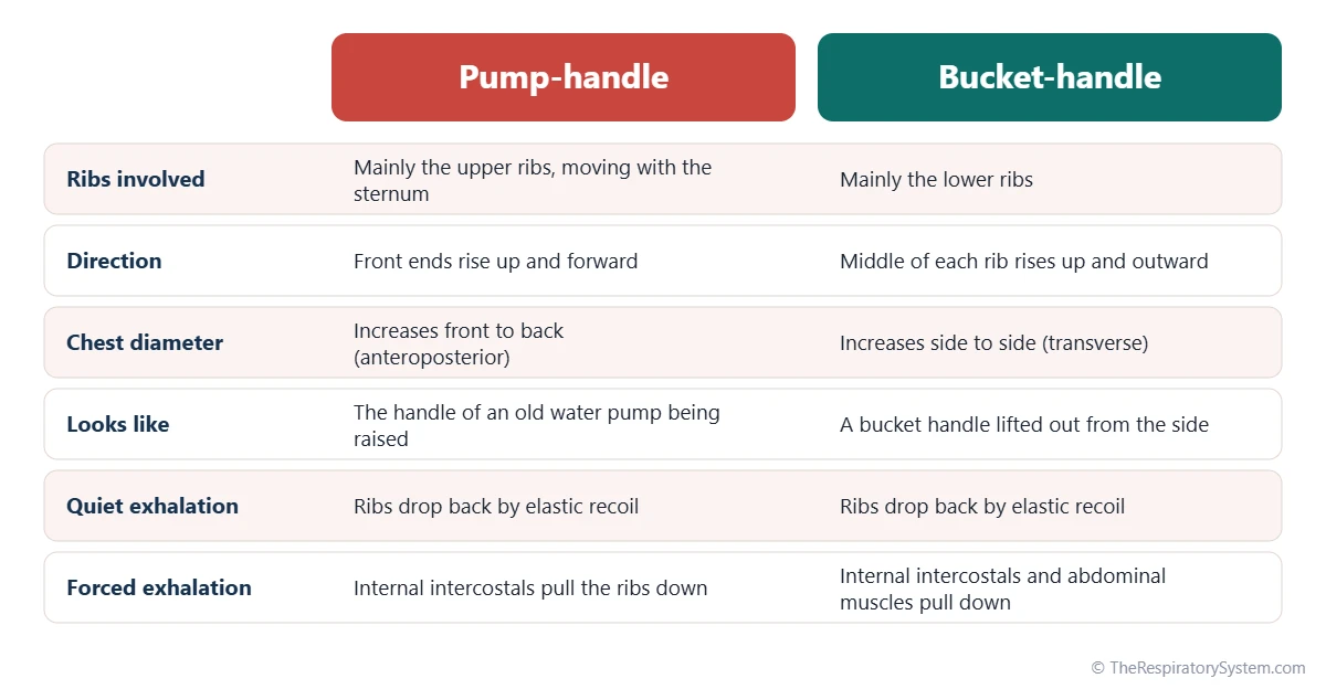
Pump-handle and bucket-handle movements
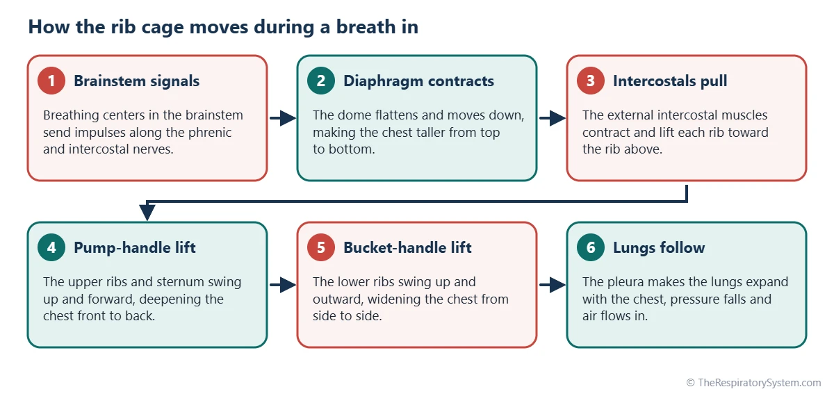
During inhalation, the external intercostal muscles and other muscles of respiration lift the ribs, which move in two ways at once. The upper ribs and the sternum swing up and forward like the handle of an old water pump, deepening the chest from front to back. The lower ribs swing up and outward like a bucket handle lifted from the side, widening the chest from left to right. At the same time the diaphragm contracts and moves down, making the chest taller.
From a bigger chest to air flow
When the chest gets bigger, the pressure inside the lungs drops slightly below the pressure of the outside air, and air flows in through the airways. This follows Boyle's law: at a constant temperature, the pressure of a gas falls as its volume rises. The lungs follow the moving chest wall because the two layers of the pleura are held together by a thin film of fluid in the pleural cavity, as explained on our mechanics of breathing page.
Breathing out
Quiet exhalation is mostly passive: the muscles relax, and the ribs drop back under the elastic recoil of the lungs, the costal cartilages and the chest wall, pushing air out. During forced breathing out, as in coughing or hard exercise, the internal intercostal muscles and the abdominal muscles pull the ribs down actively. Our inhalation and exhalation guide compares the two phases in detail.
What Is the Rib Cage Function in the Skeletal System?
In the skeletal system, the rib cage protects the organs of the chest and upper abdomen, anchors many muscles, supports the shoulder girdle and holds bone marrow that makes blood cells. Its curved, springy design lets it absorb blows that would crack a flat, rigid plate.
- Protection: it guards the heart, the lungs, the great vessels such as the aorta, and the upper parts of the liver, spleen and kidneys.
- Muscle attachment: the diaphragm and the intercostal, pectoral, serratus anterior, abdominal and back muscles all attach to the ribs or sternum.
- Support: the sternum meets the clavicles at the sternoclavicular joints, the only bony link between the arms and the trunk.
- Blood cell production: the sternum and ribs keep red bone marrow throughout adult life.
- Stability: through its many joints with the spine, the cage stiffens the thoracic part of the vertebral column.
The protective role is tied closely to breathing. The bronchi, the roots of the lungs and the large vessels of the pulmonary circulation all lie inside the cage, so a strong but flexible frame keeps the whole gas-exchange system safe while it moves.
How Does the Rib Cage Change With Age?
The rib cage starts out soft and round in a newborn, becomes longer and more sloped through childhood and gradually stiffens in older age. These changes alter how much of each breath comes from rib movement.
The ribs develop in the embryo from small outgrowths of the thoracic vertebrae, first as cartilage that later turns into bone, while the sternum forms from two strips of tissue that fuse in the midline; the wider story is told in development of the respiratory system. In infants the ribs lie almost horizontally and the chest is nearly round in cross-section, so lifting the ribs adds little volume and babies breathe mainly with the diaphragm. As children grow, the ribs slope downward, and the pump-handle and bucket-handle movements become effective.
With aging, the costal cartilages often calcify, the rib joints stiffen and the breathing muscles weaken, so the chest wall becomes less compliant. This is one reason why vital capacity tends to fall slowly over adult life, even in people with healthy lungs.
Common Conditions That Affect the Rib Cage
Conditions of the rib cage include injuries, inflammation, differences in shape present from childhood and bone changes with age, and several of them can make breathing painful or less efficient. Our respiratory diseases section covers the lung conditions linked to them, and anyone with chest pain or trouble breathing should see a doctor.
- Rib fracture: a break, most often in the middle ribs, which curve the most and take blows directly; a sharp broken end can tear the pleura and cause a pneumothorax.
- Flail chest: several neighboring ribs broken in more than one place, so a section of the chest wall moves inward while the rest moves out.
- Costochondritis: inflammation where the ribs meet their costal cartilages, causing tenderness at the front of the chest.
- Pectus excavatum and pectus carinatum: a sunken or a protruding sternum, both usually noticed in childhood.
- Cervical rib: an extra rib growing from the seventh neck vertebra, found in a small minority of people, which can press on nerves and vessels heading to the arm.
- Scoliosis and osteoporosis: a curved spine twists the cage, and thinning bones break more easily.
Painful rib injuries can limit deep breaths and coughing, which is why doctors watch for pneumonia after them.
Key Facts
- The rib cage, or thoracic cage, is made of 12 pairs of ribs, the sternum, 12 thoracic vertebrae and the costal cartilages, 37 bones in all if the sternum counts as one.
- Ribs 1 to 7 are true ribs, ribs 8 to 10 are false ribs, and ribs 11 and 12 are floating ribs.
- During inhalation the upper ribs move like a pump handle and the lower ribs like a bucket handle, enlarging the chest front to back and side to side.
- The lungs follow the moving ribs because the pleura couples the lung surface to the inside of the chest wall.
- The costal groove on the lower inner edge of each rib shelters the intercostal vein, artery and nerve.
- Men and women have the same number of ribs: 12 pairs.
- In infants the ribs are nearly horizontal, so babies rely mainly on the diaphragm to breathe.
Quick Quiz: Test What You Know About the Rib Cage and Breathing
6 questions. Tap an answer to check it.
How many pairs of true ribs does a typical adult have?
Ribs 1 to 7 are true ribs because each joins the sternum directly through its own costal cartilage.
Which ribs are called floating ribs?
Ribs 11 and 12 have no front attachment to the sternum or to another rib's cartilage.
The pump-handle movement of the upper ribs mainly increases which chest diameter?
Lifting the upper ribs and sternum up and forward deepens the chest from front to back (anteroposterior).
What type of tissue joins the true ribs to the sternum?
The costal cartilages are made of hyaline cartilage, which gives the front of the rib cage its flexibility.
What runs in the costal groove on the lower edge of each rib?
The costal groove shelters the intercostal vein, artery and nerve, in that order from top to bottom.
Which part of the sternum is the lowest?
The sternum runs from the manubrium at the top, through the body, to the small xiphoid process at the bottom.
Frequently Asked Questions
How many ribs does a human have?
Most people have 24 ribs, arranged as 12 pairs, and the number is the same in men and women; the old belief that men have one rib fewer is a myth. A small number of people have an extra rib, most often a cervical rib growing from the seventh neck vertebra, and a few have an extra or missing rib low in the chest. These variations usually cause no problems.
Why are ribs 11 and 12 called floating ribs?
Ribs 11 and 12 are called floating ribs because their front ends do not attach to the sternum or to the cartilage of another rib. They are joined only to the spine at the back, and their short, pointed tips end in the muscles of the flank. They are still firmly anchored behind, and they help protect the upper parts of the kidneys.
Is the rib cage part of the respiratory system?
Strictly, the rib cage belongs to the skeletal system, while the respiratory system is made of the airways and lungs. It is still essential for breathing: the respiratory muscles attach to it, and its movements change the size of the chest so that air flows in and out. For that reason anatomy courses usually study it together with the lungs and the muscles of respiration.
Are the lungs attached to the rib cage?
No. The lungs are not tied to the ribs. Each lung is covered by visceral pleura, and the inside of the rib cage is lined by parietal pleura. A thin film of fluid between these two layers lets them slide but resists being pulled apart, so the lung follows the chest wall outward during inhalation. The only firm attachment of each lung is at its root, beside the heart.
What organs does the rib cage protect?
The rib cage protects the heart and the two lungs, the great vessels such as the aorta, the venae cavae and the pulmonary arteries, and the esophagus and main bronchi inside the chest. Because the diaphragm domes upward, the lower ribs also cover the upper part of the liver, the stomach, the spleen and the top of each kidney.
What is the difference between the rib cage and the thoracic cavity?
The rib cage is the frame: the bones and cartilages that form the chest wall. The thoracic cavity is the space inside that frame, closed below by the diaphragm, which holds the lungs in their pleural cavities and the heart and other organs in the mediastinum. In short, the cage is the container and the cavity is the room it encloses.
Can a person breathe without moving the ribs?
Quiet breathing can rely mostly on the diaphragm, which pushes the belly out as it descends, so the ribs move only a little; this pattern is often called diaphragmatic or belly breathing. Deeper breaths, as during exercise, need the ribs as well, because their pump-handle and bucket-handle movements add much more volume to the chest than the diaphragm can add alone.
Keep Exploring


