At a Glance
Bronchi are cartilage-supported airways with mucus glands and goblet cells: two main bronchi, then lobar and segmental bronchi. Bronchioles are the narrower airways after them, about 1 mm wide or less, with no cartilage, no glands, a relatively thick layer of smooth muscle and protective club cells; they end in terminal bronchioles and then respiratory bronchioles, where gas exchange begins.
- Bronchi
- Main, lobar and segmental airways after the trachea, with cartilage rings or plates
- Bronchioles
- Smaller airways about 1 mm wide or less, with no cartilage
- Border
- An airway becomes a bronchiole when it loses its cartilage and glands
- Lining
- Bronchi: pseudostratified ciliated columnar. Bronchioles: simple columnar to cuboidal
- Gas exchange
- None in bronchi; begins only in respiratory bronchioles
- Part of
- The bronchial tree inside the lungs, lower respiratory tract
On this page
- What Is the Difference Between Bronchi and Bronchioles?
- Bronchus vs Bronchi vs Bronchioles: What Do the Words Mean?
- Where Are the Bronchi and Bronchioles Located?
- Bronchi vs Bronchioles Histology: How Do Their Walls Differ?
- What Do the Bronchi and Bronchioles Do?
- What Do Bronchi and Bronchioles Have in Common?
- How Do the Bronchi and Bronchioles Work Together?
- Common Conditions That Affect the Bronchi and Bronchioles
- Key Facts
- Quiz
- FAQ
What Is the Difference Between Bronchi and Bronchioles?
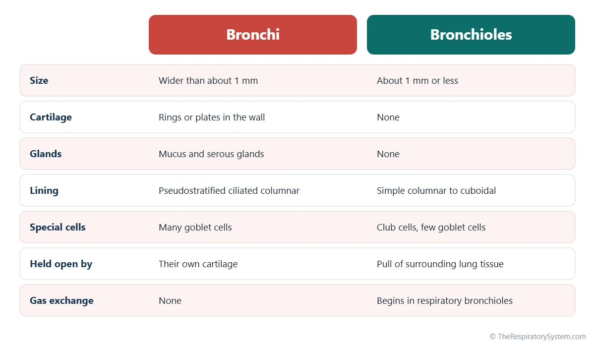
The key difference between bronchi and bronchioles is cartilage: the bronchi have cartilage in their walls, and the bronchioles do not. Size follows the same split, because airways lose their cartilage at about the point where they narrow to roughly 1 mm across.
Losing the cartilage changes everything else about the wall. Bronchi keep the mucus glands, goblet cells and tall ciliated lining of the trachea, while bronchioles have a thinner, simpler lining, few or no goblet cells, no glands and club cells that protect the small airways. Without cartilage to hold them open, bronchioles depend on the springy lung tissue around them, and their smooth muscle can narrow them much more than the muscle of a bronchus can.
The verdict in one line
If an airway has cartilage, it is a bronchus; if it has none, it is a bronchiole, and the smallest bronchioles are the gateway to the gas exchange tissue.
Explained simply
Think of the airways as a tree turned upside down. The bronchi are the thick branches with stiff, ribbed bark that keeps them round. The bronchioles are the thin twigs at the ends: soft, bendy and so many that you could never count them, and at their tips grow the tiny air sacs that act like leaves.
| Feature | Bronchi | Bronchioles |
|---|---|---|
| Types | Main (primary), lobar (secondary), segmental (tertiary) and smaller bronchi | Lobular, terminal and respiratory bronchioles |
| Typical width | More than about 1 mm, up to the width of a main bronchus | About 1 mm or less; terminal bronchioles about 0.5 mm |
| Location | From the carina, through the hilum, into each lobe and segment | Entirely inside the lung tissue, within lobules |
| Cartilage | C-shaped rings (main) or irregular plates | None |
| Glands | Submucosal mucus and serous glands | None |
| Epithelium | Pseudostratified ciliated columnar | Simple columnar to simple cuboidal |
| Goblet cells | Many | Few, then none |
| Club cells | Absent or rare | Present, most common in terminal bronchioles |
| Smooth muscle | Present, beneath the lining | Relatively thick for the airway size |
| Held open by | Cartilage in the wall | Pull of the elastic lung tissue around them |
| Gas exchange | None | Only in respiratory bronchioles |
| Main condition named after it | Bronchitis | Bronchiolitis |
| Source: Typical adult values, our editors | ||
Bronchus vs Bronchi vs Bronchioles: What Do the Words Mean?
Bronchus is the singular and bronchi the plural of the same word, while bronchiole means a small bronchus, with bronchioles as its plural. So bronchus vs bronchi is only a matter of number, but bronchi vs bronchioles is a real difference in structure.
- Bronchus (plural bronchi): a cartilage-supported airway, such as the right main bronchus.
- Bronchiole (plural bronchioles): a small airway without cartilage, from the Latin ending meaning little.
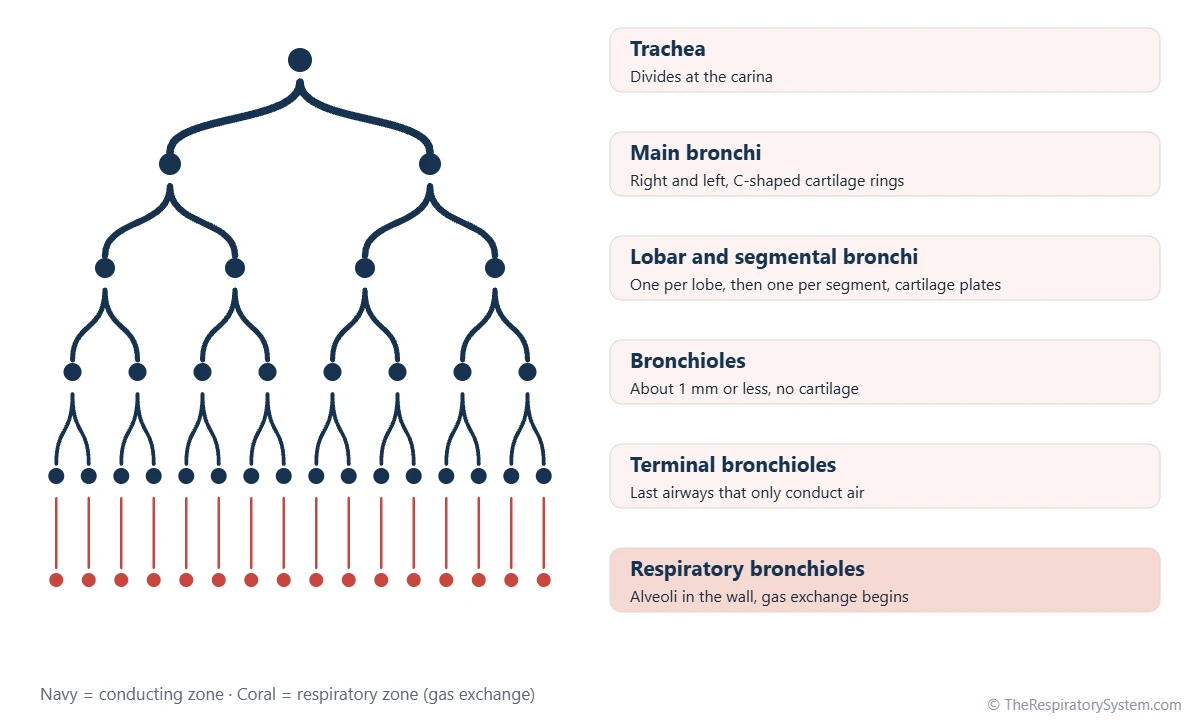
- Terminal bronchiole: the last bronchiole that only conducts air.
- Respiratory bronchiole: a bronchiole with alveoli opening from its wall, where gas exchange begins.
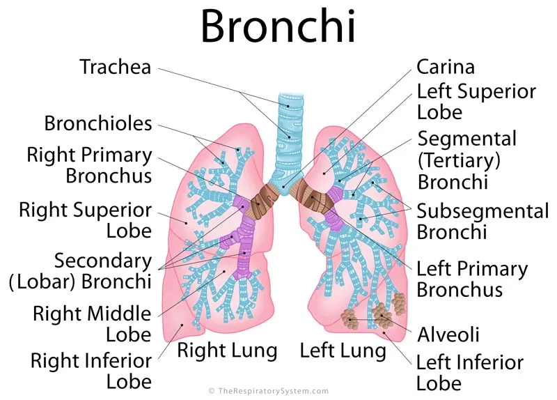
Together, these airways form the bronchial tree, which starts at the carina of the trachea and ends at the alveoli.
Where Are the Bronchi and Bronchioles Located?
The bronchi start just outside the lungs and run into them through the hilum, while the bronchioles lie entirely inside the lung tissue, within the small units called lobules.
Bronchi: from the carina into each lobe
The trachea divides at the level of the sternal angle into the right and left main bronchi, which enter each lung at the hilum. Each main bronchus splits into lobar bronchi, three on the right and two on the left, one for each of the lung lobes. The lobar bronchi divide into segmental bronchi, each supplying one of the bronchopulmonary segments, and these keep dividing into smaller bronchi.
Bronchioles: deep inside the lung
When the small bronchi have divided enough times to lose their cartilage, they become bronchioles. A bronchiole that enters a pulmonary lobule branches into several terminal bronchioles, and each terminal bronchiole gives rise to respiratory bronchioles, alveolar ducts and finally the alveoli. Because bronchioles lie so deep, they sit within a few millimeters of the gas exchange tissue they supply.
Bronchi vs Bronchioles Histology: How Do Their Walls Differ?
Under the microscope, a bronchus shows cartilage plates, submucosal glands and a tall ciliated lining full of goblet cells, while a bronchiole shows no cartilage, no glands, a short lining with club cells and a ring of smooth muscle that is thick for the size of the tube.
Wall of a bronchus
- Epithelium: pseudostratified ciliated columnar respiratory epithelium with many goblet cells.
- Lamina propria and smooth muscle: a band of smooth muscle encircles the airway beneath the lining.
- Submucosa: mucus and serous glands.
- Cartilage: C-shaped rings in the main bronchi, irregular plates in the bronchi inside the lungs.
Wall of a bronchiole
- Epithelium: simple ciliated columnar in larger bronchioles, becoming simple cuboidal in terminal bronchioles. Goblet cells fade out.
- Club cells: dome-shaped cells without cilia that release protective proteins and help repair the lining.
- Smooth muscle: a relatively thick layer for the small size of the airway.
- No cartilage and no glands: elastic fibers in the surrounding alveolar walls pull the bronchiole open from outside.
For a slide-by-slide view of the whole tract, see respiratory system histology.
What Do the Bronchi and Bronchioles Do?
The bronchi and bronchioles both conduct air to and from the alveoli, but the bronchi are built mainly to carry and clean that air, while the bronchioles are built to fine-tune where it goes and to hand it on to the gas exchange tissue.
Jobs of the bronchi
- Distribute air to each lung, lobe and segment through a branching system of open tubes.
- Clean the air: mucus traps particles, and cilia sweep them upward in the mucociliary escalator described on our cilia and mucus page.
- Finish warming and humidifying the air.
- Trigger coughing when irritants touch their sensitive lining, through the cough reflex.
Jobs of the bronchioles
- Regulate airflow: their smooth muscle widens or narrows them, adjusting how much air each region of lung receives.
- Protect the small airways: club cells release substances that defend the lining and can divide to replace damaged cells.
- Connect to the exchange zone: terminal bronchioles end the conducting airways, and respiratory bronchioles begin gas exchange, as shown on our conducting zone vs respiratory zone page.
What Do Bronchi and Bronchioles Have in Common?
Bronchi and bronchioles are consecutive parts of the same airway tree, so they share their basic purpose, their lining plan and many of their controls.
- Both conduct air: all bronchi and most bronchioles belong to the conducting zone and hold part of the anatomical dead space.
- Both are lined by ciliated epithelium: cilia continue from the bronchi through the bronchioles, though they become fewer.
- Both have smooth muscle: sympathetic nerve signals and adrenaline relax it and widen the airways, while parasympathetic signals through the vagus nerve tighten it.
- Both are fed by the bronchial arteries: the walls of both receive oxygen-rich blood from the systemic circulation, not from the pulmonary circulation.
- Both branch by division: each airway splits into two or more smaller ones.
- Both grow from the lung bud: they form by repeated branching during the early stages of respiratory system development.
How Do the Bronchi and Bronchioles Work Together?
The bronchi deliver air in bulk to each part of the lung, and the bronchioles divide it among thousands of tiny regions and pass it to the alveoli, so together they form one continuous delivery system.
Where most airway resistance lies
It might seem that the narrowest tubes resist airflow the most, but in healthy lungs most resistance comes from the medium-sized bronchi. Bronchioles are individually tiny, yet there are so many of them side by side that their combined cross-section is enormous and the air moves through them slowly and easily. This is also why disease of the small airways can go unnoticed on simple tests until it is quite advanced.
Held open in two different ways
Bronchi are held open from inside their own wall by cartilage. Bronchioles are held open from outside by the elastic alveolar walls attached around them, which pull outward more strongly as the lung expands. That is why bronchioles widen during a deep breath in and narrow a little during a breath out, a change driven by the muscles of respiration.
Bronchi vs bronchioles vs alveoli
Bronchi and bronchioles carry the air, and the alveoli use it. The bronchi end in bronchioles, the bronchioles end in alveolar ducts and sacs, and only the alveoli, with their thin walls and dense capillaries, exchange large amounts of oxygen and carbon dioxide with the blood. See terminal and respiratory bronchioles for the exact handover point.
Common Conditions That Affect the Bronchi and Bronchioles
Many airway diseases are named after the level they mainly affect, which makes the bronchi vs bronchioles distinction useful for understanding them. Our respiratory diseases section has more detail, and anyone with breathing symptoms should see a doctor.
- Bronchitis: inflammation of the bronchi, with extra mucus and cough.
- Bronchiolitis: a viral infection of the bronchioles that mainly affects babies and young children.
- Asthma: inflammation and tightening of the smooth muscle of both bronchi and bronchioles; the bronchioles narrow easily because they have no cartilage.
- Bronchiectasis: bronchi that become permanently widened and scarred, so mucus pools in them.
- COPD: long-term narrowing of the small airways, often together with emphysema.
- Cystic fibrosis: thick mucus blocks bronchi and bronchioles alike.
Key Facts
- Bronchi have cartilage in their walls, while bronchioles have none.
- An airway becomes a bronchiole when it narrows to about 1 mm and loses its cartilage and glands.
- Bronchus is the singular of bronchi; a bronchiole is literally a small bronchus.
- Bronchi are lined by pseudostratified ciliated columnar epithelium with goblet cells, while bronchioles have simple columnar to cuboidal epithelium with club cells.
- Bronchioles are held open by the elastic pull of the lung tissue around them, not by cartilage.
- In healthy lungs most airway resistance comes from the medium-sized bronchi, not from the numerous small bronchioles.
- Gas exchange begins only in the respiratory bronchioles, just before the alveoli.
Quick Quiz: Test What You Know About the Bronchi vs Bronchioles
6 questions. Tap an answer to check it.
What is the main structural feature that separates bronchi from bronchioles?
Bronchi have cartilage rings or plates; bronchioles have none. Both have cilia, smooth muscle and epithelium.
Which cells are typical of the bronchioles but rare in the bronchi?
Club cells are dome-shaped protective cells found mainly in the bronchioles, especially the terminal bronchioles.
About how wide is an airway when it becomes a bronchiole?
Airways become bronchioles when they narrow to about 1 mm and lose their cartilage.
What is the plural of bronchus?
Bronchus is singular and bronchi is plural. Bronchioles are a different, smaller type of airway.
Which airways are the first where gas exchange takes place?
Respiratory bronchioles have alveoli opening from their walls, so gas exchange begins there.
What keeps the bronchioles open?
Elastic fibers in the alveolar walls attached around each bronchiole pull it open from outside.
Frequently Asked Questions
Which is bigger, a bronchus or a bronchiole?
A bronchus is bigger. The main bronchi are about as wide as a finger, and even the smallest bronchi are wider than about 1 mm. Bronchioles begin at about 1 mm across and shrink further, with terminal bronchioles only about 0.5 mm wide. Size is a handy rule of thumb, but the true dividing line is the loss of cartilage in the wall.
Do bronchioles have cilia?
Yes, most bronchioles have cilia, although fewer than the bronchi. The lining of the larger bronchioles is ciliated columnar epithelium, and cilia continue as far as the respiratory bronchioles. They keep the mucociliary escalator running from the small airways up to the throat. The alveoli beyond them have no cilia and rely on alveolar macrophages for cleaning.
What is the difference between bronchioles and terminal bronchioles?
Terminal bronchioles are the last and smallest of the conducting bronchioles. Ordinary bronchioles branch and branch again, and the final branches before any alveoli appear are the terminal bronchioles, about 0.5 mm wide with a cuboidal lining rich in club cells. Each terminal bronchiole supplies one acinus and gives rise to the respiratory bronchioles, where gas exchange begins.
Why do bronchioles narrow so much in asthma?
Bronchioles have no cartilage and a relatively thick layer of smooth muscle for their size, so when that muscle tightens nothing stiff resists it. In asthma the lining is also inflamed and swollen and the airways fill with sticky mucus. Together these changes narrow the small airways much more than the cartilage-supported bronchi.
What is the difference between bronchitis and bronchiolitis?
Bronchitis is inflammation of the bronchi, the larger cartilage-supported airways, and is common in adults, either as a short infection or as a long-term condition linked to smoking. Bronchiolitis is inflammation of the bronchioles, the small airways, and is usually a viral infection of babies and young children. The names simply point to the level of the airway tree that is inflamed.
Are bronchioles part of the lungs?
Yes. Bronchioles lie entirely inside the lungs, within the lung lobules, surrounded by alveoli. The bronchi are partly outside the lungs at first, because the main bronchi travel from the carina to the hilum before entering lung tissue, but all of their smaller branches and every bronchiole sit within the lungs themselves.
What does the bronchi and bronchioles do in breathing?
The bronchi and bronchioles form the passageways that air travels through between the trachea and the alveoli. During inhalation they deliver fresh air to every part of each lung, and during exhalation they carry stale air back out. Along the way the bronchi clean and humidify the air, and the bronchioles adjust their width to control where the airflow goes.
How many bronchioles are in the lungs?
There is no single exact count, but the lungs contain many thousands of bronchioles. The two lungs together hold tens of thousands of terminal bronchioles alone, and each one leads to its own small cluster of respiratory bronchioles, alveolar ducts and alveoli. This huge number is what spreads incoming air across hundreds of millions of alveoli.
Keep Exploring


