At a Glance
The respiratory system is lined mostly by pseudostratified ciliated columnar epithelium with goblet cells, supported by hyaline cartilage, glands and smooth muscle in the larger airways. Toward the lungs the epithelium thins to simple cuboidal in the bronchioles and to simple squamous type I pneumocytes in the alveoli, where the air-blood barrier is only about 0.5 micrometer thick.
- Main lining
- Respiratory epithelium: pseudostratified ciliated columnar with goblet cells
- Support tissue
- Hyaline cartilage, smooth muscle, elastic fibers and seromucous glands
- Gas-exchange lining
- Simple squamous type I pneumocytes, with cuboidal type II pneumocytes
- Main trend
- Cartilage, glands and goblet cells fade out; smooth muscle and elastic fibers become more prominent
- Defense cells
- Ciliated cells, goblet cells, club cells and alveolar macrophages
- Part of
- Respiratory system anatomy and histology, from the nasal cavity to the alveoli
On this page
- What Is the Histology of the Respiratory System?
- What Tissues Make Up the Respiratory System?
- What Does the Upper Respiratory Tract Look Like Under a Microscope?
- Trachea Histology: What Is the Function of Each Layer?
- Bronchi and Bronchioles Histology: How Do the Walls Change?
- Lung Histology: What Is the Function of the Alveolar Wall?
- Common Conditions That Change Respiratory Tissue
- Key Facts
- Quiz
- FAQ
What Is the Histology of the Respiratory System?
The histology of the respiratory system is its microscopic structure: the layers, cells and fibers that make up every airway wall from the nasal cavity to the alveoli. Studying these tissues explains why the upper airways can clean and warm air, while the deepest parts of the lungs can pass oxygen into the blood.
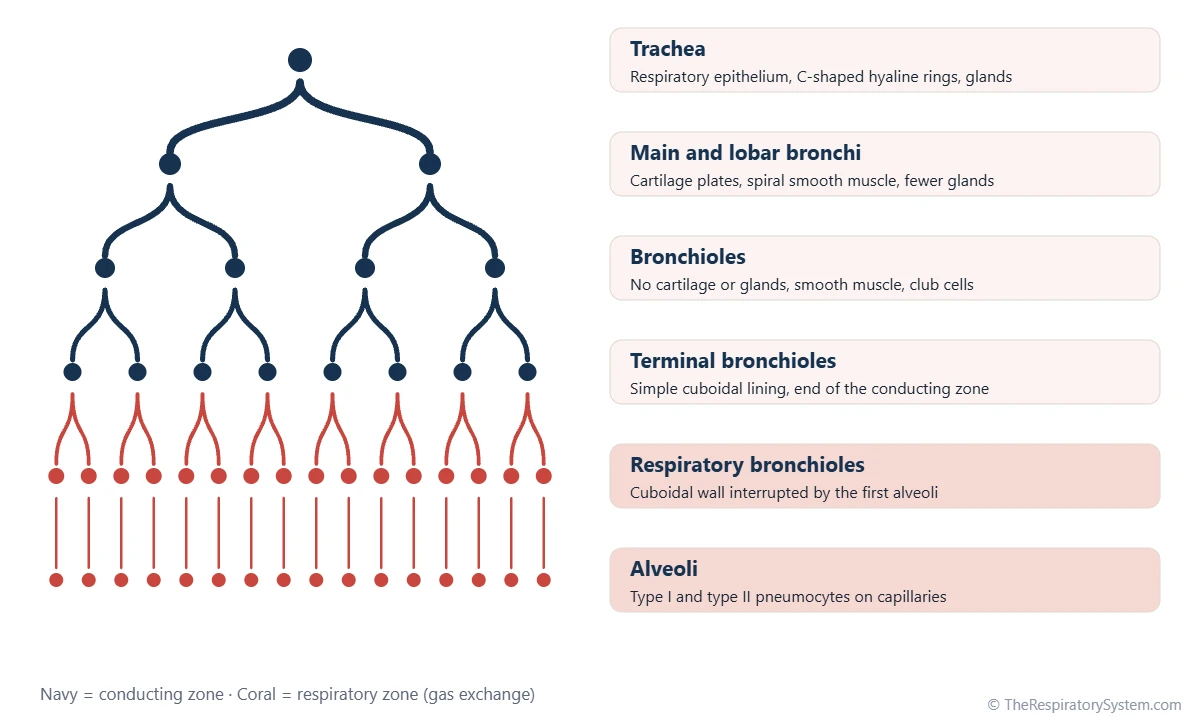
Histologists divide the airways into two zones. The conducting zone, from the nose to the terminal bronchioles, is built for moving and cleaning air, so its walls are thick and supported. The respiratory zone, from the respiratory bronchioles to the alveoli, is built for diffusion, so its walls are as thin as living tissue can be. The guide to the conducting zone vs respiratory zone shows where one ends and the other begins.
Explained simply
Picture the airways as a pipe system that turns into a sponge. The big pipes near the top have stiff rings, a wet sticky lining and tiny hairs that sweep dirt away. Deeper down the pipes get narrower and softer, and at the very end they become bubbles with walls so thin that oxygen can slip straight through them into the blood.
What Tissues Make Up the Respiratory System?
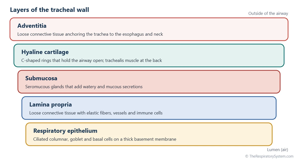
All four basic tissue types are found in the respiratory system: epithelium lines the airways, connective tissue (including cartilage) supports them, smooth muscle adjusts their width, and nerve tissue controls glands, muscle and reflexes. Most airway walls arrange these tissues in four layers.
- Mucosa: the inner lining, made of respiratory epithelium resting on a basement membrane and a loose connective tissue layer called the lamina propria, rich in elastic fibers and immune cells.
- Submucosa: denser connective tissue holding seromucous glands, which add watery and mucous secretions to the lining.
- Cartilage and smooth muscle layer: hyaline cartilage rings or plates that keep the airway open, plus smooth muscle that can narrow it.
- Adventitia: the outer connective tissue that anchors the airway to nearby organs.
The key cell of the lining is the ciliated columnar cell, which carries hundreds of motile cilia. Goblet cells between them release mucus, and the two together form the mucociliary escalator that carries trapped dust up and out of the airways. Basal cells near the basement membrane act as stem cells that replace worn-out lining cells.
What Does the Upper Respiratory Tract Look Like Under a Microscope?
The upper respiratory tract is lined by three kinds of epithelium, each matched to the wear it faces: stratified squamous where friction is high, respiratory epithelium where air must be cleaned, and olfactory epithelium where odors are sensed. The upper respiratory tract page covers the anatomy of each part.
- Nasal vestibule: the entrance of the nose is lined by skin, keratinized stratified squamous epithelium with coarse hairs, sweat glands and sebaceous glands.
- Respiratory region of the nasal cavity: pseudostratified ciliated columnar epithelium over a lamina propria packed with thin-walled veins, which warm the air passing over the nasal conchae.
- Olfactory region: a small patch in the roof of the nasal cavity with olfactory receptor neurons, supporting cells and Bowman glands, described on the sense of smell page.
- Pharynx: the nasopharynx carries respiratory epithelium, while the oropharynx and laryngopharynx, which also carry food, are lined by nonkeratinized stratified squamous epithelium.
- Larynx: mostly respiratory epithelium, but the true vocal cords and the upper part of the epiglottis are covered by stratified squamous epithelium that resists the constant rubbing of speech and swallowing.
Trachea Histology: What Is the Function of Each Layer?
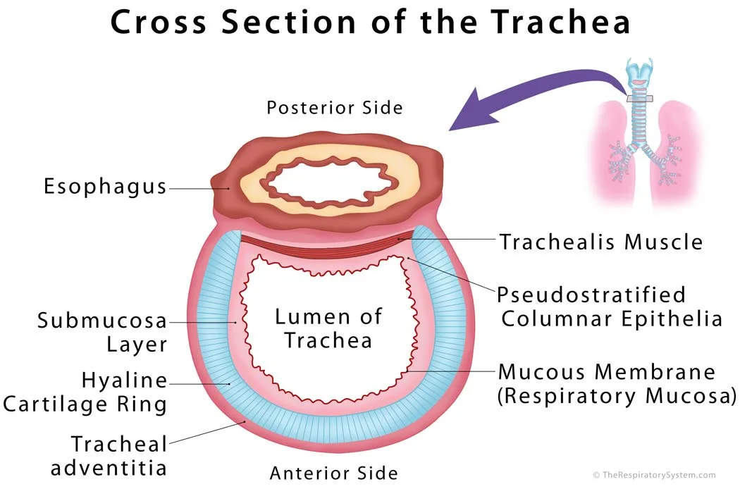
Trachea histology shows a mucosa of respiratory epithelium, a submucosa with seromucous glands, 16 to 20 C-shaped rings of hyaline cartilage, and an outer adventitia, and each layer has a clear function. The epithelium cleans the air, the glands keep it moist, and the cartilage stops the trachea from collapsing when pressure in the chest changes.
The open ends of each cartilage ring face backward toward the esophagus and are bridged by the trachealis, a band of smooth muscle. When it contracts, as in a cough, the trachea narrows and air rushes out faster. Between the rings, fibroelastic ligaments let the trachea bend and stretch when the neck moves or the lungs fill.
Under the microscope, the tracheal epithelium looks layered because its nuclei sit at different heights, but every cell actually touches the basement membrane, which is why it is called pseudostratified. The basement membrane itself is unusually thick here, and the lamina propria contains a band of elastic fibers that helps the airway recoil.
Bronchi and Bronchioles Histology: How Do the Walls Change?
As the airways branch from the bronchi into the bronchioles, the cartilage breaks up and then disappears, the glands and goblet cells fade out, and smooth muscle becomes the main tissue controlling airway width. This gradual change is the heart of bronchi and trachea histology.
Bronchi
The main bronchi look much like the trachea. Inside the lungs, the C-shaped rings are replaced by irregular plates of hyaline cartilage, and a continuous layer of spirally arranged smooth muscle appears between the mucosa and the cartilage. The epithelium is still respiratory epithelium, but it becomes shorter with each division, and small clusters of lymphoid tissue sit in the wall.
Bronchioles
The bronchioles have no cartilage and no glands, so they rely on smooth muscle and on the elastic pull of the surrounding lung to stay open. Their lining shrinks from ciliated columnar cells to simple cuboidal cells, and goblet cells give way to club cells, dome-shaped cells that release protective proteins and help repair the lining. In the terminal and respiratory bronchioles, the cuboidal wall is interrupted by the first alveoli, marking the start of gas exchange. The full branching pattern is shown on the bronchial tree page.
| Region | Epithelium | Cartilage | Glands and goblet cells | Smooth muscle |
|---|---|---|---|---|
| Nasal vestibule | Keratinized stratified squamous, with hairs | Nasal cartilages in the wall | Sweat and sebaceous glands | None in the lining |
| Nasal cavity (respiratory region) | Pseudostratified ciliated columnar | Bone and cartilage in the wall | Many glands and goblet cells | None in the lining |
| Oropharynx and laryngopharynx | Nonkeratinized stratified squamous | None in the wall | Mucous glands | Skeletal muscle in the wall |
| Larynx | Respiratory epithelium; stratified squamous on true vocal cords | Hyaline and elastic cartilages | Glands and goblet cells | Skeletal muscle in the wall |
| Trachea | Pseudostratified ciliated columnar | 16 to 20 C-shaped hyaline rings | Many glands and goblet cells | Trachealis at the back |
| Bronchi | Pseudostratified ciliated columnar, becoming shorter | Irregular hyaline plates | Glands and goblet cells, fewer with each division | Spiral layer |
| Bronchioles | Ciliated simple columnar to cuboidal, with club cells | None | Few or no goblet cells, no glands | Prominent layer |
| Terminal bronchioles | Simple cuboidal with club cells | None | None | Thin layer |
| Respiratory bronchioles | Simple cuboidal, interrupted by alveoli | None | None | Scattered bundles |
| Alveoli | Simple squamous type I and cuboidal type II pneumocytes | None | None (surfactant from type II cells) | None in the wall |
| Source: Typical adult histology, our editors | ||||
Lung Histology: What Is the Function of the Alveolar Wall?
Lung histology is dominated by the alveolar wall, a sheet of simple squamous epithelium wrapped around capillaries, whose function is to let oxygen and carbon dioxide diffuse between air and blood. A labeled section of lung looks like lace: thin walls around empty air spaces, with small airways and blood vessels scattered between them.
Three cell types line the alveoli. Type I pneumocytes are extremely flat and cover most of the alveolar surface; type II pneumocytes are plump cuboidal cells that make pulmonary surfactant and can divide to replace type I cells; and alveolar macrophages wander over the surface eating dust and microbes.
Between neighboring alveoli lies the interalveolar septum, which holds capillaries, elastic and reticular fibers and a few fibroblasts. Where a capillary presses against a type I cell, their basement membranes fuse, creating the respiratory membrane, which is about 0.5 micrometer thick in most places. Elastic fibers in the septa store energy as the lungs stretch and release it to push air out during quiet exhalation. The outer surface of each lung is covered by the visceral pleura, a layer of flat mesothelial cells over connective tissue.
Common Conditions That Change Respiratory Tissue
Many lung and airway diseases are defined by what they do to these tissues, which is why histology is the basis of pathology. Our respiratory diseases section explains each condition in more depth.
- Squamous metaplasia: long-term irritation, especially from tobacco smoke, can replace ciliated epithelium with tougher stratified squamous cells that cannot clear mucus.
- Chronic bronchitis: the submucosal glands enlarge and goblet cells multiply, producing excess mucus.
- Asthma: the airway wall thickens, the basement membrane zone thickens, smooth muscle grows and mucus can plug small airways.
- Emphysema: alveolar walls are destroyed, leaving fewer, larger air spaces.
- Pulmonary fibrosis: scar tissue thickens the interalveolar septa and the respiratory membrane.
Key Facts
- Most of the airway is lined by pseudostratified ciliated columnar epithelium with goblet cells, known as respiratory epithelium.
- The trachea is held open by 16 to 20 C-shaped rings of hyaline cartilage, closed at the back by the trachealis muscle.
- Cartilage and glands disappear at the level of the bronchioles, where smooth muscle controls the airway width.
- Club cells replace goblet cells in the bronchioles and help protect and repair the lining.
- The alveolar wall is lined by flat type I pneumocytes and surfactant-making type II pneumocytes.
- The air-blood barrier in the alveoli is about 0.5 micrometer thick in most places.
- Parts of the airway that face friction, such as the true vocal cords and oropharynx, are lined by stratified squamous epithelium instead of respiratory epithelium.
Quick Quiz: Test What You Know About the Respiratory System Histology
6 questions. Tap an answer to check it.
What type of epithelium lines most of the trachea?
The trachea is lined by respiratory epithelium, pseudostratified ciliated columnar epithelium with goblet cells.
At which level does cartilage disappear from the airway wall?
Bronchioles have no cartilage and no glands; smooth muscle and elastic lung tissue keep them open.
Which cells make pulmonary surfactant?
Cuboidal type II pneumocytes make surfactant and can divide to replace type I cells.
What type of epithelium covers the true vocal cords?
The true vocal cords face constant friction, so they are covered by tough stratified squamous epithelium.
What fills the gap at the back of each tracheal cartilage ring?
The open ends of the C-shaped rings face the esophagus and are joined by the trachealis muscle.
Which cells replace goblet cells in the bronchioles?
Dome-shaped club cells line the bronchioles, release protective proteins and help repair the lining.
Frequently Asked Questions
What is the difference between respiratory system anatomy and histology?
Anatomy describes the parts you can see with the naked eye, such as the shape of the lungs, the lobes and the branching of the bronchi. Histology looks at the same parts under a microscope and describes their tissues and cells, such as the type of epithelium, the cartilage and the glands. Together they explain both where each structure is and how it does its job.
Why is respiratory epithelium called pseudostratified?
Its nuclei sit at different heights, so in a section it looks like several layers of cells stacked on top of each other. In fact every cell rests on the basement membrane, and only some reach the surface. Because the layering is an illusion, histologists call it pseudostratified, which means falsely stratified, rather than truly stratified epithelium.
What is the function of goblet cells in the respiratory system?
Goblet cells are single-celled glands scattered among the ciliated cells of the airway lining. They release mucins, large sugar-coated proteins that absorb water and form sticky mucus. The mucus traps dust, pollen and microbes, and the cilia then sweep it upward toward the throat. Goblet cells become fewer in the smaller airways and are absent from the terminal bronchioles.
How can you identify lung tissue on a histology slide?
Lung tissue looks like a lacy net of very thin walls around many empty, rounded air spaces, the alveoli. Scattered among them are bronchioles, with a folded lining and smooth muscle but no cartilage, and branches of the pulmonary artery that travel alongside them. A bronchus is recognized by its plates of hyaline cartilage and glands in the wall.
What are club cells?
Club cells, once called Clara cells, are dome-shaped, non-ciliated cells found mainly in the bronchioles. They release proteins that protect the lining and are part of the fluid layer coating the small airways. They also break down some inhaled toxins and act as stem cells, dividing to replace damaged cells of the bronchiolar epithelium after injury.
Do the alveoli have cilia?
No. Cilia end in the bronchioles, and the alveoli have no cilia and no goblet cells, because a thick, moving mucus layer would block gas exchange. Instead, particles that reach the alveoli are cleared by alveolar macrophages, which engulf them and either carry them up to the ciliated airways or move into the lymph vessels of the lung.
Why do the airways need cartilage?
Cartilage keeps the larger airways open. During a strong breath in, pressure inside the airway falls, and during a cough the chest squeezes the airways from outside. Without stiff hyaline rings and plates, the trachea and bronchi would collapse under these changes. The smaller bronchioles have no cartilage and are instead held open by the elastic pull of the surrounding lung tissue.
Keep Exploring


