At a Glance
Pneumocytes, also called alveolar cells or alveolar epithelial cells, are the two cell types that line the alveoli. Type I pneumocytes are extremely thin squamous cells that cover about 95% of the alveolar surface and form the gas exchange barrier; type II pneumocytes are cuboidal cells that cover the remaining 5% or so, secrete pulmonary surfactant and divide to replace damaged type I cells.
- Location
- Inner lining of the alveoli and alveolar sacs in both lungs
- Types
- Type I (squamous) and type II (cuboidal, also called great alveolar cells)
- Tissue
- Simple epithelium, the alveolar epithelium
- Type I function
- Forms the thin barrier across which oxygen and carbon dioxide diffuse
- Type II function
- Makes surfactant, clears fluid and repairs the lining as stem-like cells
- Part of
- Respiratory zone of the lungs and the respiratory membrane
On this page
What Are Pneumocytes?
Pneumocytes are the epithelial cells that make up the inner wall of the alveoli, forming a continuous single layer between the air in the lungs and the tissue and capillaries around it. The name comes from the Greek word for lung, and textbooks also call them alveolar cells, alveolar epithelial cells or simply AT1 and AT2 cells.
An adult has about 300 to 500 million alveoli, and every one of them is lined by a mix of the two types. Together they form the alveolar epithelium, the air-facing layer of the respiratory membrane. Other cells live in and around the alveoli as well, including alveolar macrophages that patrol the air spaces and the endothelial cells of the capillaries, but these are not pneumocytes.
Explained simply
Think of an alveolus as a tiny balloon whose wall is tiled. Most of the wall is covered by a few huge, paper-thin tiles, the type I cells, which let gases slip straight through. Tucked into the corners are small, chunky cells, the type II cells, which spray a soapy liquid over the tiles so the balloon does not stick shut, and which grow new tiles whenever old ones break.
Where Are Pneumocytes Located?
Pneumocytes are located in the respiratory zone of the lungs, lining the alveoli that bud from the respiratory bronchioles, the alveolar ducts and alveolar sacs. They are the cells at the very end of the bronchial tree, where the airways stop conducting air and begin exchanging gases.
Type I pneumocytes spread across the flat stretches of the alveolar wall, where they lie directly on a basement membrane that is often fused with the basement membrane of a capillary. Type II pneumocytes sit mostly in the corners where neighboring alveoli meet, often bulging slightly into the air space. Small openings in the walls between alveoli, called pores of Kohn, are also edged by pneumocytes and let air pass from one alveolus to the next.
Above this level, in the terminal bronchioles, the lining is made of cuboidal club cells and ciliated cells instead, as described on the respiratory system histology page.
What Is the Function of Type 1 Pneumocytes?
Type 1 pneumocytes form the extremely thin, continuous surface across which oxygen diffuses from the air into the blood and carbon dioxide diffuses out, so their main function is gas exchange. They also form a tight seal that keeps fluid and proteins in the tissue from leaking into the air spaces.
Structure of type I cells
Each type I cell is a squamous cell with a small central nucleus and very long, flat extensions of cytoplasm that spread over a large area of the alveolar wall, often reaching into more than one alveolus. Away from the nucleus the cell is so thin that it barely adds to the distance gases must travel, which is why the whole air-blood barrier is usually only about 0.5 micrometer thick. Neighboring cells are joined by tight junctions that make the lining almost watertight.
What do type 1 pneumocytes produce?
Type 1 pneumocytes do not produce surfactant or mucus; their main product is the barrier itself. They contain many small vesicles that move fluid and small molecules across the cell, and they carry water channels called aquaporins in their membranes, which help balance the fluid in the alveoli. Mature type I cells are highly specialized and are generally thought to divide rarely, if at all, so they depend on type II cells for replacement.
What Is the Function of Type 2 Pneumocytes?
Type 2 pneumocytes secrete pulmonary surfactant, act as the stem cells of the alveolar lining by dividing and turning into new type I cells, pump excess fluid out of the air spaces and help defend the alveoli against microbes. They are sometimes called great alveolar cells or septal cells.
Making surfactant
Type II cells are cube-shaped, with short microvilli on their free surface and a cytoplasm packed with lamellar bodies, layered storage granules that hold surfactant. Surfactant is a mixture of phospholipids, mainly dipalmitoylphosphatidylcholine (DPPC), and four surfactant proteins named SP-A, SP-B, SP-C and SP-D. The cell releases it by exocytosis onto the alveolar surface, where it spreads as a thin film that lowers surface tension and keeps small alveoli from collapsing at the end of each breath out. Type II cells also take back used surfactant and recycle it. The pulmonary surfactant page explains how the film works.
Repairing the alveolar lining
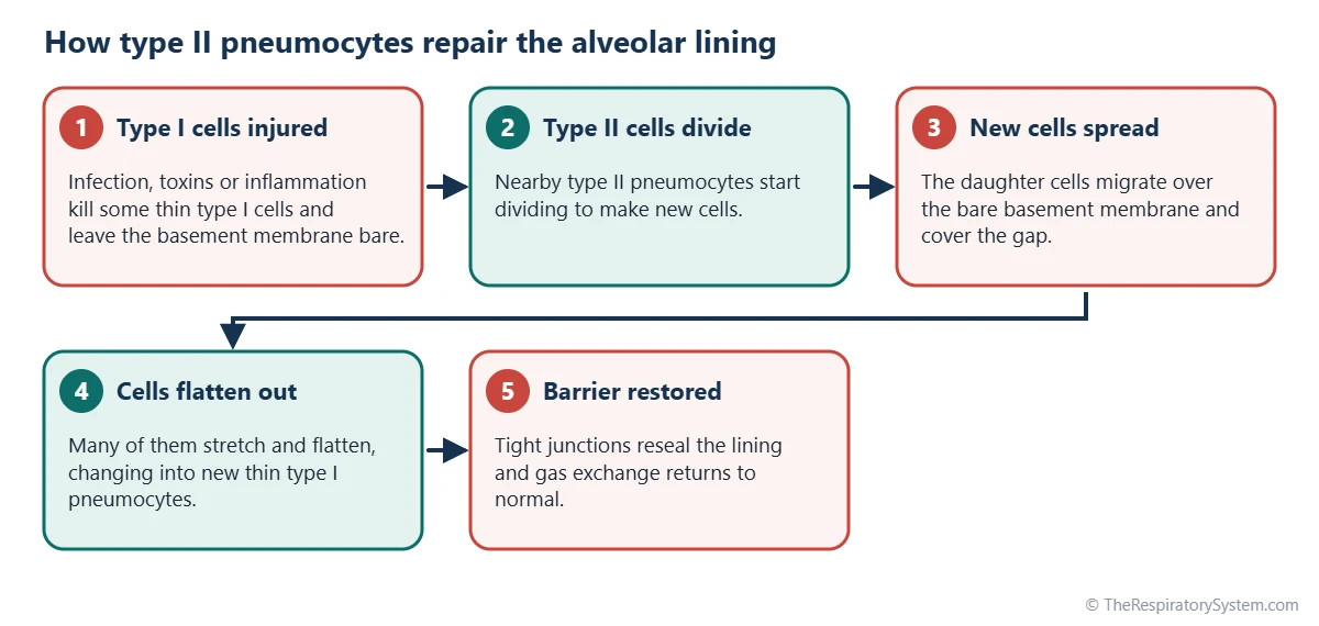
When type I cells are damaged, nearby type II cells divide, spread over the bare basement membrane and then flatten into new type I cells. This makes type II pneumocytes the main progenitor, or stem-like, cells of the adult alveolus. The steps of this repair are shown in the diagram below.
Clearing fluid and defending the lungs
Type II cells move sodium ions out of the air spaces, and water follows, which keeps the alveoli from flooding. Two of the surfactant proteins, SP-A and SP-D, bind bacteria, viruses and particles and help macrophages remove them, giving the alveoli a built-in layer of defense.
Type 1 vs Type 2 Pneumocytes: What Is the Difference?
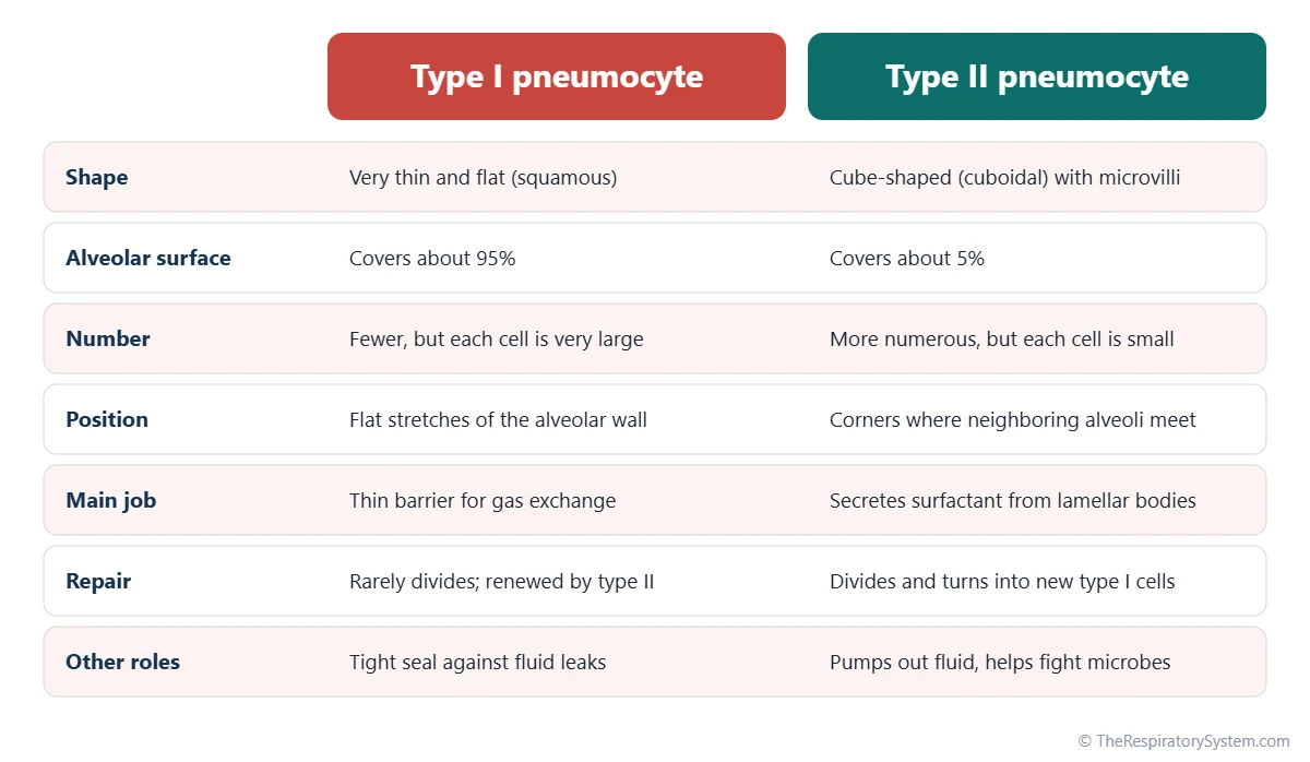
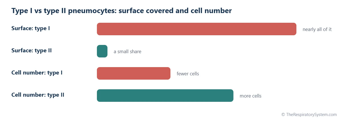
The difference is that type 1 pneumocytes are flat cells built for gas exchange, while type 2 pneumocytes are rounded cells built for making surfactant and renewing the lining. Type I cells are fewer but very large, so they cover about 95% of the alveolar surface; type II cells are more numerous but small, covering only about 5%.
The two types work as a team. The thin type I cells give the lungs their vast, fast exchange surface of about 50 to 75 square meters, but such thin cells are fragile and rarely divide. The type II cells keep that surface open with surfactant and replace lost type I cells, so the barrier stays intact over a lifetime of breathing. The table below compares them feature by feature, along with the alveolar macrophage, which is often listed beside them.
A few older textbooks also mention a type III pneumocyte. The term has been used for the rare brush cells of the alveolar wall and sometimes, loosely, for alveolar macrophages, so it is best avoided; modern anatomy recognizes only type I and type II pneumocytes.
| Feature | Type I pneumocyte | Type II pneumocyte | Alveolar macrophage |
|---|---|---|---|
| Other names | Type I alveolar cell, squamous alveolar cell, AT1 | Type II alveolar cell, great alveolar cell, septal cell, AT2 | Dust cell |
| Shape | Very thin and flat (squamous) | Cube-shaped (cuboidal) with microvilli | Rounded, moving cell |
| Share of alveolar surface | About 95% | About 5% | Not part of the lining |
| Relative number | Fewer than type II | More numerous than type I | Varies with dust and infection |
| Position | Flat parts of the alveolar wall | Corners where alveoli meet | Free in air spaces and walls |
| Typical organelles | Few, many small vesicles | Lamellar bodies storing surfactant | Lysosomes and engulfed particles |
| Main function | Gas exchange barrier | Surfactant secretion and repair | Engulfs dust and microbes |
| Divides? | Rarely, if at all | Yes, renews both cell types | Renewed from local and blood-derived cells |
| Tissue origin | Endoderm | Endoderm | Bone marrow and fetal blood-forming cells |
| Effect of loss | Leaky barrier, flooded alveoli | Too little surfactant, poor repair | Weaker clearance of particles |
| Source: Typical adult values, our editors | |||
How Do Pneumocytes Develop and Change With Age?
Pneumocytes develop from the inner layer of the embryo that lines the gut, the endoderm, as the tips of the growing airways widen into thin-walled sacs in the later months of pregnancy. Type II cells begin making surfactant in the second half of pregnancy, and in most babies the amount becomes enough for comfortable breathing by about 34 to 36 weeks.
At birth, the first breaths fill the alveoli with air, and surfactant already on their surface lets them stay open. Alveoli and their pneumocytes keep multiplying through early childhood, a stage described on the respiratory system development page. In older adults the walls between some alveoli thin and break down, so the total alveolar surface slowly shrinks, and repeated injury from smoke and pollution can wear down the repair capacity of type II cells.
Common Conditions That Affect Pneumocytes
Conditions of the pneumocytes usually involve either a shortage of surfactant or damage to the thin alveolar lining, both of which make gas exchange harder. Our respiratory diseases section covers them in more depth; anyone with breathing symptoms should see a doctor.
- Neonatal respiratory distress syndrome: babies born very early may not yet have enough surfactant from their type II cells, so their alveoli tend to collapse.
- Acute respiratory distress syndrome (ARDS): widespread injury to type I cells and capillaries floods the alveoli; recovery depends on type II cells regrowing the lining.
- Pneumonia: infection inflames the alveoli and can damage both cell types.
- Pulmonary fibrosis: repeated injury to the alveolar lining and faulty repair by type II cells are thought to drive scarring of the alveolar walls.
- Pulmonary alveolar proteinosis: a rare condition in which surfactant builds up in the alveoli because it is not cleared properly.
- Lung adenocarcinoma: some lung cancers arise from cells of the small airways and alveoli, including type II pneumocytes.
Key Facts
- Pneumocytes are the two types of epithelial cells that line the alveoli of the lungs.
- Type I pneumocytes are thin squamous cells that cover about 95% of the alveolar surface and form the gas exchange barrier.
- Type II pneumocytes are cuboidal cells that cover about 5% of the surface but are more numerous than type I cells.
- Type II pneumocytes store surfactant in lamellar bodies and release it onto the alveolar surface.
- Type II pneumocytes act as stem-like cells, dividing and turning into new type I cells after injury.
- Type I pneumocytes form part of the respiratory membrane, which is usually only about 0.5 micrometer thick.
- Alveolar macrophages live in the alveoli too, but they are immune cells, not pneumocytes.
Quick Quiz: Test What You Know About the Pneumocytes
6 questions. Tap an answer to check it.
What is the main function of type I pneumocytes?
Type I pneumocytes are thin squamous cells that form most of the alveolar surface, across which oxygen and carbon dioxide diffuse.
Which cells produce pulmonary surfactant?
Type II pneumocytes make surfactant, store it in lamellar bodies and release it onto the alveolar surface.
About how much of the alveolar surface do type I pneumocytes cover?
Although they are fewer in number, type I cells are so large and flat that they cover about 95% of the alveolar surface.
What happens to the alveolar lining when type I cells are damaged?
Type II pneumocytes are the progenitor cells of the alveolus; they divide, spread and flatten into new type I cells.
Where are type II pneumocytes usually found?
Type II cells usually sit at the junctions between neighboring alveoli, often bulging slightly into the air space.
What structures in type II pneumocytes store surfactant?
Lamellar bodies are layered granules that hold surfactant until the cell releases it by exocytosis.
Frequently Asked Questions
What are alveolar cells?
Alveolar cells is another name for pneumocytes, the cells that line the alveoli. The term usually means type I and type II pneumocytes, which together form the alveolar epithelium. Some books use alveolar cells more loosely to include every cell found in the alveolar wall, such as capillary endothelial cells and alveolar macrophages, so it helps to check which meaning a source uses.
Are type 1 pneumocytes simple squamous epithelium?
Yes. Type I pneumocytes form a simple squamous epithelium: a single layer of flat cells resting on a basement membrane. Their thinness is exactly what makes them suited to gas exchange. Because type II cells are scattered among them, the alveolar lining as a whole is sometimes described as a simple squamous epithelium with interspersed cuboidal cells.
Do type 2 pneumocytes have cilia?
No. Type II pneumocytes have short microvilli on their free surface but no cilia. Cilia stop at the level of the bronchioles, which is why particles that reach the alveoli are removed by alveolar macrophages instead of being swept away. The lack of cilia also keeps the alveolar surface smooth and thin for gas exchange.
What is the difference between pneumocytes and alveolar macrophages?
Pneumocytes are epithelial cells that are fixed in place and form the wall of each alveolus. Alveolar macrophages are immune cells that move freely across the alveolar surface and engulf dust, microbes and old surfactant. Pneumocytes build and maintain the gas exchange surface, while macrophages keep that surface clean and help defend it.
Why are type 2 pneumocytes important for premature babies?
Type II pneumocytes make the surfactant that keeps alveoli open. In babies born well before term, these cells may not yet produce enough of it, so the alveoli tend to collapse at the end of each breath and breathing becomes hard work. This is the main cause of neonatal respiratory distress syndrome, which is managed by specialist newborn care teams.
How long do pneumocytes live?
Pneumocytes turn over slowly in healthy adult lungs. Most type I and type II cells are thought to last for many months or longer, with type II cells dividing now and then to replace losses. After an injury such as a lung infection, the turnover speeds up sharply as type II cells multiply to rebuild the lining within days to weeks.
How does smoking affect pneumocytes?
Tobacco smoke contains irritants and oxidants that injure both types of pneumocytes and trigger inflammation in the alveoli. Over time this can reduce surfactant quality, weaken the repair work of type II cells and, together with damage to the alveolar walls, contribute to emphysema. Smoking is also a major cause of lung cancers that start in the small airways and alveoli.
Can pneumocytes be seen under a microscope?
Yes, but they are easier to tell apart at high magnification. In a stained lung section, type I cells show mainly as flat, dark nuclei along thin alveolar walls, since their cytoplasm is too thin to see. Type II cells look rounder, with a pale, foamy cytoplasm caused by their lamellar bodies. An electron microscope shows both types clearly.
Keep Exploring


