At a Glance
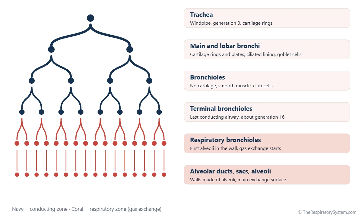
The conducting zone moves air but exchanges no gases: it runs from the nose and trachea to the terminal bronchioles and holds about 150 mL of air, the anatomical dead space. The respiratory zone starts at the respiratory bronchioles and continues through the alveolar ducts and sacs to the alveoli, where oxygen and carbon dioxide cross into and out of the blood; it makes up most of the lung, about 2.5 to 3 liters at rest.
- Conducting zone
- Nose, pharynx, larynx, trachea, bronchi, bronchioles, terminal bronchioles
- Respiratory zone
- Respiratory bronchioles, alveolar ducts, alveolar sacs and alveoli
- Border
- Between the terminal bronchiole and the first respiratory bronchiole
- Main job
- Conducting: carry and condition air. Respiratory: exchange oxygen and carbon dioxide
- Air volume
- About 150 mL (conducting) vs about 2.5 to 3 liters (respiratory) at rest
- Part of
- The airway tree of the respiratory tract, from the nose to the alveoli
On this page
- What Is the Difference Between the Conducting Zone and the Respiratory Zone?
- What Is the Conducting Zone?
- What Is the Respiratory Zone? (Respiratory Zone Anatomy)
- What Is the Function of the Conducting Zone and the Respiratory Zone?
- How Do the Airway Walls Change From the Conducting to the Respiratory Zone?
- What Do the Conducting Zone and Respiratory Zone Have in Common?
- How Do the Conducting and Respiratory Zones Work Together?
- Common Conditions That Affect the Conducting and Respiratory Zones
- Key Facts
- Quiz
- FAQ
What Is the Difference Between the Conducting Zone and the Respiratory Zone?
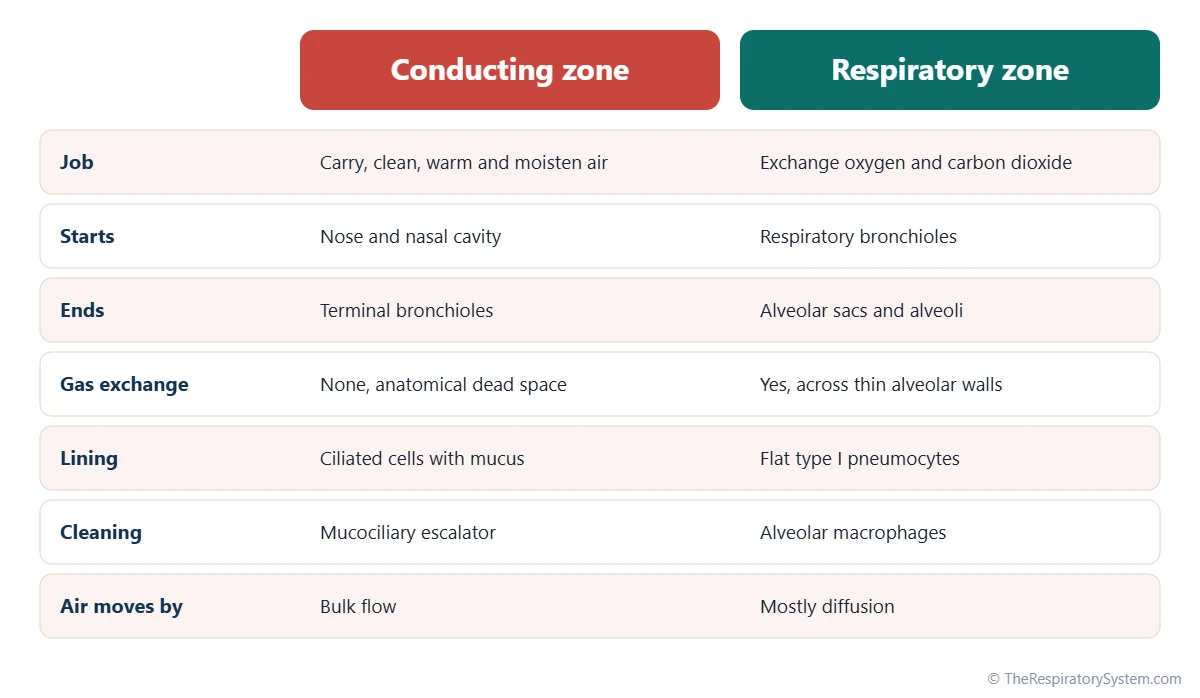
The difference is function: the conducting zone is a set of pipes that delivers air, and the respiratory zone is the surface where that air finally meets the blood. Every airway from the nostrils to the terminal bronchioles belongs to the conducting zone, and every airway that has alveoli opening from its wall belongs to the respiratory zone.
The two zones also differ in their walls, their volume and the way air moves through them. Conducting airways have thick walls lined with ciliated, mucus-covered epithelium, and air flows through them in bulk, pushed by the pressure changes of breathing. Respiratory airways have walls so thin that gases diffuse straight through them, and the air inside barely moves forward, because the total width of the airway tree becomes enormous at that depth.
The verdict in one line
If an airway can exchange oxygen and carbon dioxide with the blood, it is part of the respiratory zone; if it cannot, it is part of the conducting zone. The first airway that passes this test is the respiratory bronchiole, which is why the border between the zones sits just after the last terminal bronchiole.
Explained simply
Picture a city water system. The big mains and the street pipes only carry water, and that is the conducting zone. The taps where water is actually used are the respiratory zone. In the lungs, the taps are hundreds of millions of tiny air sacs, and what they hand over is oxygen.
| Feature | Conducting zone | Respiratory zone |
|---|---|---|
| Main function | Carries, filters, warms and humidifies air | Exchanges oxygen and carbon dioxide with the blood |
| Structures | Nose, pharynx, larynx, trachea, bronchi, bronchioles, terminal bronchioles | Respiratory bronchioles, alveolar ducts, alveolar sacs, alveoli |
| Airway generations (from the trachea) | About 0 to 16 | About 17 to 23 |
| Gas exchange | None (anatomical dead space) | Yes, across the alveolar walls |
| Air volume in an adult | About 150 mL | About 2.5 to 3 liters at rest |
| Epithelium | Pseudostratified ciliated columnar, then columnar to cuboidal | Cuboidal in respiratory bronchioles, simple squamous in alveoli |
| Cartilage | In the trachea and bronchi only | None |
| Goblet cells and glands | Present in the trachea and bronchi | Absent |
| Cleaning system | Mucus and cilia (mucociliary escalator) | Alveolar macrophages |
| How air moves | Bulk flow driven by breathing | Mostly diffusion over the last short distance |
| Blood supply of the walls | Bronchial arteries (systemic) | Pulmonary capillaries (pulmonary circulation) |
| Source: Typical adult values, our editors | ||
What Is the Conducting Zone?
The conducting zone is the part of the respiratory tract that carries air between the outside world and the gas exchange surfaces without taking part in gas exchange itself. Because the air in it never reaches the blood, its volume, about 150 mL in an adult, is called the anatomical dead space.
Conducting zone definition in anatomy
Anatomy textbooks define the conducting zone as all the airways from the nose to the terminal bronchioles. Lung physiology often uses a narrower definition that starts at the trachea: in the classic model of the airway tree, the trachea is generation 0, and each division adds one generation until the terminal bronchioles at about generation 16. Both definitions agree on where the zone ends.
Parts of the conducting zone
- Nose and nasal cavity: filter, warm and humidify incoming air over the folds of the nasal conchae.
- Pharynx: the shared throat passage for air and food.
- Larynx: guards the airway and houses the vocal folds.
- Trachea: the windpipe, held open by C-shaped rings of hyaline cartilage.
- Bronchi: main, lobar and segmental bronchi, supported by cartilage rings and plates.
- Bronchioles: small airways about 1 mm wide or less, with smooth muscle but no cartilage.
- Terminal bronchioles: the last and smallest purely conducting airways.
The whole branching system from the trachea down is described on our bronchial tree page.
What Is the Respiratory Zone? (Respiratory Zone Anatomy)
The respiratory zone is the part of the lungs where gas exchange takes place, made of the respiratory bronchioles, alveolar ducts, alveolar sacs and the alveoli. In the airway tree model it covers roughly generations 17 to 23, the last few divisions before the airways end in blind sacs.
Respiratory bronchioles
Respiratory bronchioles are short airways with a few alveoli budding from their walls, so they both conduct air and exchange gases. They are the transition point between the two zones, which is why some textbooks call them the transitional zone. Our page on terminal and respiratory bronchioles compares the two neighbors in detail.
Alveolar ducts, sacs and alveoli
Each respiratory bronchiole opens into several alveolar ducts and alveolar sacs, whose walls are made almost entirely of alveoli. An adult has about 300 to 500 million alveoli, giving a gas exchange surface of roughly 50 to 75 square meters. The alveolar wall is lined mostly by flat type I pneumocytes, with type II cells making surfactant.
The pulmonary acinus
All the respiratory tissue supplied by one terminal bronchiole forms a unit called the acinus, the basic working unit of gas exchange. Despite its importance, an acinus is tiny: the distance from a terminal bronchiole to its farthest alveolus is only a few millimeters. Acini group together into the larger pulmonary lobule.
What Is the Function of the Conducting Zone and the Respiratory Zone?
The conducting zone function is to deliver clean, warm, moist air to the lungs and carry stale air back out, while the respiratory zone function is to exchange oxygen and carbon dioxide between that air and the blood.
Conducting zone function
- Air transport: a low-resistance path for air in and out with each breath.
- Filtering: nasal hairs, mucus and the branching shape of the airways trap dust, pollen and microbes, and cilia sweep the mucus up toward the throat in the mucociliary escalator described on our cilia and mucus page.
- Warming and humidifying: by the time air reaches the deep lung, it is close to body temperature and fully saturated with water vapor.
- Control of airflow: smooth muscle in the bronchi and bronchioles can narrow or widen the airways.
- Protection and voice: the larynx closes during swallowing, triggers the cough reflex and produces sound.
Respiratory zone function
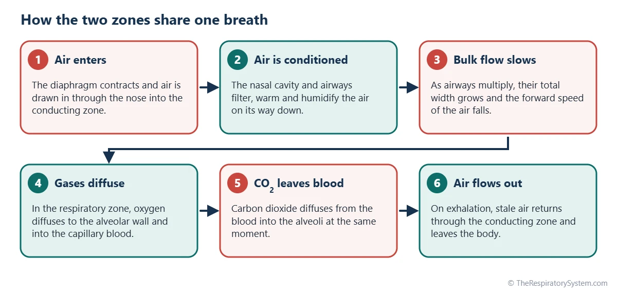
The respiratory zone has one main job: gas exchange. Oxygen diffuses from alveolar air across the respiratory membrane into the pulmonary capillaries, and carbon dioxide diffuses the opposite way. Alongside that job, alveolar macrophages clean up the fine particles that slip past the conducting zone, and type II pneumocytes make pulmonary surfactant, which keeps the small air sacs from collapsing.
How Do the Airway Walls Change From the Conducting to the Respiratory Zone?
Moving from the conducting zone to the respiratory zone, the airway walls become thinner and simpler: cartilage disappears first, then mucus glands and goblet cells, then cilia, until only a single layer of very flat cells separates air from blood.
Epithelium
The trachea and bronchi are lined by pseudostratified ciliated columnar epithelium with many goblet cells, the classic respiratory epithelium. In the bronchioles the lining becomes simple columnar and then simple cuboidal, goblet cells fade out, and club cells take over the job of protecting the lining. In the alveoli, the lining is simple squamous epithelium, formed mostly by type I pneumocytes only a fraction of a micrometer thick.
Cartilage, glands and smooth muscle
- Cartilage rings and plates support the trachea and bronchi but are absent from all bronchioles.
- Submucosal glands are found in the trachea and bronchi and are missing from bronchioles.
- Smooth muscle forms a strong layer in the bronchioles and shrinks to small bundles around the openings of the alveolar ducts.
- Cilia continue as far as the respiratory bronchioles but are absent from the alveoli, where macrophages do the cleaning.
Our respiratory system histology page shows these tissues level by level.
What Do the Conducting Zone and Respiratory Zone Have in Common?
The two zones are parts of one continuous airway tree, so they share the same air, the same branching pattern and the same aim of supplying the body with oxygen.
- One continuous tube system: every respiratory zone airway is the direct continuation of a conducting airway, with no valve or gap between them.
- Branching by division: both zones grow by repeated division of each airway into two or more smaller ones.
- Lined by epithelium: both are covered by an unbroken layer of epithelial cells, which only changes shape along the way.
- Moved by the same muscles: both fill and empty because the diaphragm and the muscles of respiration change the size of the chest.
- Develop from the same bud: both grow from the lung bud of the embryo, with the respiratory zone forming last, as described on our respiratory system development page.
- Protected by defenses: each zone has its own cleaning system, cilia and mucus in one, macrophages and surfactant in the other.
How Do the Conducting and Respiratory Zones Work Together?
The conducting zone prepares and delivers each breath, and the respiratory zone uses it, so neither works without the other. Air that is not cleaned, warmed and humidified would dry out and damage the delicate alveolar walls, and air that never reached the alveoli would be useless to the blood.
Bulk flow, then diffusion
In the conducting zone, air moves by bulk flow, like water through a hose, driven by the pressure changes of inhalation and exhalation. With every division the number of airways multiplies faster than each one narrows, so the total cross-sectional area of the airway tree grows enormously in the respiratory zone. As a result, the forward speed of the air drops so low that the final short distance to the alveolar wall is covered mainly by diffusion of the gas molecules themselves.
Why dead space matters
Of a typical resting breath of about 500 mL, about 150 mL stays in the conducting zone and is breathed out unchanged, so only about 350 mL of fresh air reaches the respiratory zone. That is why slow, deep breaths refresh the alveoli more effectively than fast, shallow ones of the same total volume; our tidal volume page explains the numbers.
Two blood supplies
The walls of the conducting airways are fed by the bronchial arteries of the systemic circulation, while the respiratory zone exchanges gases with blood from the pulmonary circulation. For the full route of a breath, see the pathway of air.
Common Conditions That Affect the Conducting and Respiratory Zones
Diseases of the conducting zone mainly narrow, inflame or widen the airways and make airflow harder, while diseases of the respiratory zone damage or flood the gas exchange surface. Our respiratory diseases section covers each one, and anyone with breathing symptoms should see a doctor.
- Asthma (conducting zone): inflamed bronchi and bronchioles tighten and fill with mucus.
- Bronchitis (conducting zone): inflammation of the bronchial lining with extra mucus and coughing.
- Bronchiectasis (conducting zone): permanently widened bronchi that clear mucus poorly.
- Emphysema (respiratory zone): alveolar walls break down, shrinking the exchange surface.
- Pneumonia (respiratory zone): infection fills alveoli with fluid and cells.
- Pulmonary fibrosis (respiratory zone): scar tissue thickens the walls between alveoli.
COPD usually affects both zones at once, combining narrowed small airways with emphysema.
Key Facts
- The conducting zone runs from the nose to the terminal bronchioles and exchanges no gases with the blood.
- The respiratory zone begins at the respiratory bronchioles and includes the alveolar ducts, alveolar sacs and alveoli.
- The air in the conducting zone, about 150 mL in an adult, is called the anatomical dead space.
- The respiratory zone makes up most of the lung, holding about 2.5 to 3 liters of air at rest.
- Cartilage and goblet cells disappear before the respiratory zone, and cilia end at the respiratory bronchioles.
- Air moves by bulk flow in the conducting zone but mostly by diffusion over the last few millimeters of the respiratory zone.
- Respiratory bronchioles are a transition: they still conduct air but already have alveoli in their walls.
Quick Quiz: Test What You Know About the Conducting Zone vs Respiratory Zone
6 questions. Tap an answer to check it.
Which structure is the last part of the conducting zone?
Terminal bronchioles are the last purely conducting airways. The respiratory zone begins with the respiratory bronchioles after them.
Which of these belongs to the respiratory zone?
Alveolar sacs are made of alveoli and exchange gases, so they are part of the respiratory zone.
About how much air does the conducting zone hold in an adult?
The conducting zone holds about 150 mL, called the anatomical dead space because none of it reaches the alveoli.
What cleans the respiratory zone, where there are no cilia?
Alveolar macrophages engulf fine particles and microbes in the alveoli, since the mucociliary escalator does not reach them.
How does air mainly move over the last short distance in the respiratory zone?
The total cross-section of the airways is so large that forward airflow becomes very slow, and gas molecules cover the last stretch by diffusion.
Which feature is found in the conducting zone but not in the respiratory zone?
Cartilage supports the trachea and bronchi in the conducting zone and is absent from all respiratory zone airways.
Frequently Asked Questions
Is the trachea part of the conducting zone or the respiratory zone?
The trachea is part of the conducting zone. Its wall is lined by ciliated epithelium and supported by C-shaped cartilage rings, and it is far too thick for gases to pass into the blood. It carries, cleans and humidifies air on its way to the bronchi, and the air it holds counts toward the anatomical dead space of about 150 mL.
Are the alveoli part of the conducting zone?
No. The alveoli are the heart of the respiratory zone. Their walls are made of very thin type I pneumocytes wrapped in capillaries, which lets oxygen and carbon dioxide diffuse between air and blood. Nothing in the conducting zone can do this, which is why the alveoli are where the conducting airways finally end.
Is the nose part of the conducting zone?
Yes. In anatomy textbooks the conducting zone begins at the nose and nasal cavity, which filter, warm and moisten incoming air before it reaches the throat. Some physiology texts count the conducting zone only from the trachea downward, because they focus on the airway tree inside the chest, but both agree that the nose exchanges no gases with the blood.
Why is the conducting zone called dead space?
It is called dead space because the air inside it never reaches the alveoli, so it cannot give oxygen to the blood. At the end of each breath out, the conducting airways are full of stale air, and this air is the first to be drawn back in with the next breath. Its volume, about 150 mL in an adult, is the anatomical dead space.
What is the transitional zone of the lungs?
The transitional zone is a name some textbooks give to the respiratory bronchioles, because they share features of both zones. They still carry air toward deeper airways, like the conducting zone, but their walls already contain scattered alveoli that exchange gases, like the respiratory zone. Most diagrams simply count them as the first part of the respiratory zone.
Do the bronchioles belong to the conducting zone?
Most bronchioles do. Ordinary and terminal bronchioles only conduct air and belong to the conducting zone. Respiratory bronchioles, the next branches, have alveoli in their walls and belong to the respiratory zone. So the word bronchiole covers airways on both sides of the border, and the full name tells you which side each one is on.
Which zone is larger, the conducting zone or the respiratory zone?
The respiratory zone is far larger. It holds about 2.5 to 3 liters of air at rest and makes up most of the lung tissue, while the conducting zone holds only about 150 mL. The conducting zone looks large on diagrams because its tubes are long and easy to draw, but the countless small airways and alveoli of the respiratory zone take up most of the space.
What is the conducting zone vs respiratory zone diagram used for?
A conducting zone vs respiratory zone diagram shows where in the airway tree air stops being carried and starts being exchanged. It usually draws the trachea and branching bronchi in one color and the respiratory bronchioles, alveolar ducts and alveoli in another, so students can see the border at the terminal bronchiole and name the parts of each zone in order.
Keep Exploring


