At a Glance
The mediastinum is the middle region of the thoracic cavity, lying between the right and left pleural cavities, from the thoracic inlet above to the diaphragm below and from the sternum in front to the thoracic vertebrae behind. A plane through the sternal angle and the T4 to T5 disc divides it into a superior and an inferior mediastinum, and the inferior part is split into anterior, middle and posterior sections; the middle one holds the heart.
- Location
- Center of the chest, between the two lungs and their pleural sacs
- Boundaries
- Thoracic inlet above, diaphragm below, sternum in front, thoracic vertebrae behind, mediastinal pleura at the sides
- Divisions
- Superior and inferior; the inferior part has anterior, middle and posterior sections
- Main contents
- Heart and pericardium, great vessels, trachea, esophagus, thymus, nerves, thoracic duct, lymph nodes
- Function
- Houses and separates the central organs of the chest and carries passages between neck, lungs and abdomen
- Part of
- Thoracic cavity
On this page
- What Is the Mediastinum?
- Where Is the Mediastinum Located?
- Mediastinum Anatomy: What Are Its Divisions?
- What Is the Location of the Trachea in the Mediastinum?
- What Is the Function of the Mediastinum?
- Mediastinum vs Pericardial Cavity: What Is the Difference?
- Common Conditions That Affect the Mediastinum
- Key Facts
- Quiz
- FAQ
What Is the Mediastinum?
The mediastinum is the central partition of the thoracic cavity, a thick wall of organs and connective tissue that separates the right and left pleural cavities and their lungs. The name comes from Latin and means roughly the thing standing in the middle.
Mediastinum definition
In anatomy, the mediastinum is defined as the region of the chest between the two pleural sacs, extending from the superior thoracic aperture to the diaphragm and from the sternum to the bodies of the thoracic vertebrae. It contains every organ in the chest except the lungs and their pleura.
Is the mediastinum a cavity?
The mediastinum is often listed as one of the three cavities of the chest, but it is not an empty, fluid-filled space like the pleural cavities. It is a compartment packed with organs, vessels, nerves and fat held together by loose connective tissue. In a living person this tissue is soft and flexible, so the mediastinum can move and change shape with breathing, swallowing and the beating heart.
Explained simply
Picture the chest as a box with two big balloons, the lungs, one on each side. The mediastinum is everything packed between the balloons: the heart in the middle, the pipes that carry blood to and from it, the windpipe and the food pipe running down from the neck, and the wires and drains that go with them.
Where Is the Mediastinum Located?
The mediastinum is located in the center of the chest, behind the breastbone and in front of the spine, filling the space between the medial surfaces of the two lungs. It extends from the base of the neck down to the diaphragm.
- Superior boundary: the superior thoracic aperture, also called the thoracic inlet, where the chest opens into the neck.
- Inferior boundary: the diaphragm, which separates it from the abdomen.
- Anterior boundary: the sternum and the costal cartilages of the rib cage.
- Posterior boundary: the bodies of the 12 thoracic vertebrae.
- Lateral boundaries: the mediastinal part of the parietal pleura on each side, with the lungs beyond it.
Because the mediastinum is open to the neck above, infections or air from the neck can spread down into it. Below, structures pass through openings in the diaphragm to reach the abdomen, as described on the openings of the diaphragm page. At the sides, each lung connects to the mediastinum through its root at the hilum of the lung.
Mediastinum Anatomy: What Are Its Divisions?
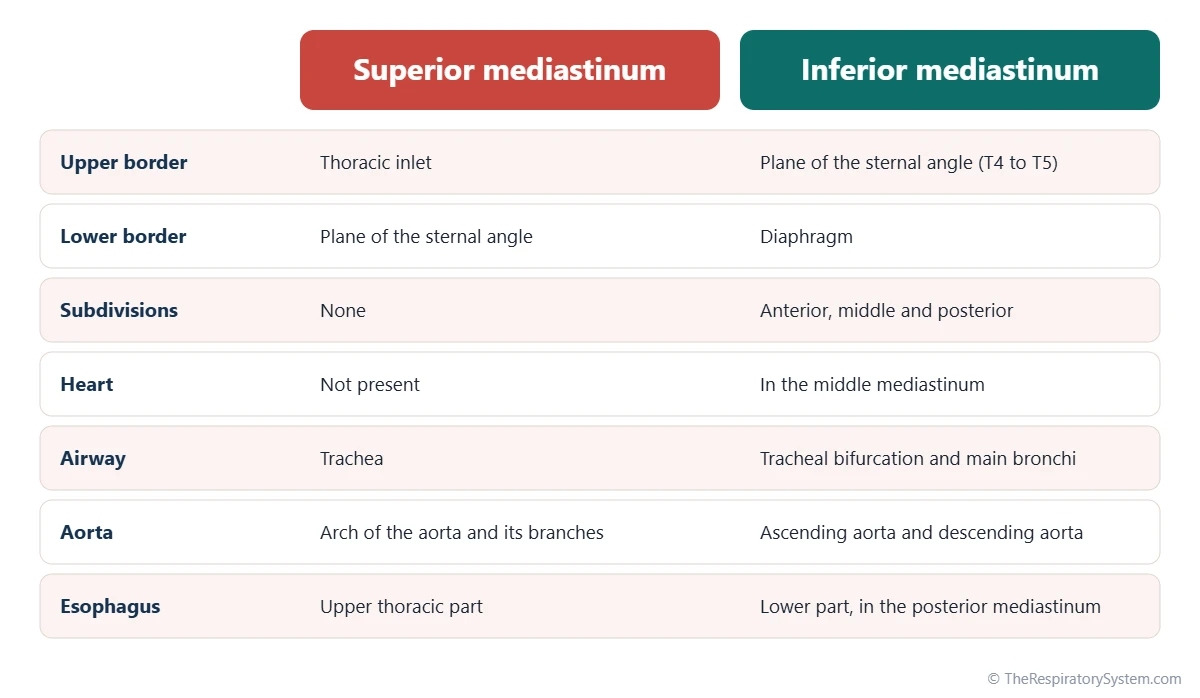
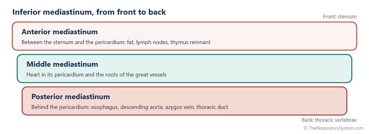
The mediastinum is divided into a superior and an inferior mediastinum by an imaginary horizontal plane, the transverse thoracic plane, which runs from the sternal angle at the front to the disc between the fourth and fifth thoracic vertebrae at the back. The inferior mediastinum is then divided into anterior, middle and posterior parts by the pericardium, the sac around the heart.
Superior mediastinum
The superior mediastinum lies above the plane of the sternal angle and below the thoracic inlet. From front to back it contains the thymus, the brachiocephalic veins and upper part of the superior vena cava, the arch of the aorta with its three large branches, the trachea, the esophagus and the thoracic duct. The vagus and phrenic nerves pass through it, and the left recurrent laryngeal nerve hooks under the aortic arch here.
Anterior mediastinum
The anterior mediastinum is the narrow space between the sternum and the front of the pericardium. In adults it holds mainly fat, loose connective tissue, small lymph nodes and branches of the internal thoracic vessels. In children it also contains the lower part of the thymus, which is large in early life and shrinks after puberty.
Middle mediastinum
The middle mediastinum contains the pericardium and the heart, together with the roots of the great vessels: the ascending aorta, the pulmonary trunk and its branches, the lower half of the superior vena cava and the ends of the pulmonary veins. The phrenic nerves run down its sides, and the tracheal bifurcation and main bronchi lie at its upper border.
Posterior mediastinum
The posterior mediastinum lies behind the pericardium and in front of the lower thoracic vertebrae. It contains the descending thoracic aorta, the esophagus, the thoracic duct, the azygos and hemiazygos veins, the vagus nerves forming a plexus on the esophagus, the thoracic splanchnic nerves and posterior lymph nodes.
Compartments used in imaging
Radiologists reading CT scans often use a simpler three-part model instead: a prevascular compartment at the front, a visceral compartment in the middle and a paravertebral compartment beside the spine. It maps the same space in a way that is easier to apply to scan images.
| Structure | Division | What it does |
|---|---|---|
| Thymus | Superior; lower part in the anterior mediastinum in children | Trains T cells in childhood, then mostly turns to fat |
| Brachiocephalic veins | Superior | Join to form the superior vena cava |
| Superior vena cava | Superior (upper half) and middle (lower half) | Returns blood from the head, neck and arms to the right atrium |
| Arch of the aorta and its branches | Superior | Sends blood to the head, neck and arms |
| Trachea | Superior, ending at the sternal angle | Carries air to the main bronchi |
| Heart and pericardium | Middle | Pumps blood through both circulations |
| Ascending aorta and pulmonary trunk | Middle | Carry blood out of the left and right ventricles |
| Phrenic nerves | Superior and middle | Control the diaphragm |
| Esophagus | Superior and posterior | Carries food from the throat to the stomach |
| Descending thoracic aorta | Posterior | Carries blood down to the chest wall and abdomen |
| Azygos and hemiazygos veins | Posterior | Drain blood from the back and chest walls |
| Thoracic duct | Superior and posterior | Returns lymph to the bloodstream |
| Vagus nerves | Superior, then posterior along the esophagus | Carry parasympathetic signals to the heart, lungs and gut |
| Source: Typical adult anatomy, our editors | ||
What Is the Location of the Trachea in the Mediastinum?
The trachea lies in the superior mediastinum, in the midline just in front of the esophagus, and it ends at about the level of the sternal angle, where it divides into the right and left main bronchi. Its lower end and the two main bronchi sit at the boundary between the superior and middle mediastinum.
As the trachea descends from the neck through the thoracic inlet, the brachiocephalic veins and the brachiocephalic trunk lie in front of it, and the arch of the aorta curves over its front and left side to reach the back of the chest. The esophagus runs directly behind it, which is why the two tubes are often compared on our trachea vs esophagus page. The point of division is marked inside the airway by a ridge called the carina, roughly at the level of the T4 to T5 vertebrae.
From there the main bronchi run outward to the hila of the lungs, beginning the bronchial tree. The left main bronchus passes under the arch of the aorta and in front of the esophagus and descending aorta, while the right main bronchus passes under the arch of the azygos vein.
What Is the Function of the Mediastinum?
The function of the mediastinum is to house and protect the central organs of the chest, keep the two lungs and their pleural cavities apart, and provide a protected passage for the vessels, airways, nerves and digestive tube that link the neck, lungs and abdomen.
- Holding the heart: the middle mediastinum cradles the heart in its pericardium, close to both lungs, so blood can flow quickly through the pulmonary circulation.
- A passage for air: the trachea and main bronchi travel through it to carry air into the lungs.
- A passage for food: the esophagus runs through the whole length of the mediastinum to the stomach.
- A route for vessels and nerves: the aorta, venae cavae, thoracic duct, vagus nerves and phrenic nerves all pass through it.
- Separating the lungs: because each lung lies in its own pleural sac, a problem on one side, such as a pneumothorax, usually affects only that lung.
- Immune function: the thymus trains T cells in early life, and mediastinal lymph nodes filter lymph from the lungs and heart.
The close packing of the heart, great vessels and airways is what lets the respiratory and circulatory systems work together so efficiently.
Mediastinum vs Pericardial Cavity: What Is the Difference?
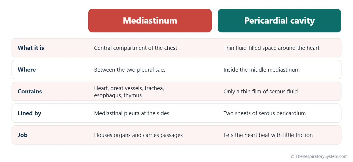
The mediastinum is the whole central compartment of the chest, while the pericardial cavity is a thin, fluid-filled space around the heart that lies inside the middle mediastinum. In other words, the pericardial cavity is one small part of the mediastinum.
The heart is wrapped in the pericardium, a sac with a tough outer fibrous layer and an inner serous layer. The serous layer has two sheets, one lining the fibrous sac and one covering the heart itself, and the narrow gap between them is the pericardial cavity. It holds only a thin film of fluid that lets the heart beat with little friction, much as the fluid in the pleural cavity lets the lungs slide.
The difference matters in practice. Fluid collecting inside the pericardial cavity squeezes the heart directly, while air, blood or infection in the rest of the mediastinum spreads through its loose connective tissue around many organs at once.
Common Conditions That Affect the Mediastinum
Conditions of the mediastinum include masses and enlarged lymph nodes, inflammation, air leaking into its tissues and shifts of its position caused by problems in the lungs. Our respiratory diseases section gives more background; anyone with chest symptoms should see a doctor.
- Mediastinal masses: growths are often grouped by compartment. Anterior masses include thymus tumors, germ cell tumors, lymphoma and a thyroid gland extending down into the chest; posterior masses are often nerve tumors.
- Enlarged mediastinal lymph nodes: seen with infections such as tuberculosis, with sarcoidosis and when lung cancer spreads.
- Mediastinitis: infection or inflammation of the mediastinal tissues, for example after a tear in the esophagus or chest surgery.
- Pneumomediastinum: air escaping into the mediastinum, sometimes after a small tear in an alveolus or the airway.
- Mediastinal shift: the mediastinum is pushed or pulled to one side by a large pleural effusion, a tension pneumothorax or collapse of a lung.
- Widened mediastinum: an X-ray finding that can reflect an enlarged aorta, bleeding or a mass.
Key Facts
- The mediastinum is the central compartment of the thoracic cavity, between the two pleural sacs.
- It extends from the thoracic inlet to the diaphragm and from the sternum to the thoracic vertebrae.
- A plane through the sternal angle and the T4 to T5 disc divides it into superior and inferior parts.
- The inferior mediastinum has anterior, middle and posterior sections, and the heart lies in the middle one.
- The trachea runs through the superior mediastinum in front of the esophagus and divides at about the level of the sternal angle.
- The pericardial cavity is a thin fluid space inside the middle mediastinum, not the same thing as the mediastinum.
- The mediastinum holds every organ of the chest except the lungs and their pleura.
Quick Quiz: Test What You Know About the Mediastinum
6 questions. Tap an answer to check it.
What lies on either side of the mediastinum?
The mediastinum is the central partition of the chest, with a pleural cavity and lung on each side.
Which landmark marks the boundary between the superior and inferior mediastinum?
A horizontal plane through the sternal angle and the T4 to T5 disc separates the superior from the inferior mediastinum.
In which division of the mediastinum is the heart found?
The middle mediastinum contains the pericardium, the heart and the roots of the great vessels.
Which structure lies in the posterior mediastinum?
The posterior mediastinum holds the descending aorta, esophagus, azygos veins and thoracic duct.
Where is the trachea located in the mediastinum?
The trachea runs through the superior mediastinum in the midline, with the esophagus directly behind it.
What is the pericardial cavity?
The pericardial cavity is the narrow, fluid-filled gap between the layers of serous pericardium, inside the middle mediastinum.
Frequently Asked Questions
Are the lungs in the mediastinum?
No. The lungs lie on either side of the mediastinum, each inside its own pleural sac, and they are the main organs of the chest that are not part of it. The mediastinum touches the medial surface of each lung, and the roots of the lungs, with their bronchi and pulmonary vessels, pass from the hila into the mediastinum to connect with the trachea and heart.
Is the esophagus in the mediastinum?
Yes. The thoracic part of the esophagus runs through the whole length of the mediastinum. It enters from the neck through the thoracic inlet, travels through the superior mediastinum behind the trachea, then continues through the posterior mediastinum behind the heart, and finally passes through an opening in the diaphragm to join the stomach in the abdomen.
What is the largest organ in the mediastinum?
The heart is the largest organ in the mediastinum. Inside its pericardial sac it fills most of the middle mediastinum and sits slightly to the left of the midline, which is why the left lung is smaller than the right. The great vessels attached to the heart, such as the aorta and the pulmonary trunk, are the next largest structures in the space.
Can the mediastinum move?
Yes. In a living person the mediastinum is flexible rather than fixed. It shifts slightly with breathing, swallowing and changes in body position. It can also be pushed or pulled sideways when the pressure in one pleural cavity changes a lot, for example with a large collection of fluid or air, which is called a mediastinal shift and can be seen on a chest X-ray.
What is a widened mediastinum?
A widened mediastinum is a chest X-ray finding in which the central shadow between the lungs looks broader than expected. It is a description, not a diagnosis. Causes range from harmless ones, such as extra fat or the way the image was taken, to an enlarged aorta, bleeding after an injury, enlarged lymph nodes or a mass, so it is usually followed up with further imaging.
What is mediastinoscopy?
Mediastinoscopy is a procedure in which a surgeon passes a thin, lighted scope through a small cut just above the sternum into the upper mediastinum. It lets doctors look at and take samples from the lymph nodes around the trachea. Today it is often used alongside, or replaced by, scope techniques passed through the airway with ultrasound guidance.
What is the mediastinal pleura?
The mediastinal pleura is the part of the parietal pleura that lines each side of the mediastinum. It forms the lateral wall of the mediastinum and the medial wall of each pleural cavity. At the root of each lung it turns outward to become continuous with the visceral pleura covering the lung, forming a sleeve around the root.
How is the mediastinum seen on imaging?
On a front chest X-ray the mediastinum appears as the white central band between the darker, air-filled lungs, formed mostly by the heart and great vessels. Because many structures overlap there, a CT scan is the main way to see its individual organs, lymph nodes and vessels. MRI is sometimes used for the heart, the great vessels and some masses.
Keep Exploring


