At a Glance
The pleural cavity, or pleural space, is the potential space between the visceral and parietal pleura on each side of the chest. It contains a film of pleural fluid only about 10 to 20 micrometers thick (roughly 0.1 to 0.3 mL per kilogram of body weight per side), and its pressure stays below atmospheric, about -5 cmH2O at rest, which holds the lungs against the chest wall.
- Location
- Inside the thoracic cavity, one on each side of the mediastinum, around each lung
- Type
- Potential space between two serous membranes, the visceral and parietal pleura
- Contains
- Only a thin film of serous pleural fluid; no air in health
- Pressure
- Slightly below atmospheric, about -5 cmH2O at the end of a quiet breath out
- Function
- Couples the lungs to the chest wall, lubricates them and keeps them expanded
- Part of
- Thoracic cavity and lower respiratory tract
On this page
- What Is the Pleural Cavity?
- Where Is the Pleural Cavity Located?
- What Is Pleural Fluid?
- What Is the Pleural Cavity Function in the Respiratory System?
- Why Is the Pressure in the Pleural Cavity Negative?
- How Does the Pleural Cavity Fit Into the Chest?
- Common Conditions That Affect the Pleural Cavity
- Key Facts
- Quiz
- FAQ
What Is the Pleural Cavity?
The pleural cavity is the narrow gap between the visceral pleura, which covers the surface of a lung, and the parietal pleura, which lines the inside of the chest. Anatomists call it a potential space: in health the two layers touch almost everywhere, separated only by a film of pleural fluid, so there is no open air gap at all. The same space is often called the pleural space or intrapleural space.
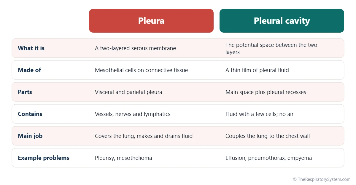
The pleura is the membrane; the pleural cavity is the space that membrane encloses. Because the lung is wrapped by the inner layer, the lung itself lies outside the pleural cavity, pressed against it, much as a fist pushed into a soft balloon stays outside the balloon's air. Each lung has its own pleural cavity, and the right and left cavities do not connect.
Pleural cavity definition
In one line, the pleural cavity is the closed, fluid-lined potential space between the parietal and visceral pleura on each side of the chest. It contains a small amount of serous pleural fluid and is kept at a pressure slightly below that of the atmosphere.
Explained simply
Press two wet panes of glass together. They slide past each other easily, but it is very hard to pull them apart. The pleural cavity is that thin wet layer between the lung and the chest wall, and it is the reason your lungs follow your ribs every time you breathe in.
Where Is the Pleural Cavity Located?
The pleural cavities lie inside the thoracic cavity, one on the right and one on the left, on either side of the central mediastinum that holds the heart, the trachea and the esophagus. Each one wraps around its lung like a thin sleeve, from the root of the neck down to the dome of the diaphragm.
- Top: the cavity rises into the base of the neck above the first rib, under the dome of cervical pleura that caps the apex of the lung.
- Front, sides and back: it follows the inner surface of the rib cage and the muscles between the ribs.
- Bottom: it rests on the upper surface of the diaphragm, which separates it from the abdomen.
- Inner side: it ends at the mediastinum and the hilum of the lung, where the pleura folds around the bronchi and vessels entering the lung.
Pleural recesses: the parts with no lung
The pleural cavity is larger than the lung inside it. At its lower edge, the costodiaphragmatic recess forms a deep, narrow gutter where the chest wall meets the diaphragm, and the lung slides partly into it only during a deep breath in. A smaller costomediastinal recess lies behind the sternum, larger on the left next to the heart.
When a person stands or sits upright, any extra fluid in the pleural cavity sinks into the costodiaphragmatic recess first, so this is where a small effusion shows up earliest on imaging. The rib levels of the pleural borders are listed on our pleura page.
What Is Pleural Fluid?
Pleural fluid is a thin, clear to pale yellow serous liquid that lines the pleural cavity as a film only about 10 to 20 micrometers thick. A healthy adult has only about 0.1 to 0.3 milliliter per kilogram of body weight on each side, a few teaspoons at most, spread over the entire surface of the lung.
What pleural fluid is made of
Pleural fluid is mostly water with dissolved salts, glucose at about the same level as in blood, and a small amount of protein, far less than in plasma. It carries a few cells, mainly macrophages, lymphocytes and mesothelial cells shed from the pleura. Glycoproteins such as hyaluronan make it slippery, and it is slightly more alkaline than blood.
How pleural fluid is made and drained
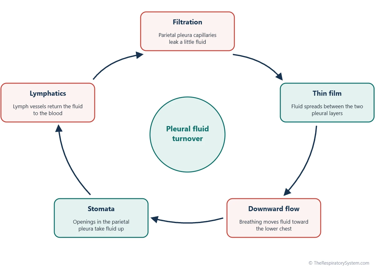
Pleural fluid is made and removed all the time. Small capillaries in the parietal pleura, fed by systemic arteries such as the intercostal arteries, filter a little fluid into the cavity because their pressure is higher than the pressure in the pleural space. Breathing movements spread the fluid and carry it toward the lower parts of the chest.
The fluid leaves through tiny openings called stomata in the parietal pleura, found mostly over the diaphragm and the lower chest wall. They lead into lymphatic vessels that return the fluid to the bloodstream. These lymphatics can increase their drainage many times over when more fluid is made, which is why the film normally stays thin.
| Feature | Typical healthy adult | Note |
|---|---|---|
| Number of pleural cavities | Two, right and left | They do not connect with each other |
| Normal contents | A film of pleural fluid only | No air, blood or lung tissue inside the space |
| Fluid volume | About 0.1 to 0.3 mL per kg of body weight per side | A few teaspoons in an adult |
| Fluid film thickness | About 10 to 20 micrometers | Thinner than a human hair |
| Fluid appearance | Clear to pale yellow | Thin, watery serous fluid |
| Fluid protein | Low, well below plasma levels | Formed by filtration from small capillaries |
| Fluid cells | Few: macrophages, lymphocytes, mesothelial cells | No red blood cells in health |
| Where fluid enters | Capillaries of the parietal pleura | Supplied by systemic arteries of the chest wall |
| Where fluid leaves | Lymphatic stomata in the parietal pleura | Mostly over the diaphragm and lower chest wall |
| Pressure at the end of a quiet breath out | About -5 cmH2O | Slightly below atmospheric pressure |
| Pressure at the end of a quiet breath in | About -8 cmH2O | More negative as the chest expands |
| Lowest point when upright | Costodiaphragmatic recess | Where extra fluid collects first |
| Source: Typical adult values, our editors | ||
What Is the Pleural Cavity Function in the Respiratory System?
The pleural cavity function in the respiratory system is to couple each lung to the chest wall so the lung expands when the chest expands, while letting the lung slide freely and keeping it in its own sealed compartment. It does all of this with a thin film of fluid and a pressure kept slightly below atmospheric.
- Coupling: surface tension in the fluid film holds the visceral and parietal pleura together, so when the diaphragm and the intercostal muscles enlarge the chest, the lung is pulled outward with it.
- Lubrication: the fluid lets the lung glide over the ribs and diaphragm with almost no friction, breath after breath.
- Keeping the lungs open: the negative pressure in the cavity stops the elastic lung from recoiling down to a small, nearly airless size.
- Separation: each lung has its own cavity, so a leak of air or fluid on one side does not directly collapse the other lung.
- Room to move: the recesses give the lungs space to expand downward during a deep breath.
Pleural cavity function in the lungs, in short
Without the pleural cavity, the lungs would hang loose inside the chest and the breathing muscles could not inflate them. The full sequence of muscle action, pressure change and airflow is explained on our mechanics of breathing page, and the muscles themselves on muscles of respiration.
Why Is the Pressure in the Pleural Cavity Negative?
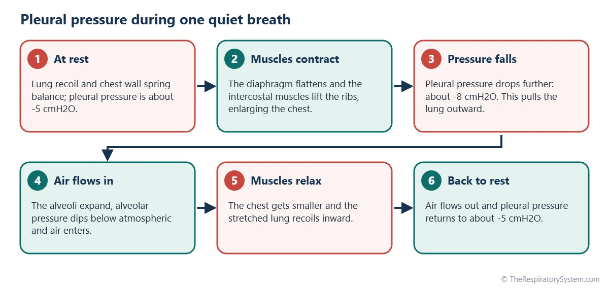
Pressure in the pleural cavity, called intrapleural or pleural pressure, is negative because two elastic structures pull in opposite directions on the thin fluid layer between them: the lungs tend to recoil inward, while the chest wall tends to spring outward. The fluid cannot stretch to fill a gap, so these opposing pulls lower its pressure below that of the air outside the body.
At the end of a quiet breath out, pleural pressure is typically about -5 cmH2O, roughly 4 mmHg below atmospheric pressure. As the diaphragm contracts and the chest enlarges, it falls to about -8 cmH2O at the end of a quiet breath in, which stretches the lungs and draws air in through the airways. During a forced breath out, such as a hard cough, it can briefly rise above atmospheric pressure.
Transpulmonary pressure
The difference between the pressure inside the alveoli and the pressure in the pleural cavity is called transpulmonary pressure. It is the pressure that actually holds the lungs open, and the larger it becomes, the more the lungs expand. Our inhalation and exhalation page shows how alveolar pressure changes alongside it, and lung volumes and capacities describes the volumes that result.
Why the top of the lung is different
In an upright person, pleural pressure is more negative at the top of the chest than at the bottom, because the weight of the lung pulls down on it. The alveoli near the apex are therefore held more open at rest than those near the base, which affects how evenly fresh air reaches different parts of the lung.
How Does the Pleural Cavity Fit Into the Chest?
The pleural cavities are two of the body's serous cavities, alongside the pericardial cavity around the heart and the peritoneal cavity around the abdominal organs. All of them are lined by the same slippery mesothelium and hold a thin film of fluid that lets an organ move without friction.
- Thoracic cavity: the whole chest space, which contains both pleural cavities plus the mediastinum.
- Pericardial cavity: lies within the mediastinum between the two pleural cavities, separated from them by the pericardium and the mediastinal pleura.
- Peritoneal cavity: lies below, separated from the pleural cavities by the diaphragm.
- Lungs: sit inside the pleural sacs but outside the cavities themselves, covered by visceral pleura.
How the pleural cavities form
In the embryo, the pleural cavities develop from a single body cavity called the intraembryonic coelom. As the lung buds grow into it on each side of the heart, folds called the pleuropericardial membranes separate the pleural cavities from the pericardial cavity. The pleuroperitoneal membranes then close them off from the abdomen as part of the forming diaphragm; when that closure fails, abdominal organs can move into the chest, a birth defect called congenital diaphragmatic hernia. The full story is on our development of the respiratory system page.
Common Conditions That Affect the Pleural Cavity
Most conditions of the pleural cavity involve something collecting in a space that should hold only a thin film of fluid: extra fluid, air, blood, pus or lymph. Any of these pushes the two pleural layers apart, breaks the seal between lung and chest wall and can press on the lung. Our respiratory diseases section covers them in more depth, and chest pain or sudden breathlessness is a reason to see a doctor.
- Pleural effusion: excess fluid in the cavity, either a thin transudate when pressures in the blood vessels change, as in heart failure, or a protein-rich exudate when the pleura is inflamed or infected.
- Pneumothorax: air in the cavity, which raises pleural pressure toward atmospheric and lets the lung recoil and collapse.
- Hemothorax: blood in the cavity, most often after a chest injury.
- Empyema: pus in the cavity, usually a complication of pneumonia.
- Chylothorax: milky lymph, called chyle, in the cavity after damage to the thoracic duct.
- Pleurisy: inflammation of the pleural layers, which roughens their surfaces so they rub together with each breath.
Key Facts
- The pleural cavity is the potential space between the visceral and parietal layers of the pleura, one on each side of the chest.
- The lungs lie outside the pleural cavities, covered by visceral pleura and pressed against the thin fluid film.
- A healthy adult has only about 0.1 to 0.3 mL of pleural fluid per kilogram of body weight on each side, spread in a film about 10 to 20 micrometers thick.
- Pleural fluid is filtered from capillaries of the parietal pleura and drained through stomata into lymphatic vessels.
- Pleural pressure is about -5 cmH2O at the end of a quiet breath out and about -8 cmH2O at the end of a quiet breath in.
- Air entering the pleural cavity, a pneumothorax, breaks the seal and lets the lung recoil and collapse.
- The costodiaphragmatic recess is the lowest part of the cavity in an upright person and the first place extra fluid collects.
Quick Quiz: Test What You Know About the Pleural Cavity and Pleural Fluid
6 questions. Tap an answer to check it.
What lies inside the pleural cavity in a healthy person?
The pleural cavity holds only a thin film of serous fluid. The lungs lie outside it, covered by visceral pleura.
Which two layers form the walls of the pleural cavity?
The pleural cavity lies between the visceral pleura on the lung and the parietal pleura on the chest wall.
What is the typical pleural pressure at the end of a quiet breath out?
Pleural pressure is about -5 cmH2O at rest because the lung recoils inward while the chest wall springs outward.
Where is most pleural fluid drained from the cavity?
Stomata in the parietal pleura, mostly over the diaphragm and lower chest, lead into lymphatic vessels that return the fluid to the blood.
What is the deepest part of the pleural cavity in an upright person?
The costodiaphragmatic recess, where the chest wall meets the diaphragm, is the lowest point and the first place extra fluid collects.
What happens when air enters the pleural cavity?
Air raises pleural pressure toward atmospheric, so nothing holds the lung out and it recoils inward. This is a pneumothorax.
Frequently Asked Questions
Are the lungs inside the pleural cavity?
Not strictly. Each lung sits inside its pleural sac, but it is wrapped by the visceral pleura, so it lies outside the pleural cavity itself. The cavity is only the thin, fluid-filled gap between the visceral pleura on the lung and the parietal pleura on the chest wall. Many textbooks loosely say the lungs are in the pleural cavities, meaning the two sides of the chest.
What is the difference between the pleura and the pleural cavity?
The pleura is the membrane, a two-layered sheet of mesothelium that covers each lung and lines the chest wall. The pleural cavity is the space between those two layers, which normally holds only a thin film of fluid. A problem of the pleura itself is pleurisy, while problems of the cavity, such as an effusion or a pneumothorax, involve something collecting in the space.
What is the difference between the pleural cavity and the thoracic cavity?
The thoracic cavity is the whole chest space inside the rib cage, above the diaphragm. It is divided into three parts: a pleural cavity on each side and the mediastinum in the middle, which holds the heart, trachea and esophagus. So each pleural cavity is a smaller compartment within the thoracic cavity, dedicated to one lung.
Is there air in the pleural cavity?
No. A healthy pleural cavity contains no air, only a few milliliters of serous fluid. Air in the pleural space is abnormal and is called a pneumothorax. It can come from a tear in the lung surface or from an injury to the chest wall, and it removes the negative pressure that normally holds the lung expanded.
What color is normal pleural fluid?
Normal pleural fluid is clear to pale yellow, similar in look to the straw-colored plasma part of blood but far lower in protein. Changes in appearance, such as cloudy, bloody or milky white fluid, point to different causes of a pleural effusion, which is why laboratories always record the color and clarity of any fluid removed from the chest.
Do the right and left pleural cavities connect?
No. In humans the right and left pleural cavities are completely separate, divided by the mediastinum in the middle of the chest. This separation means that air or fluid collecting on one side usually affects only that lung. The two cavities come closest behind the sternum, in front of the heart, where their pleural edges almost meet.
How much pleural fluid is normal?
A healthy adult has only about 0.1 to 0.3 milliliter of pleural fluid per kilogram of body weight on each side, only a few teaspoons in total. It is spread as a film just 10 to 20 micrometers thick. Because the lymphatics can drain much more than is normally made, the volume stays small unless production rises or drainage is blocked.
What happens to the lungs if the pleural cavity is opened?
If the pleural cavity is opened to the outside air, as during chest surgery or after a penetrating injury, air rushes in until the pressure equals atmospheric pressure. With no negative pressure holding it out, the elastic lung recoils toward its root at the hilum, while the chest wall springs slightly outward. This is why surgeons use special ventilation during open chest operations.


