At a Glance
Pulmonary ventilation works by Boyle's law: at a constant temperature, the pressure of a gas rises as its volume falls. Contraction of the diaphragm and external intercostal muscles enlarges the chest, lowering the pressure in the alveoli by about 1 mmHg below atmospheric pressure so air flows in; in quiet breathing, relaxation and elastic recoil then raise it about 1 mmHg above atmospheric pressure so air flows out.
- Also called
- Pulmonary ventilation, the mechanism of breathing
- Physical law
- Boyle's law: gas pressure and volume change in opposite directions
- Main muscles
- Diaphragm and external intercostals for inspiration; quiet expiration is passive
- Key pressures
- Atmospheric, alveolar (intrapulmonary) and intrapleural pressure
- Resisted by
- Elastic recoil, surface tension in the alveoli and airway resistance
- Part of
- The first step of respiration, before gas exchange and gas transport
On this page
- What Are the Mechanics of Breathing?
- What Is Boyle's Law in the Lungs?
- Which Pressures Drive Pulmonary Ventilation?
- What Is the Mechanism of Breathing in Human Beings?
- How Do the Lungs Follow the Chest Wall?
- What Are Lung Compliance and Elastic Recoil?
- What Is Airway Resistance?
- How Does Quiet Breathing Differ From Forced Breathing?
- Common Conditions That Affect the Mechanics of Breathing
- Key Facts
- Quiz
- FAQ
What Are the Mechanics of Breathing?
The mechanics of breathing are the forces and pressure changes that move air between the atmosphere and the alveoli: muscles change the volume of the chest, that volume change creates pressure differences, and air flows from higher to lower pressure. This process, called pulmonary ventilation, is the first of the steps of respiration, followed by gas exchange in the lungs, gas transport in the blood and exchange in the tissues.
Air never needs to be pushed or sucked in actively. The lungs simply follow the chest wall, and air rushes in or out until the pressure in the alveoli equals the pressure outside. Every breath is a cycle of two phases, which our inhalation and exhalation page describes phase by phase; this page explains the physics underneath both.
Breathing process in humans at a glance
At rest an adult takes about 12 to 20 breaths per minute, each moving roughly 500 milliliters of air, the tidal volume. That adds up to about 6 liters of air per minute. The pressure differences that drive this flow are remarkably small: during a quiet breath the pressure in the alveoli changes by only about 1 mmHg from the pressure of the outside air.
Explained simply
Squeeze an empty plastic bottle and air puffs out; let it spring back and air rushes in. Your chest works the same way, except that your muscles make it bigger first. When the chest grows, the air inside has more room, its pressure drops, and outside air flows in to fill the space. When the muscles relax, the chest springs back, the pressure rises and air flows out.
What Is Boyle's Law in the Lungs?
Boyle's law states that, at a constant temperature, the pressure of a fixed amount of gas is inversely proportional to its volume, so when the lungs get bigger the pressure of the air inside them falls, and when they get smaller it rises. It is usually written P1V1 = P2V2.
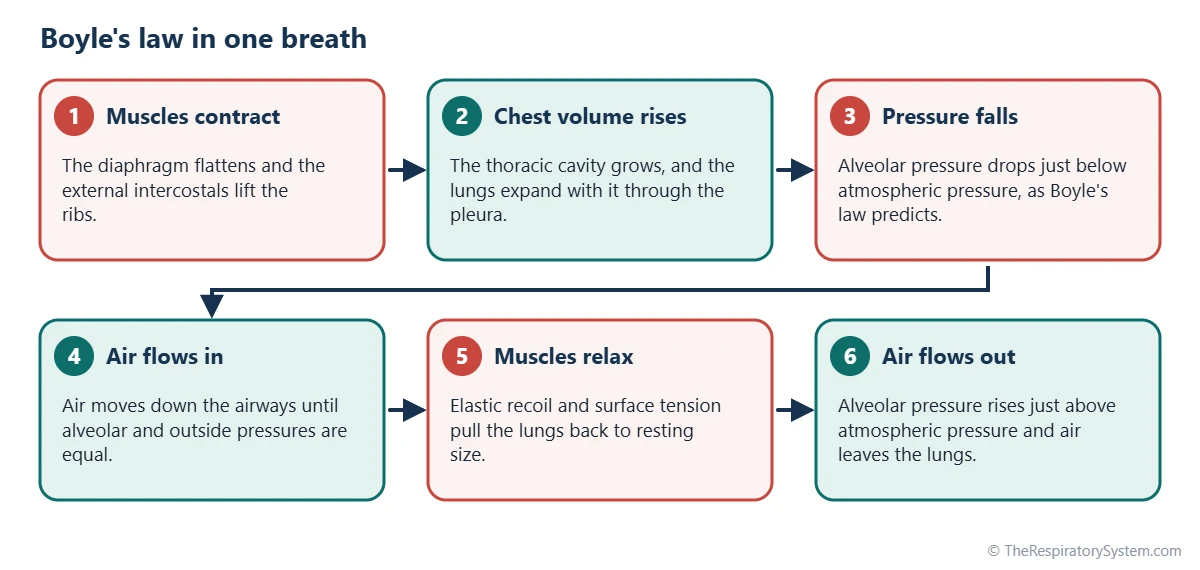
In breathing, Boyle's law explains why changing the size of the chest is enough to move air. If the volume of the lungs increases, the same number of gas molecules is spread through a larger space, they strike the walls less often, and the pressure falls. Outside air, now at a higher pressure, flows down the airways until the two pressures are equal. The reverse happens when the lungs get smaller.
Boyle's law breathing in steps
- The diaphragm and external intercostal muscles contract.
- The volume of the thoracic cavity increases, and the lungs expand with it.
- The alveolar pressure falls slightly below atmospheric pressure.
- Air flows into the lungs until the pressures are equal again.
- The muscles relax, and the elastic lungs and chest wall recoil.
- The lung volume decreases, alveolar pressure rises above atmospheric pressure, and air flows out.
Temperature is not perfectly constant, because inhaled air is warmed to body temperature, but the change is small and Boyle's law describes breathing well. A balloon lung model, described in how to make a lung model, shows the principle with a stretchy sheet standing in for the diaphragm.
Which Pressures Drive Pulmonary Ventilation?
Pulmonary ventilation is driven by the difference between atmospheric pressure and alveolar pressure, while a third pressure, the intrapleural pressure, keeps the lungs stretched against the chest wall. Physiologists usually express these pressures relative to atmospheric pressure, which is counted as zero.
- Atmospheric pressure: the pressure of the air around the body, about 760 mmHg at sea level.
- Alveolar (intrapulmonary) pressure: the pressure of the air inside the alveoli. It equals atmospheric pressure between breaths, falls about 1 mmHg below it during a quiet inspiration and rises about 1 mmHg above it during expiration.
- Intrapleural pressure: the pressure of the thin film of fluid in the pleural cavity. It is always slightly below atmospheric pressure in quiet breathing, about 4 mmHg lower at rest and about 6 mmHg lower at the end of a quiet inspiration.
- Transpulmonary pressure: the difference between alveolar and intrapleural pressure. It is the pressure that holds the lungs open, and the larger it is, the more the lungs are stretched.
Why intrapleural pressure is negative
The lungs are elastic and constantly try to recoil inward, while the chest wall tends to spring outward. These opposing pulls act on the pleural fluid between them, lowering its pressure below atmospheric pressure. This negative pressure is what keeps the lungs from collapsing. If air enters the pleural cavity, the pressure equalizes with the atmosphere and the lung on that side pulls away from the chest wall, a condition called pneumothorax.
The table below lists typical values during quiet breathing in a healthy adult.
| Pressure | Between breaths | During quiet inspiration | During quiet expiration |
|---|---|---|---|
| Atmospheric pressure | Reference (about 760 mmHg at sea level) | Unchanged | Unchanged |
| Alveolar (intrapulmonary) pressure | Equal to atmospheric (0) | About 1 mmHg below atmospheric | About 1 mmHg above atmospheric |
| Intrapleural pressure | About 4 mmHg below atmospheric | Falls to about 6 mmHg below atmospheric | Returns toward about 4 mmHg below |
| Transpulmonary pressure | About 4 mmHg, holding the lungs open | Rises as the lungs are stretched | Falls as the lungs recoil |
| Airflow | None (pressures equal) | Into the lungs | Out of the lungs |
| Lung volume | Resting volume (functional residual capacity) | Increases by about one tidal volume | Returns to resting volume |
| Muscle activity | None | Diaphragm and external intercostals contract | None; passive recoil |
| Source: Typical values for a healthy adult at rest at sea level, our editors | |||
What Is the Mechanism of Breathing in Human Beings?
The mechanism of breathing in human beings is the enlargement of the chest in three directions by the breathing muscles, followed by its return to resting size by elastic recoil. Each direction is produced by a different movement.
Vertical enlargement by the diaphragm
The diaphragm is a dome-shaped sheet of muscle under the lungs. When its fibers contract, the dome flattens and moves down, lengthening the chest from top to bottom. In quiet breathing it descends only about 1 to 2 centimeters, yet this produces most of the air movement, roughly two-thirds to three-quarters of each quiet breath. It is driven by the phrenic nerve.
Front-to-back and side-to-side enlargement by the ribs
The external intercostal muscles lift the ribs. The upper ribs move forward and up, pushing the sternum out like an old water-pump handle, which deepens the chest from front to back. The lower ribs swing up and out like the handle of a bucket, which widens the chest from side to side. Our rib cage and breathing page shows these pump-handle and bucket-handle movements in detail.
Return to resting size
At the end of a quiet inspiration the muscles relax. The stretched elastic fibers of the lungs and the surface tension in the alveoli pull the lungs inward, the diaphragm rises back to its dome, and the ribs drop. No muscle work is needed for quiet expiration. The full list of muscles used for normal and forced breathing is on our muscles of respiration page.
How Do the Lungs Follow the Chest Wall?
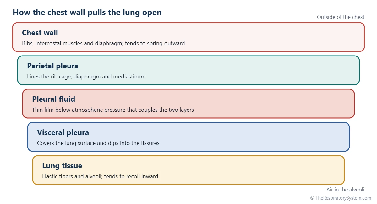
The lungs follow the chest wall because they are linked to it by the pleura, two thin membranes held together by a film of fluid under negative pressure, so any movement of the chest wall is passed straight to the lung surface. The lungs themselves have no muscle that could make them expand.
The pleura has two layers. The parietal pleura lines the inside of the rib cage, the upper surface of the diaphragm and the sides of the mediastinum. The visceral pleura covers each lung and dips into its fissures. Between them lies the pleural cavity, which is not really a space but a very narrow gap filled with a small amount of pleural fluid.
The wet glass effect
Two wet sheets of glass slide easily over each other but are very hard to pull apart. The pleural fluid works the same way: it lets the lung slide smoothly against the chest wall during each breath while keeping the two surfaces stuck together. Combined with the negative intrapleural pressure, this coupling means that when the diaphragm descends or the ribs rise, the lungs are pulled outward with them.
When the coupling fails
If air or excess fluid enters the pleural cavity, the coupling is lost. Air between the layers, a pneumothorax, lets the lung recoil and collapse away from the chest wall. A large pleural effusion compresses the lung from outside. In both cases the chest wall may still move, but the lung on that side no longer follows it fully.
What Are Lung Compliance and Elastic Recoil?
Lung compliance is how easily the lungs stretch when pressure is applied, and elastic recoil is their tendency to spring back to a smaller size; together they decide how much effort inspiration takes and how easily expiration happens. A compliant lung inflates with little effort, while a stiff lung needs more muscle force for the same breath.
Compliance is measured as the change in lung volume for each unit change in transpulmonary pressure. Healthy adult lungs have a compliance of about 200 milliliters per centimeter of water, which means a small pressure change produces a generous breath. Compliance falls when the lung tissue is stiffened by scarring and rises when its elastic fibers are destroyed.
Two sources of recoil
- Elastic fibers: a network of elastin and collagen in the lung tissue and around the airways is stretched during inspiration and stores energy like a rubber band.
- Surface tension: the thin layer of water lining each alveolus pulls inward, because water molecules attract each other strongly. This is the larger of the two forces in a normal lung.
Why surfactant matters
Surface tension alone would make small alveoli collapse. By the law of Laplace, the pressure needed to keep a bubble open rises as its radius falls, so tiny air sacs would empty into larger ones. Pulmonary surfactant, a mix of lipids and proteins made by type II pneumocytes, lowers surface tension, more so as an alveolus shrinks. This keeps alveoli of different sizes stable and greatly reduces the work of breathing in.
What Is Airway Resistance?
Airway resistance is the friction that air meets as it flows through the airways, and it depends most of all on their radius: halving the radius of a tube increases its resistance about sixteen times. In healthy lungs, resistance is low, so a pressure difference of about 1 mmHg is enough for a normal breath.
Where resistance is highest
Surprisingly, the tiniest airways are not the main source of resistance. The greatest resistance lies in the medium-sized bronchi, because each small bronchiole works in parallel with thousands of others, giving them an enormous combined cross-section. During nose breathing, the nose and upper airway also add a large share of the total, which is one reason people switch to mouth breathing during hard exercise.
What changes airway resistance
- Smooth muscle tone: parasympathetic nerve signals narrow the bronchioles, while sympathetic signals and adrenaline widen them.
- Lung volume: airways are pulled wider as the lungs inflate and become narrower at low lung volumes.
- Mucus and swelling: secretions and inflamed airway walls reduce the space for airflow.
- Dynamic compression: during a forced exhalation, intrapleural pressure becomes positive and can squeeze the airways, limiting how fast air can be blown out.
These effects are measured with spirometry, which records how much air can be breathed out and how fast.
| Factor | What it is | Effect on breathing | Changed by |
|---|---|---|---|
| Lung compliance | Volume change per unit of pressure | High compliance makes inspiration easy | Lower in fibrosis; higher in emphysema |
| Elastic recoil | Tendency of stretched lungs to spring back | Powers quiet expiration | Reduced when elastic fibers are destroyed |
| Surface tension | Inward pull of the alveolar fluid layer | Main source of lung recoil | Lowered by pulmonary surfactant |
| Airway radius | Width of the airways | Halving the radius raises resistance about 16 times | Bronchoconstriction, mucus, swelling |
| Intrapleural pressure | Pressure in the pleural fluid | Keeps lungs stretched against the chest wall | Lost in pneumothorax |
| Chest wall compliance | How easily the rib cage and abdomen move | Limits how far the chest can expand | Spinal curvature, obesity, rib injury |
| Muscle strength | Force of the diaphragm and intercostals | Sets the maximum pressure change | Nerve injury, muscle disease, fatigue |
| Source: Standard physiology, our editors | |||
How Does Quiet Breathing Differ From Forced Breathing?
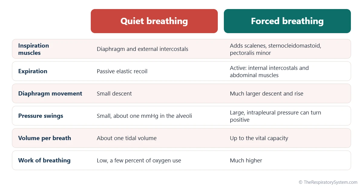
Quiet breathing uses only the diaphragm and external intercostals for inspiration and relies on passive recoil for expiration, while forced breathing adds accessory muscles to both phases and produces much larger pressure swings. The switch happens whenever the body needs more ventilation, for example during exercise or coughing.
In forced inspiration, muscles of the neck and chest such as the sternocleidomastoid, scalenes and pectoralis minor lift the upper ribs and sternum higher, and the diaphragm descends much further, by several centimeters. In forced expiration, the internal intercostals pull the ribs down and the abdominal muscles squeeze the abdomen, pushing the diaphragm up. This lets a person breathe out well below the resting volume, down to the residual volume described on our lung volumes and capacities page.
Work of breathing
The work of breathing is the energy the muscles spend overcoming elastic recoil and airway resistance. At rest it is small, only a few percent of the oxygen the body uses, because healthy lungs are compliant and the airways are wide. It rises steeply when the lungs stiffen, when the airways narrow, or when ventilation must increase greatly. How the brain sets the depth and rate of each breath is covered on our control of breathing page.
Common Conditions That Affect the Mechanics of Breathing
The mechanics of breathing are disturbed when the airways narrow, when the lungs or chest wall become stiff, when the pleural coupling is lost, or when the breathing muscles are weak. Doctors often group these problems as obstructive or restrictive, as explained on our obstructive vs restrictive lung disease page. The respiratory diseases section has more detail, and anyone with breathing symptoms should see a doctor.
- Asthma: inflamed airways tighten and fill with mucus, raising airway resistance, especially during exhalation.
- COPD and emphysema: loss of elastic recoil makes the lungs overly compliant, so small airways collapse during exhalation and air is trapped.
- Pulmonary fibrosis: scar tissue lowers compliance, so more effort is needed for each breath and breaths become smaller.
- Pneumothorax and pleural effusion: air or fluid in the pleural cavity breaks the link between the lung and the chest wall.
- Chest wall problems: severe curvature of the spine, broken ribs or marked obesity limit how far the chest can expand.
- Neuromuscular weakness: conditions affecting the nerves or muscles, including injury to the phrenic nerve, reduce the force available to enlarge the chest.
- Surfactant deficiency: babies born very early may lack enough surfactant, so their alveoli tend to collapse and breathing takes great effort.
Key Facts
- Pulmonary ventilation follows Boyle's law: when the chest enlarges, alveolar pressure falls and air flows in.
- During a quiet breath, alveolar pressure changes by only about 1 mmHg above or below atmospheric pressure.
- Intrapleural pressure stays below atmospheric pressure, about 4 mmHg lower at rest, which keeps the lungs from collapsing.
- The diaphragm produces most of the air movement in quiet breathing, while the external intercostals lift the ribs.
- Quiet expiration is passive, powered by elastic recoil and surface tension; forced expiration uses the internal intercostals and abdominal muscles.
- Surfactant lowers surface tension in the alveoli, keeping small air sacs open and reducing the work of breathing.
- Halving the radius of an airway raises its resistance about sixteen times, which is why narrowed airways make breathing so hard.
Quick Quiz: Test What You Know About the Mechanics of Breathing
6 questions. Tap an answer to check it.
Which gas law explains how air moves into and out of the lungs?
Boyle's law states that gas pressure falls as volume rises at constant temperature, so enlarging the chest lowers alveolar pressure.
During quiet inspiration, alveolar pressure is:
A small fall of about 1 mmHg below atmospheric pressure is enough to draw air into the lungs.
Why is intrapleural pressure normally below atmospheric pressure?
The opposite pulls of the lungs and chest wall on the pleural fluid lower its pressure, keeping the lungs stretched open.
Which muscle produces most of the air movement during quiet breathing?
The diaphragm flattens as it contracts and accounts for most of each quiet breath.
What is the main role of pulmonary surfactant in breathing mechanics?
Surfactant lowers surface tension, keeping small alveoli open and making inspiration easier.
What happens to the lung on one side in a pneumothorax?
Air in the pleural cavity equalizes the pressure with the atmosphere, so the lung recoils and collapses.
Frequently Asked Questions
What is the difference between breathing and pulmonary ventilation?
In everyday language they mean the same thing. In physiology, pulmonary ventilation is the precise term for the mechanical movement of air into and out of the lungs, while respiration is the broader process that also includes gas exchange, gas transport in the blood and the use of oxygen by cells. Breathing is the common word for ventilation, not for the chemistry that follows it.
Is exhalation active or passive?
Quiet exhalation is passive. When the diaphragm and external intercostals relax, the stretched elastic fibers of the lungs and the surface tension in the alveoli pull the lungs back to their resting size, pushing air out without muscle work. Exhalation becomes active during exercise, coughing, singing or blowing hard, when the internal intercostals and abdominal muscles contract to push air out faster and further.
Do the lungs have muscles to inflate themselves?
No. The lungs have no skeletal muscle that could expand them. They contain smooth muscle in the airway walls, but this only changes the width of the airways. The lungs inflate passively because they are coupled to the chest wall by the pleura and pleural fluid, so they follow the movements made by the diaphragm and the muscles between the ribs.
Why does air flow into the lungs?
Air flows into the lungs because the pressure inside the alveoli falls below the pressure of the outside air. Gases always move from higher to lower pressure. When the breathing muscles enlarge the chest, the volume of the lungs grows, and by Boyle's law the pressure of the air inside falls slightly. Air then flows in until the two pressures are equal again.
What is negative pressure breathing?
Negative pressure breathing is the normal way humans breathe: the chest enlarges and lowers the pressure inside the lungs below atmospheric pressure, so air is drawn in. Positive pressure breathing works the opposite way, pushing air into the lungs from outside at a pressure above atmospheric, which is how most mechanical ventilators in hospitals work. Old iron lung machines used negative pressure around the chest.
How does posture affect the mechanics of breathing?
Posture changes how freely the diaphragm and ribs can move. When a person lies flat, the abdominal organs push up against the diaphragm, which slightly reduces the resting volume of the lungs. Sitting upright or leaning forward lets the diaphragm descend more easily and allows the accessory muscles to work from a fixed shoulder girdle, which is why people who are short of breath often prefer to sit up.
What is the role of the abdominal muscles in breathing?
The abdominal muscles are the main muscles of forced exhalation. When they contract, they press the abdominal organs upward against the diaphragm and pull the lower ribs down, shrinking the chest and raising alveolar pressure. They are used during exercise, coughing, sneezing, shouting and playing wind instruments. In quiet breathing they stay largely relaxed, because elastic recoil is enough to empty the lungs.
What is functional residual capacity?
Functional residual capacity is the volume of air left in the lungs at the end of a quiet exhalation, when the inward recoil of the lungs exactly balances the outward spring of the chest wall. In an adult it is roughly 2 to 3 liters. This leftover air keeps the alveoli partly open between breaths and smooths out changes in blood gases from one breath to the next.
Keep Exploring


