At a Glance
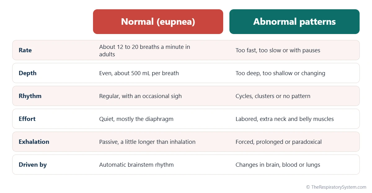
A breathing pattern describes how fast, how deep, how regular and how effortful breathing is. Normal adult breathing at rest (eupnea) is about 12 to 20 quiet, even breaths a minute, driven mainly by the diaphragm, with exhalation a little longer than inhalation. Abnormal patterns include tachypnea (too fast), bradypnea (too slow), apnea (pauses), Cheyne-Stokes breathing (waxing and waning with pauses) and Kussmaul breathing (deep, labored breaths in metabolic acidosis).
- Definition
- The rate, depth, rhythm and effort of breathing taken together
- Normal pattern
- Eupnea: about 12 to 20 quiet, regular breaths a minute in a resting adult
- Set by
- Breathing centers in the medulla and pons, adjusted by chemoreceptors and lung reflexes
- Main features
- Rate, tidal volume (depth), rhythm, effort and the inhalation to exhalation ratio
- Well-known abnormal patterns
- Tachypnea, bradypnea, apnea, Cheyne-Stokes, Kussmaul, Biot's, ataxic, apneustic, agonal
- Part of
- Respiratory physiology and the control of breathing
On this page
- What Is a Breathing Pattern?
- What Is a Normal Breathing Pattern?
- What Are the Abnormal Breathing Patterns?
- What Is Cheyne-Stokes Breathing?
- What Is Kussmaul Breathing and What Is Its Function?
- Breathing Patterns Linked to the Brain, Chest and Airways
- What Are the Breathing Patterns at End of Life?
- What Changes a Breathing Pattern?
- Key Facts
- FAQ
What Is a Breathing Pattern?
A breathing pattern is the way a person breathes over time, described by four features: the rate, the depth of each breath, the rhythm or regularity, and the effort or muscles used. Clinicians watch these features together because each one can change for a different reason.
- Rate: the number of breaths per minute, covered on our respiratory rate page.
- Depth: the volume of air in each breath, the tidal volume, about 500 milliliters at rest in an adult.
- Rhythm: whether breaths come at even intervals or in clusters, cycles or random bursts.
- Effort: whether breathing is quiet and driven mainly by the diaphragm, or labored, with the accessory muscles of respiration in the neck and abdomen joining in.
Who sets the pattern
The basic rhythm comes from groups of neurons in the medulla of the brainstem, shaped by centers in the pons. Chemoreceptors that sense carbon dioxide, acid and oxygen, and stretch receptors in the lungs, constantly adjust the rate and depth, as described on our control of breathing page. An abnormal pattern therefore often tells observers where in this control system something has changed.
Explained simply
Think of breathing as music. The rate is the tempo, the depth is the volume, the rhythm is the beat and the effort is how hard the musicians are working. A normal pattern is a calm song played at an even tempo, and each abnormal pattern changes the tempo, the volume or the beat in its own recognizable way.
What Is a Normal Breathing Pattern?
A normal breathing pattern at rest, called eupnea, is quiet, regular and effortless breathing at about 12 to 20 breaths a minute in an adult, with each breath moving about 500 milliliters of air. Most people are not aware of it at all.
Features of eupnea
In normal quiet breathing, the diaphragm does most of the work, so the belly rises gently with each breath in, while the chest moves only a little. Inhalation is active, because the muscles contract, but exhalation is passive, as the elastic lungs and chest wall recoil. Exhalation usually lasts a little longer than inhalation, an inhalation to exhalation ratio of roughly 1 to 2, and air normally enters through the nose, as compared on our nose vs mouth breathing page.
Normal variations
A normal pattern is not perfectly mechanical. Healthy people take an occasional deeper breath, a sigh, every few minutes, which helps reopen small groups of alveoli that tend to close during shallow breathing. Breathing also becomes slower and more regular in deep sleep and less regular in dreaming (REM) sleep. Children breathe faster than adults, and newborns faster still, so the normal range always depends on age.
What is the correct breathing pattern?
From a physiology point of view, the pattern the body settles into at rest, quiet diaphragm-driven breathing through the nose at a steady rate, is the efficient one. It moves enough air for gas exchange with little muscle work and keeps blood carbon dioxide close to its normal level.
What Are the Abnormal Breathing Patterns?
Abnormal breathing patterns are changes from eupnea in rate, depth, rhythm or effort, and they fall into three broad groups: changes in rate or depth, changes in rhythm, and changes in effort or movement. The table below lists the main named patterns.
Changes in rate and depth
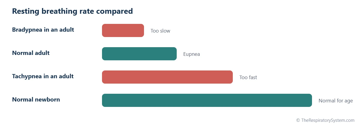
- Tachypnea: breathing faster than normal, more than about 20 breaths a minute in a resting adult, often with shallow breaths.
- Bradypnea: breathing slower than normal, fewer than about 12 breaths a minute in a resting adult.
- Apnea: a pause in breathing; in sleep studies a pause of 10 seconds or more counts as an apnea.
- Hyperpnea: deeper and often faster breathing that matches a higher demand, as in exercise.
- Hypopnea: abnormally shallow breathing, often during sleep.
Hyperventilation and hypoventilation sound similar but are defined by blood carbon dioxide rather than by appearance, as explained on our hyperventilation and hypoventilation page.
| Pattern | Rate and depth | Rhythm | Often linked with |
|---|---|---|---|
| Eupnea (normal) | About 12 to 20 breaths a minute in adults, even depth | Regular | Quiet breathing at rest |
| Tachypnea | Fast, often shallow | Regular | Fever, lung infection, anxiety, exertion |
| Bradypnea | Slow | Regular | Sedating drugs, sleep, some brain conditions |
| Apnea | No breaths for a period | Pauses | Sleep apnea, brainstem conditions |
| Hyperpnea | Deeper, often faster | Regular | Exercise, higher metabolic demand |
| Hypopnea | Shallow | Regular or interrupted | Sleep-related breathing problems |
| Cheyne-Stokes | Breaths deepen, then fade | Cyclic, with pauses | Heart failure, stroke, sleep at altitude, end of life |
| Kussmaul | Deep and labored, often fast | Regular | Metabolic acidosis such as diabetic ketoacidosis |
| Biot's | Quick breaths of similar depth | Clusters separated by pauses | Medulla damage, raised pressure in the skull, sedating drugs |
| Ataxic | Varying depth | Completely irregular | Serious injury to the medulla |
| Apneustic | Long, deep breaths in | Pause at full inspiration | Damage to the pons |
| Agonal | Slow gasps | Irregular | Cardiac arrest, extreme oxygen lack |
| Obstructive | Prolonged exhalation | Regular | Asthma, COPD |
| Paradoxical | Chest or abdomen moves inward on inhalation | Regular | Diaphragm weakness, broken ribs |
| Source: Standard clinical physiology descriptions, our editors | |||
What Is Cheyne-Stokes Breathing?
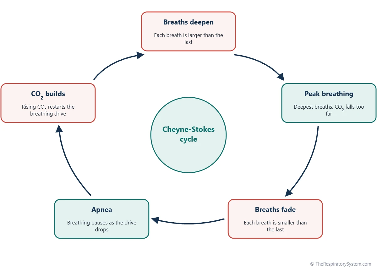
Cheyne-Stokes breathing is a cyclic pattern in which breaths grow steadily deeper and then steadily shallower, followed by a pause with no breathing at all, after which the cycle repeats. It is named after two physicians working in Dublin, the Scottish-born John Cheyne and the Irish William Stokes, who described it in the nineteenth century.
Why it happens
Cheyne-Stokes breathing is a fault in the feedback loop that controls breathing. When blood flow is slow, as in heart failure, the chemoreceptors learn about a change in blood carbon dioxide late, so the brainstem overcorrects: it drives breathing too hard, carbon dioxide falls too far, breathing stops, carbon dioxide builds up again and the cycle restarts. An oversensitive response to carbon dioxide makes the swings larger.
When it is seen
It is most often seen in people with heart failure, especially during sleep, and after a stroke or other brain injury. Healthy people can show a similar periodic breathing pattern while sleeping at high altitude, where low oxygen makes the control loop unstable. It is also one of the common patterns in the final days of life.
What Is Kussmaul Breathing and What Is Its Function?
Kussmaul breathing is deep, labored and regular breathing, often also fast, that the body uses to blow off carbon dioxide when the blood has become too acidic from a metabolic cause. Its function is compensation: removing carbon dioxide limits the fall in blood pH.
How it works
In metabolic acidosis, acids other than carbon dioxide build up in the blood. The most familiar example is diabetic ketoacidosis, in which ketone acids accumulate; kidney failure and some poisonings can do the same. The rising acid stimulates the chemoreceptors, and the brainstem responds with large, deep breaths that lower arterial carbon dioxide. Because carbon dioxide forms carbonic acid in the blood, breathing it off partly offsets the extra acid, as explained in our guide to respiratory system homeostasis.
Kussmaul vs Cheyne-Stokes breathing
The two are easy to tell apart. Kussmaul breathing is deep and even, with no pauses, and it is driven by blood chemistry. Cheyne-Stokes breathing waxes and wanes and includes pauses, and it reflects instability in the control of breathing. The pattern is named after Adolf Kussmaul, a German physician who described it in people with diabetes in the nineteenth century.
Breathing Patterns Linked to the Brain, Chest and Airways
Several other abnormal patterns point to the part of the body that has changed: irregular rhythms usually reflect the brainstem, while changes in effort or movement usually reflect the airways, chest wall or breathing muscles.
Patterns from the brainstem
- Biot's breathing: clusters of quick breaths of similar depth separated by irregular pauses, seen with damage to the medulla, raised pressure inside the skull or the effects of sedating drugs.
- Ataxic breathing: completely irregular breaths and pauses with no pattern at all, a sign of serious injury to the medulla.
- Apneustic breathing: long, gasping breaths in with a pause at full inspiration, linked to damage in the pons.
- Central neurogenic hyperventilation: sustained, deep and rapid breathing caused by injury to the upper brainstem.
- Agonal breathing: slow, irregular gasps during cardiac arrest or extreme oxygen lack; these gasps are a brainstem reflex and do not provide effective breathing.
Patterns from the chest and airways
- Obstructive breathing: a long, slow exhalation, often with wheezing, because narrowed airways empty slowly, as in asthma and COPD.
- Paradoxical breathing: part of the chest or abdomen moves inward during inhalation instead of outward, as with a weak or paralyzed diaphragm or a broken segment of the rib cage.
- Obstructive sleep apnea: repeated pauses during sleep while the chest keeps trying to breathe, because the throat collapses; see sleep apnea.
What Are the Breathing Patterns at End of Life?
Breathing patterns at end of life commonly include long pauses between breaths, Cheyne-Stokes cycles, very shallow or irregular breaths, and noisy breathing caused by secretions in the throat, followed in the final minutes by a few gasping breaths. These changes are a natural part of the dying process as the brainstem, circulation and muscles slow down.
As circulation weakens, the feedback loop that controls breathing becomes unstable, which produces waxing and waning cycles and pauses that can grow longer over hours or days. Muscles that normally clear the throat relax, so saliva and mucus collect and make a rattling sound with each breath, often called terminal respiratory secretions. The jaw may move with each breath as the breathing muscles weaken.
Hospice and palliative care teams describe these patterns to families because they can look distressing, even though they are an expected part of the body shutting down. Our cough reflex page explains why clearing the airways depends on muscle strength and reflexes that fade at this stage.
What Changes a Breathing Pattern?
A breathing pattern changes whenever the body's need for gas exchange changes, whenever the control centers in the brain are affected, or whenever the lungs, airways or chest wall make breathing harder. Many of these changes are completely normal.
- Exercise: breathing becomes deeper and faster to match the extra carbon dioxide made by working muscles, as described on our exercise and the respiratory system page.
- Sleep: breathing slows and becomes more regular in deep sleep and more variable in dreaming sleep.
- Emotion and speech: talking, singing, laughing and stress all reshape the pattern, through the brain's voluntary and emotional pathways.
- Altitude: low oxygen increases breathing and can cause periodic breathing during sleep; see breathing at high altitude.
- Reflexes: sighs, yawns, hiccups and sneezes briefly interrupt the normal rhythm.
- Disease and drugs: lung disease, heart failure, brain injury, acid-base problems and sedating drugs produce the abnormal patterns described above.
Because the same pattern can have very different causes, a noticeably changed breathing pattern is a reason to see a doctor. The Hering-Breuer reflex, triggered by lung stretch receptors, is one of the built-in brakes that normally keeps each breath from becoming too deep.
Key Facts
- A breathing pattern combines rate, depth, rhythm and effort; the normal resting pattern is called eupnea.
- A resting adult normally breathes about 12 to 20 times a minute, and children and newborns breathe faster, as shown on our respiratory rate page.
- In quiet breathing, inhalation is active and exhalation is passive, so exhalation usually lasts a little longer.
- Cheyne-Stokes breathing waxes and wanes with pauses and reflects an unstable breathing control loop, often in heart failure.
- Kussmaul breathing is deep, labored breathing that blows off carbon dioxide to offset metabolic acidosis, as in diabetic ketoacidosis.
- Biot's, ataxic and apneustic patterns point to damage in specific parts of the brainstem.
- Agonal gasps during cardiac arrest are a brainstem reflex, not effective breathing.
Frequently Asked Questions
What is the breathing pattern definition?
A breathing pattern is the overall way a person breathes, defined by four features: how many breaths are taken each minute, how deep each breath is, how regular the timing is and how much muscle effort is needed. Doctors and nurses describe patterns with names such as eupnea, tachypnea or Cheyne-Stokes breathing so that changes can be recorded clearly and compared over time.
What is the difference between Cheyne-Stokes and Kussmaul breathing?
Cheyne-Stokes breathing is cyclic: breaths grow deeper, then shallower, then stop for a while before the cycle repeats. It reflects an unstable control loop, often in heart failure or after a stroke. Kussmaul breathing is deep, labored and steady, with no pauses, and it is a response to acid in the blood from a metabolic cause such as diabetic ketoacidosis.
Is Cheyne-Stokes breathing always a sign of dying?
No. Cheyne-Stokes breathing is common near the end of life, but it also occurs in people with heart failure, especially during sleep, after strokes and other brain injuries, and in healthy people sleeping at high altitude. In those settings it reflects instability in the breathing control loop rather than the dying process, and it may come and go over months or years.
What is the difference between shallow breathing and normal breathing?
Normal breathing at rest moves about 500 milliliters of air per breath. Shallow breathing moves much less, so a larger share of each breath only fills the airways that take no part in gas exchange, called dead space. To keep the same alveolar ventilation, shallow breathing must be faster, which is why tachypnea and shallow breaths often appear together.
Why does breathing change during sleep?
During sleep the brain's wakeful drive to breathe disappears, chemoreceptors respond less strongly to carbon dioxide and the muscles that hold the throat open relax. Breathing therefore becomes slightly slower and shallower, and blood carbon dioxide rises by a few mmHg. In deep sleep breathing is very regular, while in dreaming (REM) sleep it becomes irregular, with variable rate and depth.
What is periodic breathing in babies?
Periodic breathing is a pattern seen in many healthy newborns, especially premature babies, in which a few seconds of regular breathing alternate with short pauses. It is linked to the immaturity of the brainstem breathing centers and usually fades during the first months of life. Pediatricians distinguish it from longer pauses, which are evaluated differently.
What is a sigh in breathing?
A sigh is a spontaneous breath about twice as deep as a normal one, often followed by a slightly longer exhalation. Healthy people sigh every few minutes without noticing. Sighs stretch the lungs and help reopen small groups of alveoli that slowly collapse during shallow breathing, so they are part of the normal pattern. Emotional sighs, of relief or sadness, use the same reflex.
How is a breathing pattern measured?
The simplest method is observation: counting breaths for a full minute while watching depth, rhythm and effort. In hospitals, chest movement sensors, capnography that tracks carbon dioxide in each breath and pulse oximetry add continuous data. Sleep studies record airflow, chest and abdominal movement, oxygen levels and brain activity overnight to detect pauses and patterns that occur only during sleep.
Keep Exploring


