At a Glance
Breathing is controlled by groups of neurons in the medulla oblongata and pons, called the respiratory center, which fire automatically and send signals through the phrenic and intercostal nerves to the diaphragm and rib muscles. The main chemical signal is the carbon dioxide level of the blood, normally held near 40 mmHg, while low oxygen becomes a strong extra stimulus mainly when arterial oxygen falls below about 60 mmHg.
- Main controller
- Respiratory center in the medulla oblongata and pons of the brainstem
- Rhythm generator
- Pre-Bötzinger complex in the ventral medulla
- Main chemical signal
- Arterial carbon dioxide (via pH), normally about 40 mmHg
- Sensors
- Central and peripheral chemoreceptors, lung stretch and irritant receptors, joint and muscle receptors
- Effectors
- Diaphragm (phrenic nerve, C3 to C5) and intercostal and accessory muscles
- Part of
- Homeostasis of blood gases and acid-base balance
On this page
- What Is Control of Breathing?
- How Is Breathing Controlled?
- What Is the Respiratory Center?
- What Is Neural Control of Breathing?
- What Is Chemical Control of Breathing?
- Can You Control Your Breathing Voluntarily?
- How Does Breathing Control Change During Sleep, Exercise and at Altitude?
- What Is Disordered Control of Breathing?
- Key Facts
- Quiz
- FAQ
What Is Control of Breathing?
Control of breathing is the way the nervous system decides when to breathe, how deeply and how fast, so that the air moved in and out of the lungs matches what the body needs at that moment. It runs automatically, day and night, without any conscious effort, yet it can also be overridden for a short time when we speak, sing, sniff or hold our breath.
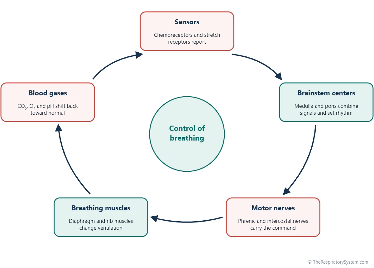
Physiologists describe it as a feedback system with three parts. Sensors measure the chemistry of the blood and the stretch of the lungs. A controller in the brainstem combines that information and produces a breathing rhythm. Effectors, the breathing muscles, carry out the command and change ventilation, which in turn changes the blood gases the sensors are measuring.
Why breathing needs to be controlled
Body cells use oxygen and produce carbon dioxide at rates that change from minute to minute. If ventilation were fixed, carbon dioxide would build up during activity and the blood would become acidic, while at rest the blood would be over-ventilated. The control system keeps arterial carbon dioxide close to about 40 mmHg and arterial oxygen close to about 95 to 100 mmHg, which also keeps blood pH near 7.4. This makes breathing one of the main tools of respiratory homeostasis.
Explained simply
Think of a thermostat. A thermometer checks the room, the thermostat compares it with the setting, and the heater turns up or down. In breathing, sensors check the carbon dioxide in your blood, your brainstem compares it with its setting, and your breathing muscles work harder or easier until the level is right again.
How Is Breathing Controlled?
Breathing is controlled by a loop in which sensors report the state of the blood and lungs to the brainstem, the brainstem sends rhythmic signals to the breathing muscles, and the resulting change in ventilation feeds back on the sensors. The loop runs continuously, adjusting each breath to the one before.
- Sensing. Respiratory chemoreceptors in the brainstem, the carotid bodies and the aortic bodies track carbon dioxide, hydrogen ions and oxygen. Stretch receptors in the airways report how full the lungs are.
- Integrating. Neurons in the medulla and pons combine these inputs with signals from higher brain areas, such as the cortex during speech or the hypothalamus during fever.
- Generating a rhythm. A small network in the medulla fires in repeating bursts that set the basic pace of breathing.
- Sending the command. Motor neurons in the spinal cord pass the signal along the phrenic nerve to the diaphragm and along the intercostal nerves to the intercostal muscles.
- Moving air. The muscles expand the chest, air flows in, and then the muscles relax for exhalation. The physical side of this step is covered on our mechanics of breathing page.
- Feedback. Fresh air changes the gas levels in the alveoli and then in the arterial blood, and the sensors report the new values.
The controller can change two things: the depth of each breath, measured as tidal volume, and the number of breaths per minute, the respiratory rate. Their product is minute ventilation, about 6 liters per minute in a resting adult.
What Is the Respiratory Center?
The respiratory center is the set of neuron groups in the medulla oblongata and pons of the brainstem that generates the breathing rhythm and adjusts it to the body's needs. It is not a single lump of tissue but several paired clusters on both sides of the brainstem that work as one network.
Medullary respiratory centers
- Dorsal respiratory group (DRG): lies in the dorsal medulla, near the nucleus of the solitary tract. It receives sensory input from the vagus and glossopharyngeal nerves, including signals from the peripheral chemoreceptors and lung stretch receptors, and drives mainly inspiration.
- Ventral respiratory group (VRG): a long column of neurons in the ventral medulla. It contains both inspiratory and expiratory neurons and becomes important for active, forceful breathing out, as during exercise or coughing.
- Pre-Bötzinger complex: a small cluster within the ventral respiratory group, widely regarded as the main generator of the basic breathing rhythm. Its neurons can produce rhythmic bursts on their own.
Pontine respiratory centers
The pontine respiratory group, once called the pneumotaxic center, sits in the upper pons and helps switch inspiration off, fine-tuning the rate and smoothing the change from breathing in to breathing out. An apneustic area in the lower pons has been described in animal studies as promoting long inspirations, though its exact role in humans is less certain. Together, the pons and medulla turn a raw rhythm into a smooth, adaptable pattern.
The table below lists the main parts of the control system and what each one does.
| Part | Location | Type | Main role |
|---|---|---|---|
| Dorsal respiratory group | Dorsal medulla, near the nucleus of the solitary tract | Controller | Receives vagal and glossopharyngeal input; drives inspiration |
| Ventral respiratory group | Ventral medulla | Controller | Inspiratory and expiratory neurons; active, forceful breathing |
| Pre-Bötzinger complex | Within the ventral respiratory group | Controller | Main generator of the basic breathing rhythm |
| Pontine respiratory group | Upper pons | Controller | Helps end inspiration; fine-tunes rate and pattern |
| Motor cortex | Frontal lobe of the cerebrum | Controller | Voluntary control for speech, singing and breath-holding |
| Central chemoreceptors | Near the ventral surface of the medulla | Sensor | Respond to hydrogen ions in brain fluid produced from carbon dioxide |
| Carotid bodies | Fork of each common carotid artery | Sensor | Respond to low oxygen, high carbon dioxide and acid; glossopharyngeal nerve |
| Aortic bodies | Near the arch of the aorta | Sensor | Respond mainly to low oxygen; vagus nerve |
| Pulmonary stretch receptors | Smooth muscle of the airways | Sensor | Signal lung inflation; Hering-Breuer reflex |
| Irritant and J receptors | Airway lining and alveolar walls | Sensor | Respond to irritants and lung congestion; cough and rapid shallow breathing |
| Diaphragm | Floor of the thoracic cavity | Effector | Main muscle of inspiration; supplied by the phrenic nerve (C3 to C5) |
| Intercostal and accessory muscles | Between the ribs, neck and abdomen | Effector | Raise or lower the ribs; active exhalation and deep breathing |
| Source: Standard physiology, our editors | |||
What Is Neural Control of Breathing?
Neural control of breathing is the part of the system made of nerve signals: the rhythm generated in the brainstem, the motor nerves that carry it to the muscles, and the reflexes from the lungs, airways and body that adjust it. It sets the basic pattern even before any chemical change occurs.
The breathing rhythm
During quiet breathing, inspiratory neurons fire with a gradually rising signal for about two seconds, so the diaphragm contracts smoothly rather than in a jerk. The signal then stops, the muscles relax, and the elastic recoil of the lungs and chest wall pushes air out without any active effort. Exhalation becomes active only when ventilation must rise, when expiratory neurons recruit the internal intercostals and abdominal muscles.
Motor pathways
The diaphragm is supplied by the phrenic nerve, which arises from spinal segments C3 to C5 in the neck. The intercostal muscles are supplied by the intercostal nerves from the thoracic spinal cord, and accessory muscles in the neck join in during heavy breathing, as described on our muscles of respiration page. Because the phrenic nerve leaves the cord high in the neck, an injury below about C5 usually spares the diaphragm.
Reflexes from the lungs and body
- Pulmonary stretch receptors in the airway smooth muscle signal through the vagus nerve when the lungs inflate. The Hering-Breuer reflex uses this signal to end inspiration, which matters most in infants and during large breaths in adults.
- Irritant receptors in the airway lining respond to dust, smoke and cold air and can trigger the cough reflex, airway narrowing or rapid shallow breathing.
- J receptors (juxtacapillary receptors) lie in the alveolar walls near the capillaries and respond to fluid or congestion in the lung tissue, producing rapid, shallow breathing.
- Proprioceptors in muscles and joints signal movement, which helps raise breathing as soon as exercise begins.
What Is Chemical Control of Breathing?
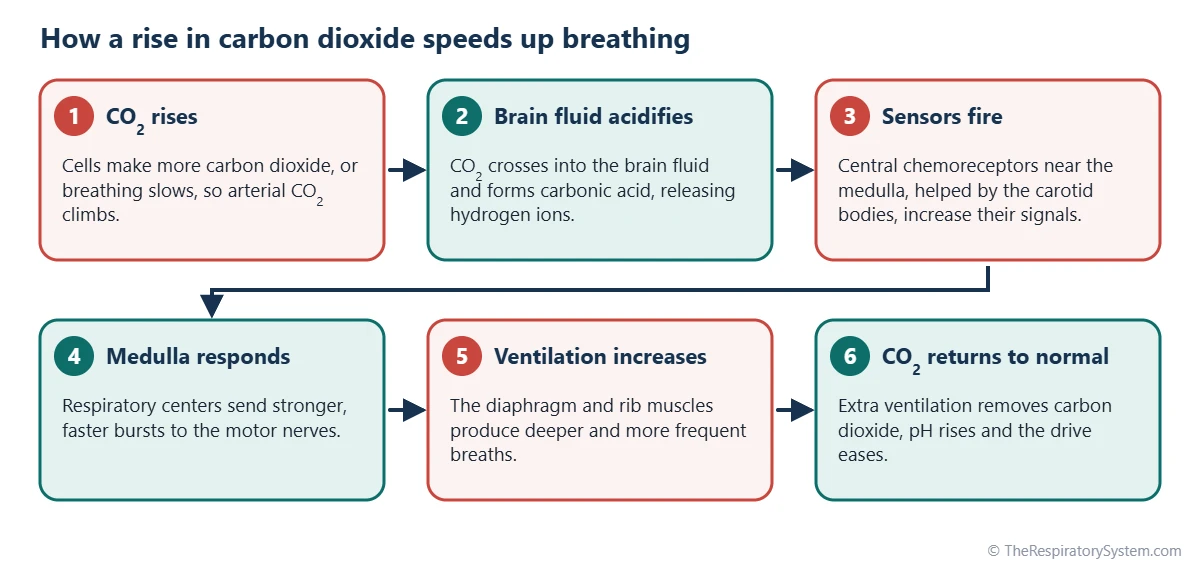
Chemical control of breathing is the adjustment of ventilation to the levels of carbon dioxide, hydrogen ions and oxygen in the blood and brain fluid, sensed by chemoreceptors. Of the three, carbon dioxide is by far the strongest everyday driver: a small rise in arterial carbon dioxide causes a large rise in ventilation.
Carbon dioxide and pH
Central chemoreceptors lie near the ventral surface of the medulla, bathed in cerebrospinal fluid. Carbon dioxide crosses the blood-brain barrier easily and combines with water to form carbonic acid, which releases hydrogen ions. The receptors respond to those hydrogen ions, so a rise in blood carbon dioxide quickly becomes a more acidic brain fluid and a stronger drive to breathe. Hydrogen ions from the blood itself cross the barrier poorly, which is why carbon dioxide acts faster than other acids on these receptors. The chemistry of carbon dioxide in the blood is described on our carbon dioxide transport page.
Oxygen
Peripheral chemoreceptors in the carotid bodies, at the fork of each common carotid artery, and in the aortic bodies, near the arch of the aorta, are the only sensors that respond directly to low oxygen. Their signal stays modest until arterial oxygen falls below about 60 mmHg, then rises steeply. This fits the oxygen-hemoglobin dissociation curve, because hemoglobin stays almost fully loaded until oxygen pressure falls into that range. The carotid bodies report through the glossopharyngeal nerve and the aortic bodies through the vagus nerve.
How the signals combine
The chemical signals add to each other. Low oxygen makes the response to carbon dioxide stronger, and acid from exercising muscles adds a further push through the carotid bodies. Our page on respiratory chemoreceptors explains how each sensor works in detail, and partial pressure of gases explains the units used here.
Can You Control Your Breathing Voluntarily?
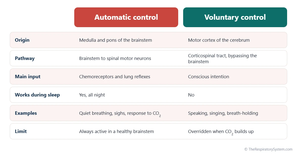
Yes, breathing can be controlled voluntarily for short periods, because the motor cortex sends its own signals down the spinal cord to the breathing muscles, bypassing the automatic centers in the brainstem. This is what allows speech, singing, laughing, sniffing and breath-holding.
Voluntary control has limits. During a breath-hold, carbon dioxide builds up and oxygen falls, and the chemical drive grows until the automatic system takes over and breathing restarts, a moment called the breaking point. That is why a healthy person cannot simply decide to stop breathing for good. Our page on how long you can hold your breath looks at the factors that set this point.
Speech and other behaviors
Speaking requires a long, controlled exhalation through the narrowed larynx, so the cortex lengthens expiration and shortens inspiration while we talk; our page on how the voice is produced covers the vocal side. Swallowing briefly stops breathing so food does not enter the airway. Reflex patterns such as yawning, sighing, hiccups and sneezing are generated by the brainstem as well; see why we yawn and why we hiccup.
Emotions and the limbic system
Fear, anxiety, excitement and pain speed up breathing through the limbic system and hypothalamus, which project to the brainstem centers. Body temperature has a similar effect: fever raises breathing rate. These inputs explain why breathing can change sharply even when blood gases are normal, which can lead to the over-breathing described on our hyperventilation and hypoventilation page.
How Does Breathing Control Change During Sleep, Exercise and at Altitude?
The control system shifts its settings with the body's state: it relaxes during sleep, rises sharply with exercise and gradually resets at high altitude. In each case the aim is the same, keeping blood gases within a safe range.
During sleep
In sleep the wakefulness drive to breathe disappears, the sensitivity to carbon dioxide falls and ventilation drops slightly, so arterial carbon dioxide rises by a few mmHg. Breathing is most regular in deep non-REM sleep and least regular in REM sleep, when the rib muscles lose much of their tone and the diaphragm does most of the work. The muscles that hold the upper airway open also relax, which is the background to sleep apnea.
During exercise
Ventilation rises within seconds of starting exercise, before blood gases have had time to change. This fast rise comes from signals sent by the motor cortex alongside the commands to the limbs and from proprioceptors in the moving joints. A slower rise follows as the body settles into the work, and in moderate exercise arterial oxygen and carbon dioxide stay close to their resting values. During very hard exercise, acid from the muscles drives breathing even higher; see how exercise affects the respiratory system.
At high altitude
At altitude the air holds less oxygen, so the carotid bodies sense low oxygen and drive faster, deeper breathing. This lowers blood carbon dioxide, which at first restrains breathing, but over several days the kidneys adjust bicarbonate and the controller resets, allowing ventilation to stay higher. This process, called ventilatory acclimatization, is described on our breathing at high altitude page.
What Is Disordered Control of Breathing?
Disordered control of breathing is a problem in which the lungs and airways may be normal but the drive or rhythm of breathing is abnormal, so ventilation is irregular, too little or too much. Pediatric emergency teaching uses it as one of four broad categories of respiratory problems, alongside upper airway obstruction, lower airway obstruction and lung tissue disease. Our respiratory diseases section covers lung conditions in more depth, and anyone with breathing symptoms should see a doctor.
- Brain injury and raised pressure inside the skull: damage to or pressure on the brainstem can produce irregular or very slow breathing.
- Depressant drugs: sedating drugs and alcohol reduce the activity of the respiratory centers and their response to carbon dioxide, which can cause dangerous hypoventilation.
- Central sleep apnea: the brainstem briefly stops sending signals to the breathing muscles during sleep, unlike obstructive apnea, where the airway closes.
- Cheyne-Stokes breathing: a cycle of gradually deeper breaths followed by gradually shallower ones and a pause, often linked to heart failure or stroke; see breathing patterns.
- Congenital central hypoventilation syndrome: a rare genetic condition in which the automatic response to carbon dioxide is weak or absent, so breathing becomes inadequate, especially during sleep.
- Neuromuscular conditions: diseases of the nerves or muscles weaken the effectors, so the command from the brainstem is not carried out fully.
- Chronic carbon dioxide retention: in some people with severe COPD, the response to carbon dioxide is blunted and low oxygen becomes a larger part of the drive to breathe.
When ventilation falls short, oxygen in the blood drops, a state described on our hypoxia and hypoxemia page, and carbon dioxide rises.
Key Facts
- Breathing is controlled automatically by the respiratory center in the medulla oblongata and pons of the brainstem.
- The pre-Bötzinger complex in the ventral medulla is regarded as the main generator of the breathing rhythm.
- Carbon dioxide, sensed as hydrogen ions by central chemoreceptors, is the strongest minute-to-minute driver of breathing.
- Low oxygen strongly stimulates breathing mainly when arterial oxygen falls below about 60 mmHg, sensed by the carotid and aortic bodies.
- The phrenic nerve, from spinal segments C3 to C5, carries the breathing command to the diaphragm.
- Quiet exhalation is passive; expiratory muscles are recruited only when ventilation must rise.
- The motor cortex can override automatic breathing for speech or breath-holding, but rising carbon dioxide eventually restarts breathing.
Quick Quiz: Test What You Know About the Control of Breathing
6 questions. Tap an answer to check it.
Where is the main respiratory center located?
The respiratory center consists of neuron groups in the medulla oblongata and pons of the brainstem.
Which chemical is the strongest everyday stimulus for breathing?
A small rise in arterial carbon dioxide causes a large rise in ventilation, mainly through central chemoreceptors.
Central chemoreceptors respond most directly to which change?
Carbon dioxide enters the brain fluid and forms carbonic acid; the receptors respond to the hydrogen ions released.
Which nerve carries the breathing command to the diaphragm?
The phrenic nerve, from spinal segments C3 to C5, supplies the diaphragm.
Which group of neurons is regarded as the main generator of the breathing rhythm?
The pre-Bötzinger complex, within the ventral respiratory group of the medulla, produces the basic rhythm.
Which reflex ends inspiration when the lungs are well inflated?
Stretch receptors in the airways signal through the vagus nerve to end inspiration, the Hering-Breuer reflex.
Frequently Asked Questions
What part of the brain controls breathing?
The brainstem controls breathing, specifically the medulla oblongata and the pons, which sit at the base of the brain where it joins the spinal cord. The medulla generates the rhythm and drives inspiration, while the pons smooths the pattern and helps switch between breathing in and out. The cerebral cortex can take over briefly for speech or a breath-hold, and emotions act through the limbic system and hypothalamus.
Is breathing voluntary or involuntary?
Breathing is both. Most of the time it is involuntary, run by the brainstem without any thought, including throughout sleep. It can also be controlled voluntarily, because the motor cortex has its own pathway to the breathing muscles. Voluntary control is temporary: when carbon dioxide builds up during a breath-hold, the automatic system overrides the conscious one and breathing restarts.
Why is carbon dioxide more important than oxygen in controlling breathing?
Carbon dioxide changes the acidity of the blood and brain fluid within seconds, and the central chemoreceptors are very sensitive to that acidity, so even a rise of a few mmHg raises ventilation clearly. Oxygen, by contrast, barely affects breathing until it falls quite low, because hemoglobin stays almost fully loaded across a wide range of oxygen pressures. Holding carbon dioxide steady also keeps blood pH steady.
What happens if the respiratory center is damaged?
Damage to the respiratory center in the brainstem, from injury, stroke, swelling or pressure, can make breathing irregular, very slow or absent, because the rhythm and drive come from there. Depending on the level and extent of the damage, a person may show abnormal breathing patterns or need a machine to breathe for them. Injuries to the spinal cord above the phrenic nerve roots can also stop the diaphragm.
What is the difference between central and peripheral chemoreceptors?
Central chemoreceptors sit near the surface of the medulla and respond to hydrogen ions in the cerebrospinal fluid, which mostly reflect carbon dioxide. Peripheral chemoreceptors sit in the carotid and aortic bodies and respond to low arterial oxygen, high carbon dioxide and acid in the blood. Central receptors provide most of the carbon dioxide drive, while peripheral receptors are the only ones that sense low oxygen.
Why do we breathe faster when we exercise?
Breathing speeds up at the start of exercise through nerve signals from the motor cortex and from receptors in moving joints and muscles, even before blood gases change. As exercise continues, the respiratory center keeps ventilation matched to the extra carbon dioxide being produced. During very hard work, acid released by the muscles stimulates the carotid bodies and pushes ventilation higher still.
Does breathing change when you sleep?
Yes. During sleep the alerting drive from the waking brain disappears, so ventilation falls slightly and arterial carbon dioxide rises by a few mmHg. Breathing is steadiest in deep non-REM sleep and more irregular in REM sleep, when most rib muscles relax and the diaphragm does the work. The throat muscles also relax, which makes the upper airway narrower than during the day.
What does the pneumotaxic center do?
The pneumotaxic center, now usually called the pontine respiratory group, lies in the upper pons. It helps end each inspiration and so influences how long each breath lasts and how fast breathing is. When its influence is strong, breaths become shorter and more frequent. It works together with the medullary centers and with stretch signals from the lungs to produce a smooth breathing pattern.
Keep Exploring


