At a Glance
The pleura is a double serous membrane around each lung. The visceral pleura sticks to the lung surface, the parietal pleura lines the chest wall, diaphragm and mediastinum, and the two layers meet at the root of the lung. A thin film of pleural fluid between them lubricates breathing movements and holds the lung against the chest wall so it expands with the chest.
- Location
- Around each lung, lining the inside of the thoracic cavity
- Layers
- Visceral pleura on the lung; parietal pleura on the chest wall, diaphragm and mediastinum
- Tissue
- Serous membrane: mesothelium (simple squamous cells) on thin connective tissue
- Function
- Lubricates lung movement and couples the lungs to the chest wall
- Connects to
- The two layers join at the hilum, the root of the lung
- Part of
- Lower respiratory tract; encloses the pleural cavity
On this page
What Is the Pleura?
The pleura is the serous membrane that wraps each lung and lines the chest cavity around it, forming a closed sac with two layers. The inner layer, the visceral pleura, is fixed to the surface of the lung, while the outer layer, the parietal pleura, is attached to the inner surface of the rib cage, the diaphragm and the side of the mediastinum. The two layers are one continuous sheet that folds back on itself at the hilum of the lung, where the bronchi and vessels enter.
The narrow space between the layers is the pleural cavity, which normally holds only a thin film of pleural fluid. Each lung has its own pleural sac, so the right and left sides are completely separate. Anatomy courses group the pleura with the other organs of the lower respiratory tract because breathing depends on it.
Lung vs pleura
The lung is the organ that exchanges gases, made of airways, alveoli and blood vessels. The pleura is only its wrapping: a membrane a fraction of a millimeter thick that covers the lung and lines the space it sits in. A disease of the pleura, such as an effusion, affects the space around the lung rather than the lung tissue itself.
Explained simply
Push your fist into a soft, partly filled balloon. The part of the balloon touching your fist is like the visceral pleura, the outer part is like the parietal pleura, and the tiny gap of air in between is like the pleural cavity. In the body, that gap holds a few drops of slippery fluid instead of air.
Where Is the Pleura Located?
The pleura lines both halves of the thoracic cavity: the visceral pleura covers every surface of the lung, and the parietal pleura covers the inside of the chest wall, the upper surface of the diaphragm and the side of the mediastinum. The parietal pleura is named after the surface it lines, as shown in the table below.
Parietal pleura location
- Costal pleura: lines the inner surfaces of the ribs, costal cartilages, intercostal muscles and sternum.
- Diaphragmatic pleura: covers the upper, chest-facing surface of the diaphragm.
- Mediastinal pleura: covers the side of the mediastinum and turns outward at the root of the lung to become the visceral pleura.
- Cervical pleura (cupula): the dome that rises over the apex of the lung into the root of the neck, a few centimeters above the inner part of the clavicle.
Pleural recesses
The parietal pleura reaches lower than the lungs, so in quiet breathing parts of the pleural sac contain no lung, only two layers of parietal pleura facing each other. The largest of these gaps is the costodiaphragmatic recess, where the costal pleura meets the diaphragmatic pleura around the lower edge of the chest; the smaller costomediastinal recess lies behind the sternum. In a typical adult, the lower border of the lung crosses the 6th rib at the midclavicular line, the 8th at the midaxillary line and the 10th near the spine, while the pleura reaches about two ribs lower at each line.
| Part | Where it lies | Nerve supply | Pain sensitive? |
|---|---|---|---|
| Visceral pleura | On the whole surface of each lung and inside its fissures | Autonomic fibers of the pulmonary plexus | No, senses stretch only |
| Costal pleura | Inner surface of the ribs, intercostal muscles and sternum | Intercostal nerves | Yes, felt in the chest wall |
| Diaphragmatic pleura, outer rim | Edges of the upper surface of the diaphragm | Lower intercostal nerves | Yes, felt in the lower chest wall |
| Diaphragmatic pleura, central part | Middle of the upper surface of the diaphragm | Phrenic nerve | Yes, often felt in the shoulder |
| Mediastinal pleura | Side wall of the mediastinum | Phrenic nerve | Yes, often felt in the neck or shoulder |
| Cervical pleura (cupula) | Dome over the lung apex, rising into the root of the neck | Intercostal nerves | Yes |
| Pulmonary ligament | Double fold of pleura hanging below the root of the lung | Continuous with the mediastinal pleura | Shares the mediastinal supply |
| Costodiaphragmatic recess | Lowest gutter of the sac, between costal and diaphragmatic pleura | Intercostal nerves | Yes |
| Costomediastinal recess | Behind the sternum, between costal and mediastinal pleura | Intercostal and phrenic nerves | Yes |
| Source: Typical adult anatomy, our editors | |||
What Is the Pleura Made Of?
The pleura is made of a single layer of flat mesothelial cells resting on a thin bed of connective tissue that contains collagen and elastic fibers, small blood vessels, lymphatic vessels and nerves. The mesothelial cells carry tiny microvilli on their surface, which help hold a slippery coat of fluid and glycoproteins.
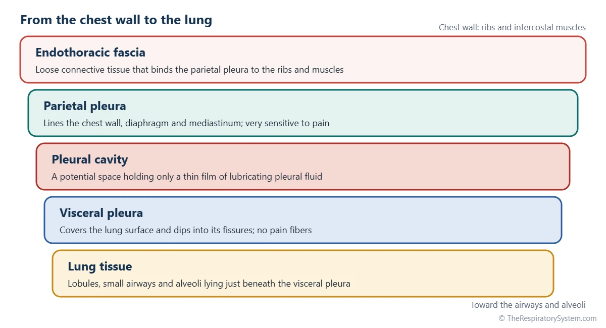
Both layers share this plan, but they differ in what lies underneath. The visceral pleura is bound tightly to the lung and blends with the connective tissue that separates the pulmonary lobules, so it cannot be peeled away easily. The parietal pleura is attached to the chest wall through a layer of loose connective tissue called the endothoracic fascia, which lets surgeons separate it from the ribs.
How the pleura develops
The pleura forms early in the embryo, when the growing lung buds push into the spaces on each side of the developing heart, which become the pleural cavities. The tissue covering the lung bud, the splanchnic mesoderm, becomes the visceral pleura, while the somatic mesoderm lining the body wall becomes the parietal pleura. Thin folds of tissue later seal each pleural cavity off from the heart's cavity and from the abdomen, as described in development of the respiratory system.
What Is the Visceral Pleura Function?
The visceral pleura gives each lung a smooth, slippery outer surface, so the lung can glide against the chest wall and its lobes can slide against each other as they expand and shrink. It follows every curve of the lung and dips deep into the lung fissures, so each of the lung lobes has its own covering.
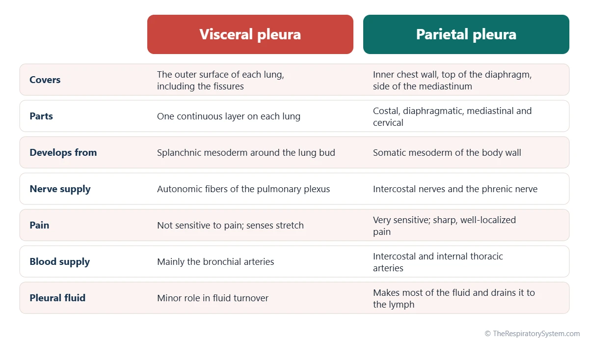
The elastic fibers in its connective tissue add a little to the natural recoil that helps the lung shrink during exhalation. Its blood comes mainly from the bronchial arteries, and part of its venous blood drains into the pulmonary veins. The visceral pleura has no pain fibers: its nerves come from the autonomic pulmonary plexus and respond to stretch, which is why disease confined to the lung surface is often painless.
Visceral pleura function simple version
The visceral pleura is the lung's own slick skin. It keeps the lung smooth, lets it slide without rubbing and helps it spring back after each breath in.
What Is the Parietal Pleura Function?
The parietal pleura lines the walls of each pleural cavity, produces and drains most of the pleural fluid and acts as a pain-sensing alarm for the chest. Its smooth inner surface faces the visceral pleura across the thin fluid film, so the lung slides along it with almost no friction.
Small capillaries from the chest wall arteries, such as the intercostal and internal thoracic arteries, filter a little fluid into the pleural cavity. Tiny openings in the parietal pleura called stomata, found mainly over the diaphragm and the lower chest, lead into lymphatic vessels that carry the fluid away again. This steady turnover keeps the film thin; the details belong to our pleural cavity and pleural fluid page.
Why the parietal pleura hurts
The parietal pleura is supplied by somatic nerves, so it is very sensitive to pain, which is usually sharp and well localized. The costal pleura and the outer rim of the diaphragmatic pleura are supplied by the intercostal nerves, so their pain is felt in the chest wall. The mediastinal pleura and the central diaphragmatic pleura are supplied by the phrenic nerve, so their pain is often referred to the neck and the tip of the shoulder.
Parietal pleura function simple version
The parietal pleura is the lining of the room the lung lives in. It makes the slippery fluid, drains the extra away and sends a pain signal when something irritates it.
What Is the Pleural Membrane Function in the Respiratory System?
In the respiratory system, the pleural membranes link each lung to the chest wall so the lungs inflate when the chest expands, while letting them move without friction and keeping each lung in its own sealed compartment. Without the pleura, the lungs would not follow the movements of the ribs and diaphragm.
- Coupling: the thin fluid film works like a drop of water between two panes of glass; the layers slide easily but resist being pulled apart, so the lung stays pressed against the chest wall.
- Lubrication: the fluid lets the lung slide over the ribs and diaphragm thousands of times a day without wear.
- Pressure: because the lungs tend to recoil inward and the chest wall tends to spring outward, the pressure in the pleural space stays slightly below atmospheric pressure, which keeps the lungs expanded.
- Separation: each lung has its own sac, so air, fluid or infection on one side does not spread directly to the other.
When the intercostal muscles and diaphragm enlarge the chest, the parietal pleura moves outward and pulls the visceral pleura and the lung with it. The steps of this process are covered on our mechanics of breathing page.
Common Conditions That Affect the Pleura
Conditions of the pleura include inflammation, the build-up of fluid, air or blood in the pleural space, infection and tumors, and many of them cause chest pain or breathlessness. Our respiratory diseases section describes them in more depth, and anyone with these symptoms should see a doctor.
- Pleurisy (pleuritis): inflammation of the pleura, often after a viral infection, causing sharp pain that worsens with breathing as the rough layers rub together.
- Pleural effusion: too much fluid in the pleural cavity, which can squeeze the lung beneath it.
- Pneumothorax: air entering the pleural space, which breaks the seal between the layers and lets part or all of the lung collapse.
- Hemothorax and empyema: blood or pus collecting in the pleural space, after chest injury or a pneumonia.
- Pleural plaques and mesothelioma: thickened patches and a rare cancer of the mesothelium, both strongly linked to past asbestos exposure.
Key Facts
- The pleura is a serous membrane with two layers: the visceral pleura on the lung and the parietal pleura on the chest wall, diaphragm and mediastinum.
- The two layers are continuous with each other at the hilum, the root of the lung, and below it they form the pulmonary ligament.
- The parietal pleura has four named parts: costal, diaphragmatic, mediastinal and cervical (the cupula).
- The parietal pleura is sensitive to pain through the intercostal and phrenic nerves, while the visceral pleura has no pain fibers.
- A thin film of fluid in the pleural cavity lets the layers slide but holds the lung against the chest wall.
- The right and left pleural sacs are completely separate, so a pneumothorax on one side usually leaves the other lung working.
- The pleura extends about two ribs lower than the lower border of the lung, forming the costodiaphragmatic recess.
Quick Quiz: Test What You Know About the Pleura
6 questions. Tap an answer to check it.
Which layer of the pleura covers the surface of the lung?
The visceral pleura is fixed to the lung surface; the parietal pleura lines the chest wall, diaphragm and mediastinum.
Where do the visceral and parietal pleura meet?
The two layers are one continuous sheet that folds back at the hilum, where the bronchi and vessels enter the lung.
Which part of the pleura is sensitive to pain?
The parietal pleura is supplied by somatic intercostal and phrenic nerves, so it feels sharp pain; the visceral pleura does not.
What is the dome of parietal pleura above the lung apex called?
The cervical pleura, or cupula, rises over the apex of the lung into the root of the neck.
What type of cells forms the surface of the pleura?
The pleura is a serous membrane lined by a single layer of flat mesothelial cells.
Pain from the central diaphragmatic pleura is often felt in which area?
The central diaphragmatic pleura is supplied by the phrenic nerve, so its pain is often referred to the neck and shoulder tip.
Frequently Asked Questions
Is the pleura part of the lung?
The visceral pleura is attached so closely to the lung that it is usually counted as the lung's outer covering, while the parietal pleura belongs to the chest wall. Together they form a separate membrane system rather than lung tissue. The lung itself is made of airways, alveoli and blood vessels; the pleura only wraps it and lines the cavity it sits in.
How thick is the pleura?
The pleura is extremely thin, much less than a millimeter in healthy adults, which is why normal pleura is barely visible on a chest X-ray. Most of its thickness comes from the connective tissue beneath the single layer of mesothelial cells. The pleura becomes visibly thick only when it is inflamed, scarred or affected by conditions such as pleural plaques.
Why does pleurisy hurt when you breathe?
Pleurisy hurts with breathing because the inflamed layers lose their smooth, slippery surface and rub against each other as the lung moves. The parietal pleura is supplied by pain-sensing somatic nerves, so this rubbing causes sharp, stabbing pain that is worse on a deep breath, a cough or a sneeze. A doctor may hear the rubbing with a stethoscope as a pleural friction rub.
What is the difference between the pleura and the pleural cavity?
The pleura is the membrane itself, made of the visceral and parietal layers. The pleural cavity is the thin space enclosed between those two layers, which normally holds only a small amount of lubricating pleural fluid. In other words, the pleura forms the walls of a sealed sac and the pleural cavity is the inside of that sac.
Do both lungs share one pleural sac?
No. Each lung has its own pleural sac, and the right and left pleural cavities are separated by the mediastinum, the central compartment holding the heart. Because of this separation, air or fluid that collects on one side, as in a pneumothorax or an effusion, usually stays on that side, and the other lung can keep working normally.
What is the pulmonary ligament?
The pulmonary ligament is a thin double fold of pleura hanging down from the root of each lung toward the diaphragm. It forms where the mediastinal pleura wraps around the root and the front and back layers come together below it. Despite its name it is not a strong ligament; it gives the root room to move as the lung expands.
What is the endothoracic fascia?
The endothoracic fascia is a thin layer of loose connective tissue between the inner surface of the chest wall and the costal pleura. It works like a glue layer that holds the parietal pleura to the ribs and intercostal muscles. Above the lung apex it thickens into a firmer sheet, the suprapleural membrane, which helps protect the dome of the pleura at the root of the neck.


