At a Glance
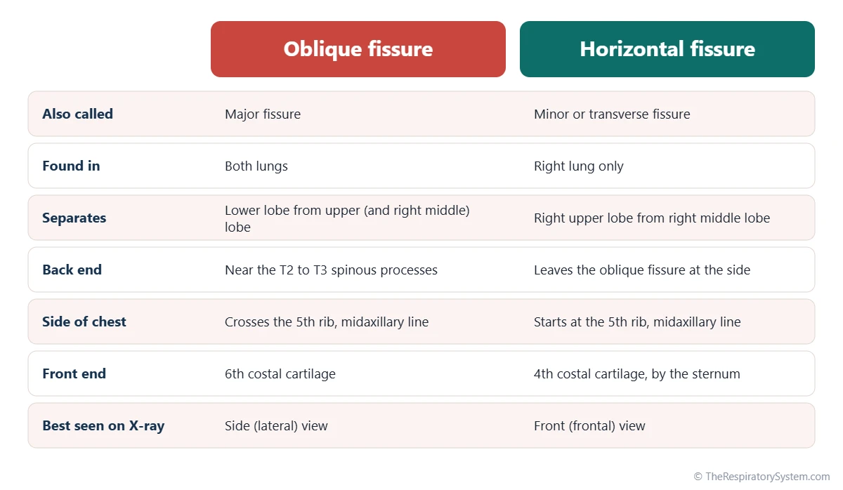
The lung fissures are pleura-lined grooves that cut deep into the lungs and separate the lobes. The oblique fissure, present in both lungs, runs from about the level of the T2 to T3 spinous processes at the back, down and forward to the 6th costal cartilage at the front. The horizontal fissure, found only in the right lung, follows the 4th rib and costal cartilage and separates the upper lobe from the middle lobe.
- Definition
- Deep clefts in the lung, lined by visceral pleura, that separate the lobes
- Number
- Right lung: 2 (oblique and horizontal); left lung: 1 (oblique)
- Lining
- Two layers of visceral pleura facing each other, with a thin film of pleural fluid
- Oblique fissure
- T2 to T3 level at the back to the 6th costal cartilage at the front
- Horizontal fissure
- Along the 4th rib and costal cartilage, right lung only
- Part of
- The lungs and pleura; they define the lung lobes
On this page
What Are the Fissures of the Lungs?
The fissures of the lungs are deep clefts that cut from the outer surface of each lung inward toward its root and divide the lung into lobes: the oblique and horizontal fissures in the right lung, and the oblique fissure alone in the left. Each lobe has its own lobar bronchus and blood vessels, as described on our lung lobes page; this page covers the clefts between them.
Lung fissure definition
A lung fissure is a narrow space between two neighboring lobes, lined on both sides by the visceral layer of the pleura, the thin membrane that covers the surface of the lung. The visceral pleura dips into the cleft, covers the facing surfaces of the two lobes, called the interlobar surfaces, and turns back out again. The parietal pleura, which lines the chest wall, does not enter the fissures.
How deep are the fissures?
A complete fissure cuts all the way through the lung tissue to the hilum, so the two lobes touch only where their bronchi and vessels enter. The narrow gap inside the fissure is part of the pleural cavity and holds a thin film of pleural fluid, just like the space between the lung and the chest wall.
Explained simply
Picture a loaf of bread sliced almost to the bottom crust. The slices are the lobes, the cuts between them are the fissures, and the bottom crust that still holds the slices together is the hilum, where the airways and blood vessels enter the lung.
Where Is the Oblique Fissure of the Lung?
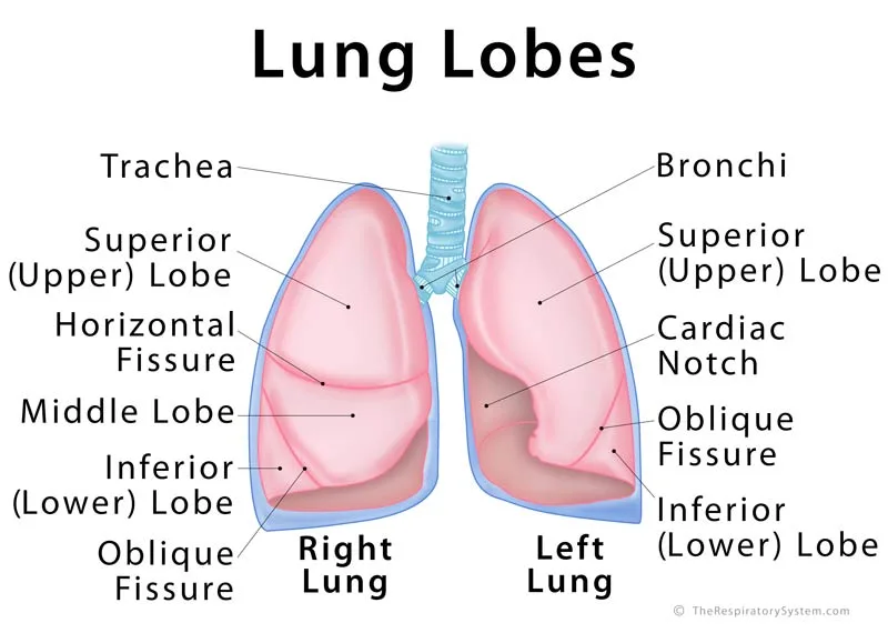
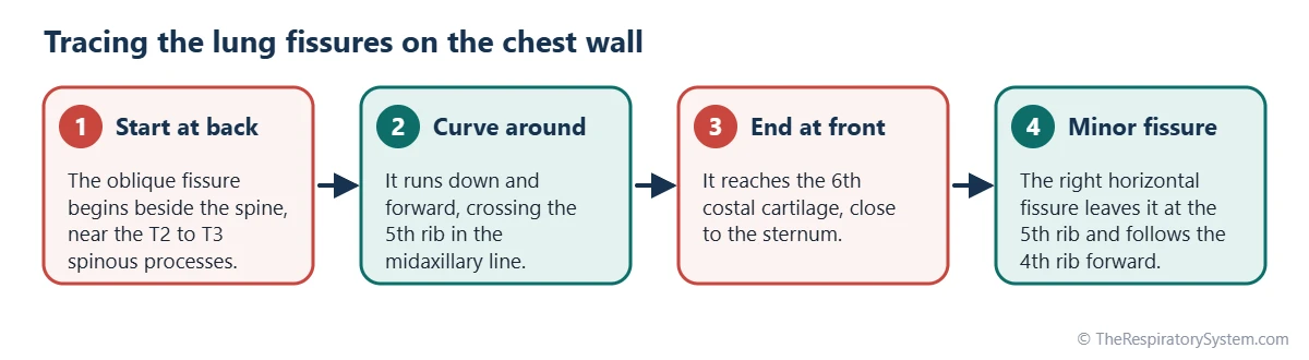
The oblique fissure runs around each lung from the back, near the spine at about the level of the T2 to T3 spinous processes, downward and forward around the side of the chest, to end at the front near the 6th costal cartilage. It is also called the major fissure, and it is the only fissure the left lung normally has.
On the lung itself, the oblique fissure begins on the medial surface just above and behind the hilum, climbs to the posterior border, and then sweeps down and forward across the outer, costal surface. It reaches the lower border near the front of the lung and turns back onto the medial surface toward the lower part of the hilum. Because of this slanted path, the lower lobe lies mostly at the back of the chest and the upper lobe mostly at the front.
- Right oblique fissure: separates the inferior lobe from the superior and middle lobes of the right lung.
- Left oblique fissure: separates the superior lobe, including the lingula, from the inferior lobe of the left lung.
A simple surface guide is the medial border of the shoulder blade: when the arm is raised fully above the head, that border lies roughly along the line of the oblique fissure.
Where Is the Horizontal Fissure?
The horizontal fissure is found only in the right lung, where it runs almost straight forward from the oblique fissure at the side of the chest, along the 4th rib and 4th costal cartilage, to the front edge of the lung near the sternum. It separates the right superior lobe above from the right middle lobe below, and it is also called the minor or transverse fissure.
The horizontal fissure begins where it leaves the oblique fissure, at about the level of the 5th rib in the midaxillary line, the vertical line down the middle of the armpit. On the medial surface of the lung it passes back toward the hilum. Because it is short and lies almost flat, it is the fissure most often seen on a front-view chest X-ray, as a thin horizontal line in the right half of the chest.
The left lung normally has no horizontal fissure. Its lingula, the tongue-shaped lower front part of the left superior lobe below the cardiac notch, occupies the position that the right middle lobe holds on the other side, but without a fissure to separate it.
Why Do Lungs Have Fissures?
Lungs have fissures because the lobes grow from separate branches of the embryonic airway, and the clefts between them let each lobe move, slide and change shape independently as the chest expands during breathing.
Lung fissures function
- Smooth expansion: during a deep breath, the diaphragm pulls the lower lobes downward while the rib cage lifts the upper lobes forward and up. The fissures let the lobes glide past one another instead of stretching as one solid block, which helps air spread evenly through the lungs.
- Low-friction sliding: the pleural fluid inside each fissure lubricates the interlobar surfaces, just as it does between the lung and the chest wall.
- Separate compartments: a complete fissure divides a lung into units with their own airways and vessels, which is what makes it possible to remove a single lobe surgically.
How the fissures develop
In the embryo, the lung buds branch into lobar buds, three on the right and two on the left, and each bud grows into a lobe. As the lobes grow into the surrounding tissue, the pleura covers the clefts between them, and these clefts become the fissures. When the clefts fail to deepen fully, the fissure is incomplete, which is why incomplete fissures are so common. Our page on development of the respiratory system describes the stages of lung growth.
Incomplete and Accessory Lung Fissures
Many people have at least one incomplete fissure, in which lung tissue bridges the two lobes, and some have accessory fissures that set apart an extra lobe or segment; both are normal variants that usually cause no symptoms.
Incomplete fissures
In an incomplete fissure, the cleft stops short of the hilum and a band of lung tissue, sometimes containing small airways and vessels, joins neighboring lobes. The horizontal fissure is the one most often incomplete or missing. Through these bridges, air can pass from one lobe to the next, a pathway called collateral ventilation.
Accessory fissures
An accessory fissure is an extra cleft, usually running along the border between two bronchopulmonary segments. The table below lists the normal fissures and the best-known accessory ones. The most famous is the azygos fissure, formed when the azygos vein cuts down through the top of the right upper lobe during development; because the vein pulls the pleura in with it, this fissure is lined by four pleural layers instead of two.
| Fissure | Lung | Where it runs | What it separates | Status |
|---|---|---|---|---|
| Oblique (major) fissure | Right | From the T2 to T3 level at the back to the 6th costal cartilage at the front | Inferior lobe from the superior and middle lobes | Normal |
| Oblique (major) fissure | Left | From the T2 to T3 level at the back to the 6th costal cartilage at the front | Superior lobe (with the lingula) from the inferior lobe | Normal |
| Horizontal (minor) fissure | Right | From the oblique fissure at the 5th rib, forward along the 4th rib and costal cartilage | Superior lobe from the middle lobe | Normal, often incomplete |
| Azygos fissure | Right | Curves down through the top of the right upper lobe, around the azygos vein | A small azygos lobe from the rest of the upper lobe | Rare variant |
| Inferior accessory fissure | Either | Across the base of the lower lobe | Medial basal segment from the other basal segments | Variant |
| Superior accessory fissure | Either | Across the upper part of the lower lobe | Superior segment from the basal segments of the lower lobe | Variant |
| Left minor fissure | Left | Roughly level with the right horizontal fissure | Lingula from the rest of the left superior lobe | Variant |
| Source: Typical adult anatomy, our editors | ||||
How Do Lung Fissures Look on X-Ray and CT?
On imaging, lung fissures appear as thin, hairline white lines where the X-ray beam or CT slice passes along the pleural layers, and their position helps doctors tell which lobe a shadow belongs to.
On a front-view chest X-ray, the right horizontal fissure is often visible as a fine line at about the level of the 4th rib, while the oblique fissures, which slant away from the viewer, are seen best on a side view. On CT scans, the fissures show as thin curved lines or narrow bands with fewer vessels, which makes it possible to judge whether each fissure is complete.
Changes in the fissures are useful clues. A fissure that bows toward a lobe suggests that the lobe has lost volume, for example when it collapses; a fissure that bulges away from a lobe suggests that the lobe is swollen. Fluid trapped inside a fissure can look like a rounded mass on an X-ray, and because it often disappears once the underlying fluid buildup clears, it has been nicknamed a vanishing tumor.
Are Lung Fissures Dangerous?
No, lung fissures are a normal part of the anatomy of every healthy lung, not cracks or damage, and incomplete or extra fissures are common variants that rarely cause problems. The word fissure on a scan report usually just names this landmark. Some conditions do involve the fissures, and our respiratory diseases section explains them; anyone with chest symptoms should see a doctor.
- Interlobar fluid: a pleural effusion can track into a fissure and collect there, often with heart failure.
- Lobar pneumonia: infection may fill a whole lobe and stop sharply at a complete fissure, outlining the lobe on an X-ray.
- Lobe collapse: when a lobe loses its air, the fissures shift toward it, which helps identify the collapsed lobe.
- Thickened fissures: fluid or scarring in the lung tissue, as in pulmonary edema or pulmonary fibrosis, can make the fissure lines look thicker.
- Planning procedures: before lung surgery or some airway treatments for emphysema, specialists check whether the fissures are complete, because collateral ventilation across an incomplete fissure changes how the lobes behave.
Key Facts
- Lung fissures are clefts lined by visceral pleura that divide the lungs into lobes.
- The right lung has two fissures, oblique and horizontal, and three lobes; the left lung has one oblique fissure and two lobes.
- The oblique fissure runs from about the T2 to T3 level at the back to the 6th costal cartilage at the front, crossing the 5th rib at the side.
- The horizontal fissure follows the 4th rib and costal cartilage and separates the right upper lobe from the right middle lobe.
- Fissures let the lobes slide against each other, lubricated by pleural fluid, as the lungs expand.
- Incomplete fissures are common, and the horizontal fissure is the one most often incomplete.
- The azygos fissure is a rare variant, lined by four layers of pleura, that sets apart a small azygos lobe at the top of the right lung.
Quick Quiz: Test What You Know About the Lung Fissures
6 questions. Tap an answer to check it.
How many fissures does the right lung normally have?
The right lung has an oblique fissure and a horizontal fissure, which divide it into three lobes.
Which fissure is found only in the right lung?
The horizontal (minor) fissure is found only in the right lung, between the upper and middle lobes.
What lines the surfaces of the lobes inside a fissure?
The visceral pleura dips into the fissure and covers the facing surfaces of the neighboring lobes.
Along which rib does the horizontal fissure run at the front of the chest?
The horizontal fissure follows the 4th rib and 4th costal cartilage forward to the sternum.
What does the left oblique fissure separate?
The left lung has only two lobes, and its oblique fissure separates the superior lobe, including the lingula, from the inferior lobe.
What is an incomplete fissure?
In an incomplete fissure, a bridge of lung tissue connects neighboring lobes; it is a common, usually harmless variant.
Frequently Asked Questions
What is the difference between a lung fissure and a lung lobe?
A lobe is a section of lung tissue with its own lobar bronchus and blood vessels, while a fissure is the pleura-lined cleft between two lobes. The fissures define the borders of the lobes: two fissures give the right lung three lobes, and one fissure gives the left lung two. The lobes do the work of breathing; the fissures are the gaps that let them move separately.
Can you feel or see lung fissures from the outside?
No, the fissures lie inside the chest under the ribs and muscles, so they cannot be felt or seen. Their positions can be traced on the skin using bony landmarks, such as the spine, the 4th and 5th ribs and the 6th costal cartilage. Clinicians use these surface markings to know which lobe lies under the stethoscope or under a shadow on an X-ray.
Why does the left lung have only one fissure?
The left lung develops from a lung bud that divides into two lobar branches instead of three, so it forms two lobes and needs only one fissure between them. The left side of the chest also has to make room for the heart, which tilts to the left. The lingula of the left upper lobe sits roughly where the right middle lobe would be.
Is an incomplete lung fissure a health problem?
In most people, no. Incomplete fissures are common anatomical variants that cause no symptoms and are usually noticed only on CT scans. They matter mainly to specialists planning lung surgery or certain airway procedures, because the tissue bridges allow air to move between lobes and make it harder to separate one lobe cleanly from its neighbor.
What is the major fissure of the lung?
The major fissure is another name for the oblique fissure, the long, slanted cleft found in both lungs. It is called major because it is the longest and deepest fissure, separating the lower lobe from the rest of the lung on each side. The horizontal fissure of the right lung is correspondingly called the minor fissure.
What does fluid in the lung fissure mean?
Fluid in a fissure usually means that fluid has collected in the pleural space and tracked into the cleft between two lobes. This is often linked to conditions that cause pleural effusion, such as heart failure. On an X-ray it can look like a rounded shadow, but it typically follows the shape of the fissure. A doctor interprets such findings together with the rest of the scan.
Do the lung fissures move during breathing?
Yes. As the diaphragm descends and the rib cage expands, the lobes slide along their fissure surfaces, and the fissures themselves shift slightly in position. The lower lobes move down the most, while the upper lobes move forward and upward with the ribs. The thin film of pleural fluid inside each fissure lets this sliding happen with very little friction.
Keep Exploring


