At a Glance
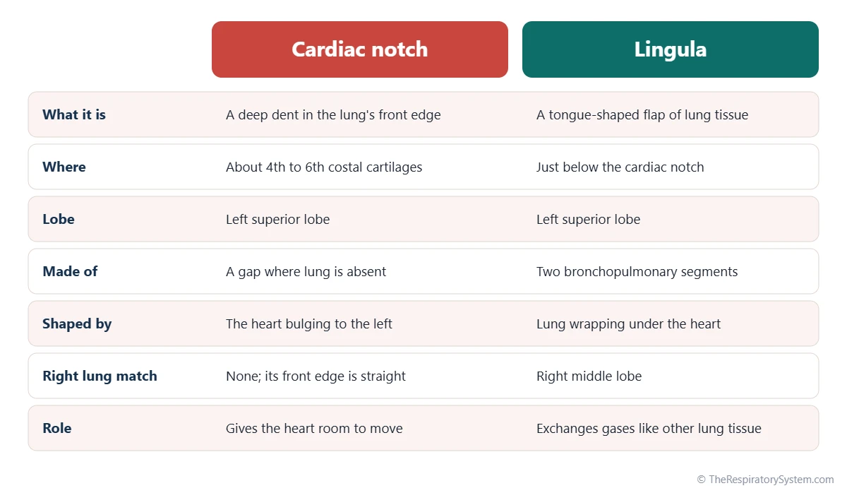
The cardiac notch is a deep, curved indentation on the anterior border of the left lung, at about the level of the 4th to 6th costal cartilages, where the heart and its pericardium bulge to the left. Below the notch, the superior lobe of the left lung forms a tongue-like projection called the lingula, which has two bronchopulmonary segments and matches the right lung's middle lobe.
- Location
- Anterior border of the left lung's superior lobe, behind the 4th to 6th costal cartilages
- What it is
- Cardiac notch: an indentation in the lung edge; lingula: a tongue-shaped flap below it
- Formed by
- The heart and pericardium lying toward the left side of the chest
- Lingula segments
- Superior lingular and inferior lingular segments
- Right lung match
- The lingula corresponds to the right middle lobe; the right lung has no cardiac notch
- Part of
- The superior (upper) lobe of the left lung
On this page
- What Is the Cardiac Notch of the Lung?
- Where Is the Cardiac Notch on the Left Lung?
- What Is the Lingula of the Lung?
- Left Lung Anatomy: How Do the Cardiac Notch and Lingula Fit Together?
- What Is the Function of the Cardiac Notch?
- How Do the Cardiac Notch and Lingula Develop?
- Common Conditions That Affect the Lingula
- Key Facts
- Quiz
- FAQ
What Is the Cardiac Notch of the Lung?
The cardiac notch of the lung is a deep, curved indentation in the anterior border of the left lung, where the lung edge pulls back to the left to leave space for the heart. It is one of the main features that make the left lung look different from the right lung.
Cardiac notch definition in anatomy
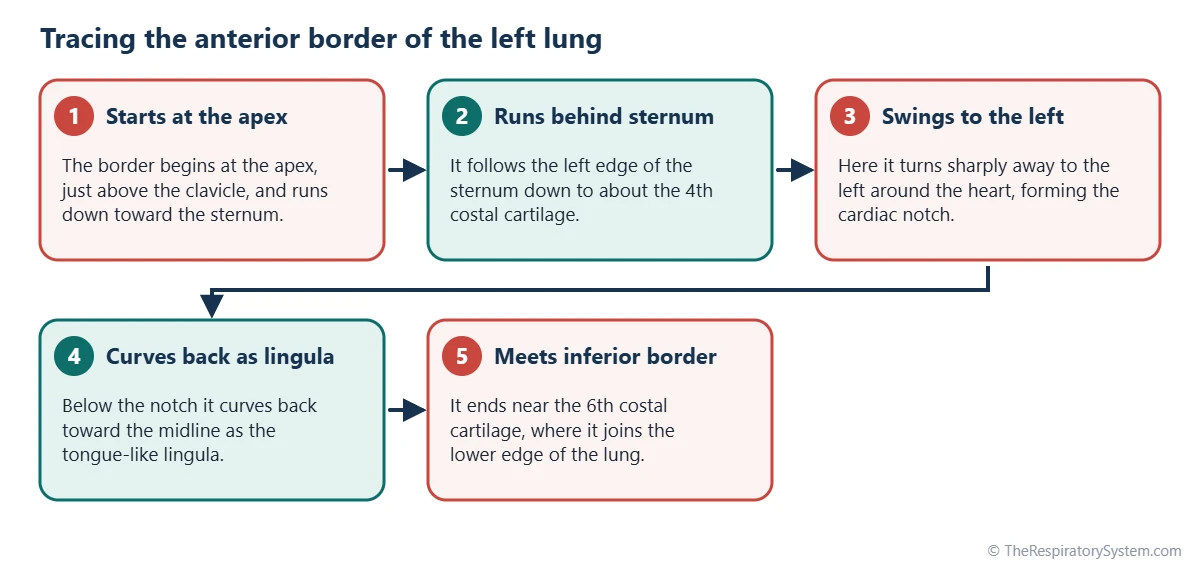
In anatomy, the cardiac notch (Latin incisura cardiaca pulmonis sinistri) is defined as the concave gap in the anterior border of the superior lobe of the left lung, lying in front of the heart and pericardium. Above the notch, the anterior border runs straight down behind the sternum; at the notch, it swings sharply away to the left; below it, the lingula curves back toward the midline.
Why only the left lung has one
The heart does not sit exactly in the middle of the chest. About two-thirds of it lies to the left of the midline, with its apex pointing down and to the left. The left lung grows around this bulge, so its front edge is cut away, while the right lung, with less of the heart beside it, has a straight anterior border. This is also one reason the left lung is slightly smaller, as covered on our which lung is bigger page.
Explained simply
Imagine two pillows pressed against either side of a ball that sits a little to the left. The left pillow needs a scooped-out curve to fit around the ball. That curve is the cardiac notch, and the part of the pillow that wraps underneath the ball is the lingula.
Where Is the Cardiac Notch on the Left Lung?
The cardiac notch is on the anterior border of the left lung, behind the left side of the sternum and the 4th to 6th costal cartilages, directly in front of the heart. It belongs to the superior lobe, above the lingula and in front of the oblique fissure.
Surface anatomy
On the front of the chest, the anterior border of the left lung runs down close to the midline behind the sternum until about the level of the 4th costal cartilage. There it turns outward to the left, curves around the heart, and descends to the 6th costal cartilage a few centimeters to the left of the sternum, where it meets the inferior border. The exact depth of the notch varies from person to person and changes slightly with each breath.
The cardiac notch of the pleura
The parietal pleura lining the chest wall has its own, shallower cardiac notch in the same place. Because the lung's notch is deeper than the pleura's, a narrow strip of pleural space in front of the heart holds no lung at rest. This pocket is part of the costomediastinal recess, which the lingula slides into during a deep breath in.
A small window to the heart
Where the lung is notched, the pericardium lies against the chest wall with no lung tissue in front of it. Doctors call this region the bare area of the pericardium, and when they tap on the chest it gives a dull note instead of the hollow sound of air-filled lung.
What Is the Lingula of the Lung?
The lingula is the thin, tongue-shaped lower front part of the superior lobe of the left lung, lying just below the cardiac notch and curving over the front of the heart. Its name is Latin for "little tongue."
Where is the left lung lingula?
The lingula sits at the front of the left side of the chest, below the cardiac notch and above the diaphragm, roughly behind the 5th and 6th costal cartilages. It lies in front of the oblique fissure and rests against the pericardium over the left ventricle. On a frontal chest X-ray it lies right next to the left border of the heart.
Segments, airway and vessels
The lingula is made of two bronchopulmonary segments: the superior lingular segment and the inferior lingular segment. They are aired by the lingular bronchus, a branch of the left upper lobe bronchus that divides into one segmental bronchus for each. A lingular branch of the left pulmonary artery runs with the airway, and lingular veins drain into the left superior pulmonary vein, all entering or leaving through the hilum.
Lingula vs right middle lobe
The lingula is the left lung's counterpart of the right middle lobe. Both lie at the front of the chest beside the heart, both have two segments, and both receive one bronchus from the main airway of their side. The difference is that the right middle lobe is a separate lobe, cut off by the horizontal fissure, while the lingula usually has no fissure of its own and stays part of the superior lobe.
Left Lung Anatomy: How Do the Cardiac Notch and Lingula Fit Together?
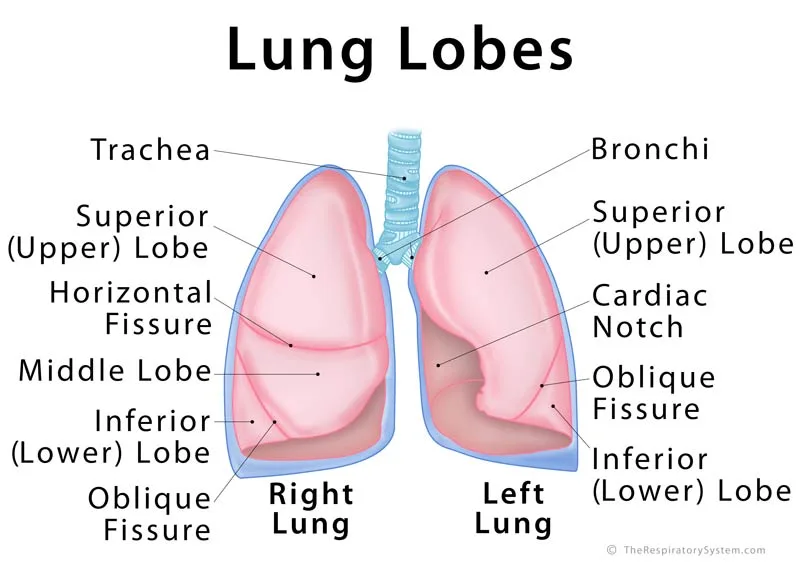
The cardiac notch and the lingula are two halves of the same shape: the notch is the gap cut into the superior lobe by the heart, and the lingula is the tongue of that lobe that wraps around the gap from below. Together they form the lower part of the anterior border of the left lung.
The left lung has two lobes, superior and inferior, separated by the oblique fissure. The superior lobe holds the apex, most of the anterior border, the cardiac notch and the lingula. The inferior lobe forms most of the base and the back of the lung. The table below lists the structures around the notch and what each one does.
Cardiac notch vs cardiac impression
The cardiac notch is easy to confuse with the cardiac impression. The notch is a gap in the lung's front edge, seen from the front. The cardiac impression is a hollow on the inner (mediastinal) surface of the lung, seen from the side, where the heart presses against it. Both lungs have a cardiac impression, but the left one is deeper, and only the left lung has a notch. The other parts of that inner surface are described on our mediastinum page.
| Structure | Where it is | What it is or does |
|---|---|---|
| Cardiac notch | Anterior border of the left superior lobe, about 4th to 6th costal cartilages | Indentation that leaves room for the heart and pericardium |
| Lingula | Below the cardiac notch, lower front of the superior lobe | Tongue-shaped flap; counterpart of the right middle lobe |
| Superior lingular segment | Upper part of the lingula | Bronchopulmonary segment aired by its own segmental bronchus |
| Inferior lingular segment | Lower part of the lingula, above the diaphragm | Bronchopulmonary segment aired by its own segmental bronchus |
| Lingular bronchus | Branch of the left upper lobe bronchus | Splits into the two lingular segmental bronchi |
| Cardiac impression | Inner (mediastinal) surface of the lung | Hollow where the heart presses; deeper on the left |
| Oblique fissure | Runs from the back, downward and forward | Separates the superior lobe from the inferior lobe |
| Left superior lobe | Upper and front part of the left lung | Holds the apex, cardiac notch and lingula |
| Left inferior lobe | Lower and back part of the left lung | Forms most of the base resting on the diaphragm |
| Costomediastinal recess | Pleural space behind the sternum, in front of the heart | Space the lingula slides into on a deep breath |
| Source: Typical adult anatomy, our editors | ||
What Is the Function of the Cardiac Notch?
The function of the cardiac notch is to give the heart and pericardium room on the left side of the chest without squeezing the lung or limiting the heart's movement. It is a shape that results from where the heart sits, rather than a structure that does active work.
- Room for the heart: the heart changes shape with every beat, and the notch lets it move freely against the chest wall.
- Spare space for breathing: during a deep breath in, the lingula and the edge of the notch slide forward into the pleural recess in front of the heart, so the lung can still expand fully.
- Gas exchange in the lingula: the lingula is ordinary lung tissue, so its two segments take part in gas exchange like the rest of the lung.
- A landmark: the area of dull sound over the notch helps doctors outline the heart on examination, and it is the region where the pericardium can be reached without passing through lung.
In short, the notch is the lung's way of sharing the left side of the thoracic cavity with the heart, and the lingula makes sure that space is still used for breathing.
How Do the Cardiac Notch and Lingula Develop?
The cardiac notch and lingula form before birth, as the left lung grows around the developing heart and its airway buds divide into lobes and segments. The shape is set by the heart's leftward position.
Early in development, the airway tube divides into right and left lung buds. The right bud branches into three lobar bronchi and the left into two, which is why the right lung ends up with three lobes and the left with two. The airway that would form a middle lobe on the left instead becomes the lingular bronchus inside the superior lobe. Meanwhile, the growing heart turns toward the left, and the expanding left lung molds itself around it, leaving the notch. Our respiratory system development page traces these stages in order.
Variations are common. The depth of the notch differs between people, and a small extra fissure sometimes separates the lingula from the rest of the superior lobe, making it look like a small middle lobe on the left side.
Common Conditions That Affect the Lingula
The cardiac notch itself is rarely affected by disease, but the lingula can be involved in the same conditions as other parts of the lung, and its position next to the heart makes these easy to spot on imaging. Our respiratory diseases section explains each condition in depth.
- Lingular pneumonia: infection that fills the lingula with fluid blurs the left border of the heart on a frontal chest X-ray, a clue known as the silhouette sign.
- Bronchiectasis: long-lasting widening of the airways often involves the lingula and the right middle lobe, partly because their long, narrow bronchi drain poorly.
- Collapse (atelectasis): a blocked lingular bronchus can make the lingula collapse, again changing the outline of the heart on X-ray.
- Nontuberculous mycobacterial infection: one form of this infection tends to affect the lingula and the right middle lobe, especially in older women.
Anyone with ongoing cough, fever or breathing problems should see a doctor, since these conditions need medical assessment.
Key Facts
- The cardiac notch is a deep indentation in the anterior border of the left lung's superior lobe, made by the heart.
- It lies behind the 4th to 6th costal cartilages, just left of the sternum, in front of the heart and pericardium.
- Only the left lung has a cardiac notch; the anterior border of the right lung is nearly straight.
- The lingula, Latin for "little tongue," is the tongue-shaped part of the left superior lobe below the notch.
- The lingula has two bronchopulmonary segments, superior and inferior lingular, aired by the lingular bronchus.
- The lingula is the left lung's counterpart of the right middle lobe but normally has no fissure of its own.
- The cardiac notch is different from the cardiac impression, a hollow on the inner surface of both lungs where the heart presses.
Quick Quiz: Test What You Know About the Cardiac Notch and Lingula of the Left Lung
6 questions. Tap an answer to check it.
Which lung has a cardiac notch?
Only the left lung has a cardiac notch, because most of the heart lies to the left of the midline.
Which lobe contains the cardiac notch and the lingula?
Both are parts of the superior (upper) lobe of the left lung.
What does the word lingula mean?
Lingula is Latin for little tongue, describing its tongue-like shape below the notch.
Which part of the right lung corresponds to the lingula?
The lingula matches the right middle lobe: both lie beside the heart and have two segments.
At about which costal cartilages is the cardiac notch found?
The anterior border of the left lung swings left at about the 4th costal cartilage and returns near the 6th.
How many bronchopulmonary segments make up the lingula?
The lingula has a superior lingular and an inferior lingular segment.
Frequently Asked Questions
Does the right lung have a cardiac notch?
No. The right lung has no cardiac notch, because much less of the heart lies on the right side of the chest. Its anterior border runs almost straight down behind the sternum. The right lung does have a shallow cardiac impression on its inner surface where the right side of the heart touches it, but this hollow is much less deep than the one on the left lung.
Is the lingula a separate lobe of the lung?
Usually not. The lingula is part of the superior lobe of the left lung and is normally joined to the rest of that lobe without a fissure between them. It is often described as the left lung's equivalent of a middle lobe because it occupies the same position as the right middle lobe and has two segments, but in most people it is not cut off as a lobe of its own.
Is the cardiac notch of the lung the same as the cardiac notch of the stomach?
No. The two share a name but are unrelated. The cardiac notch of the lung is the indentation in the left lung for the heart. The cardiac notch of the stomach, also called the cardiac incisure, is the sharp angle between the esophagus and the top of the stomach, near the stomach opening known as the cardia. Context makes it clear which one is meant.
Can you see the lingula on a chest X-ray?
The healthy lingula is not seen as a separate shape on a chest X-ray, because it is air-filled lung just like the tissue around it. It becomes visible when it fills with fluid or collapses. Because the lingula lies against the left border of the heart, disease there blurs that border on a frontal X-ray, which helps doctors tell lingular problems apart from those in the left lower lobe.
Does the cardiac notch change during breathing?
Yes, slightly. During a deep breath in, the lungs expand, and the edge of the left lung around the notch and the lingula slide forward into the pleural recess in front of the heart, making the notch a little smaller. During a breath out they slide back again. The heart also moves with each heartbeat and with changes in posture, so the notch is never a fixed outline.
Can a person live without the lingula?
Yes. The lingula holds only two of the lung's bronchopulmonary segments, so the rest of the left lung and the whole right lung can still supply the body with oxygen if it is lost to disease or removed in an operation called a lingulectomy. Because each segment has its own airway and blood vessels, the lingula can be separated from the rest of the superior lobe along these natural borders.
Are there other structures called lingula in the body?
Yes. The name means little tongue and is used for several tongue-shaped parts. There is a lingula on the inner side of the lower jaw, next to the opening for the nerve to the lower teeth, a lingula of the cerebellum in the brain, and a lingula of the sphenoid bone in the skull. In respiratory anatomy, lingula always refers to the part of the left lung.
Why is the left lung smaller than the right lung?
The left lung is smaller mainly because it shares the left side of the chest with most of the heart. The cardiac notch and the deep cardiac impression take space that the right lung can use for lung tissue, and the left lung has two lobes instead of three. The right lung is also a little shorter, because the liver pushes the right side of the diaphragm higher.


