At a Glance
Alveolar macrophages are white blood cells of the macrophage family that sit on the inner surface of the alveoli, bathed in surfactant. Their role is to engulf and digest inhaled particles and microbes by phagocytosis, clear old surfactant and dead cells, and send chemical signals that call in other immune cells when a threat is too large to handle alone.
- Location
- Inside the alveoli, on the surfactant-coated surface of the alveolar wall
- Cell type
- Tissue macrophage, a white blood cell of the monocyte-macrophage family
- Function
- Engulf dust and microbes, recycle surfactant, clear dead cells, signal the immune system
- Also called
- Dust cells, alveolar phagocytes
- Works with
- Type I and type II pneumocytes, surfactant proteins, the mucociliary escalator
- Part of
- The innate immune defense of the lower respiratory tract
On this page
What Are Alveolar Macrophages?
Alveolar macrophages are immune cells that live on the inside surface of the alveoli, the tiny air sacs of the lungs, where they swallow up particles and germs that the airways failed to stop. They belong to the macrophage family, large white blood cells whose name means "big eaters" in Greek.
Alveolar macrophages definition
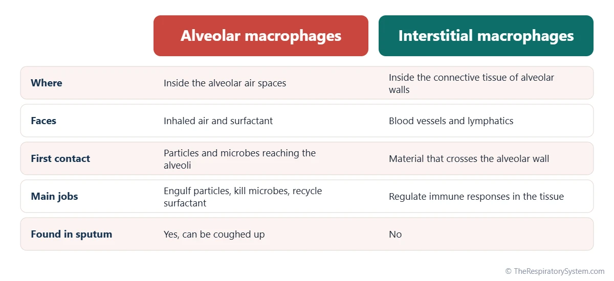
In histology, an alveolar macrophage is defined as a phagocytic cell found free in the alveolar air spaces and on the alveolar walls, often containing engulfed particles in its cytoplasm. They are not part of the alveolar wall itself, which is built by the two kinds of pneumocytes; instead they wander over its surface.
Why they are called dust cells
The nickname dust cells comes from the dark specks of inhaled dust, soot and carbon that can be seen inside them under a microscope. In city dwellers and smokers, many alveolar macrophages are filled with this pigment, a visible record of what the lungs have breathed in.
Explained simply
Think of the alveoli as millions of tiny rooms that must stay spotless so oxygen can pass through their walls. Alveolar macrophages are the cleaning crew that patrols every room, picking up dust and germs, eating what they can and carrying the rest out the door.
Where Are Alveolar Macrophages Found?
Alveolar macrophages are found inside the alveoli and alveolar sacs, resting on or crawling over the thin lining layer of fluid and pulmonary surfactant that coats the alveolar wall. They sit in the respiratory zone, beyond the terminal bronchioles, in the part of the lung where gas exchange takes place.
Why they are needed here
The larger airways are protected by mucus and beating cilia, the mucociliary escalator, which traps most inhaled particles long before they reach the air sacs. The alveoli have no cilia and no mucus, because a thick, wet layer would block gas exchange. Fine particles only a few micrometers across can slip past the airway defenses, and the alveolar macrophages are the main cells that deal with them.
How they move around
Alveolar macrophages move by extending and pulling in parts of their cell body, crawling across the surface of type I and type II pneumocytes. They can pass from one alveolus to the next through small openings in the walls between neighboring air sacs, the pores of Kohn, and they travel along the alveolar ducts and sacs. A separate group, the interstitial macrophages, lives inside the connective tissue of the alveolar walls rather than in the air spaces.
What Is the Role of Alveolar Macrophages?
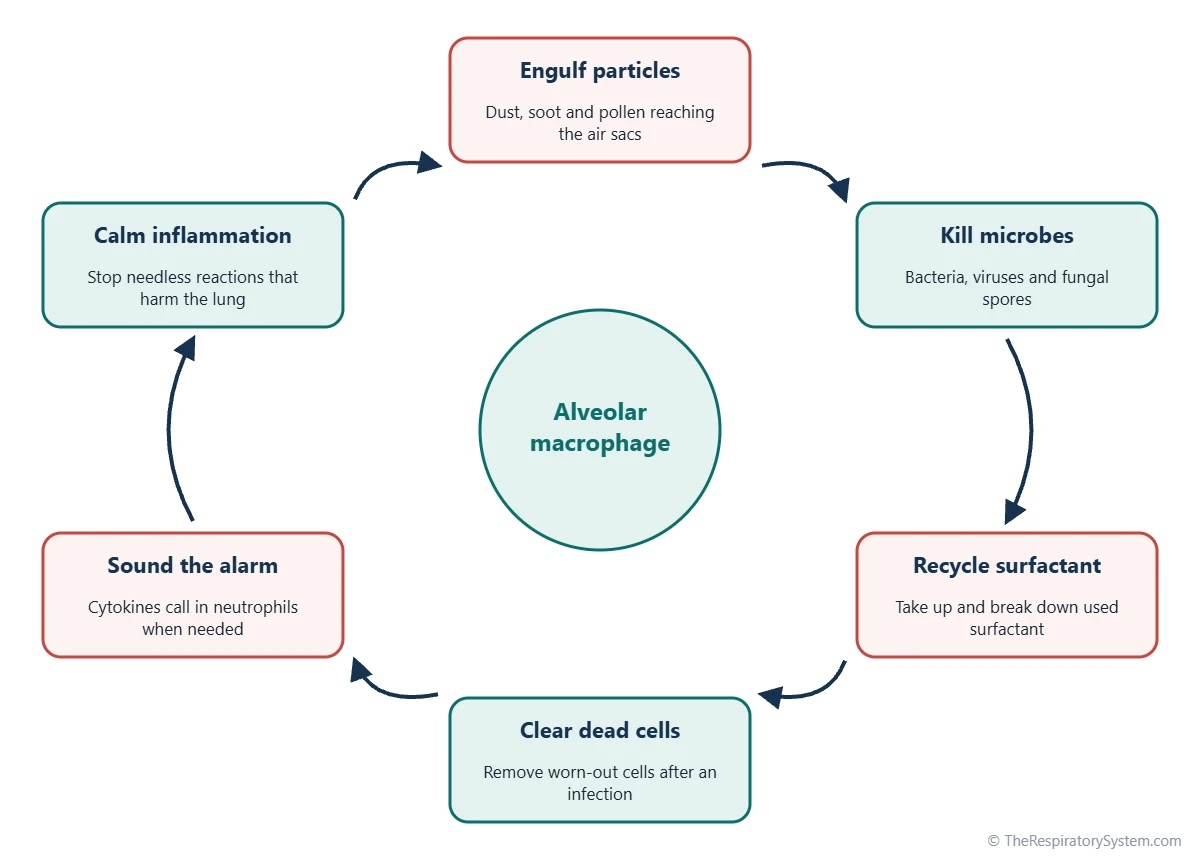
The role of alveolar macrophages is to keep the alveoli clean, sterile and quiet: they remove inhaled particles and microbes, recycle used surfactant, clear away dead cells, and decide when to raise or calm an immune response. Each job protects the thin respiratory membrane where oxygen enters the blood.
- Removing particles: dust, soot, pollen and other fine particles are engulfed and either digested or stored.
- Killing microbes: bacteria, viruses and fungal spores that reach the alveoli are taken in and destroyed.
- Recycling surfactant: they take up and break down old surfactant, helping keep the right amount on the alveolar surface.
- Cleaning up: they eat worn-out and dying cells, including spent white blood cells after an infection.
- Sounding the alarm: they release signaling proteins called cytokines that call in neutrophils and other immune cells.
- Keeping the peace: they also release calming signals, so harmless particles do not set off constant inflammation that would damage the lung.
Together with the airway defenses, these jobs form a large part of the protective functions of the respiratory system.
How Do Alveolar Macrophages Work?
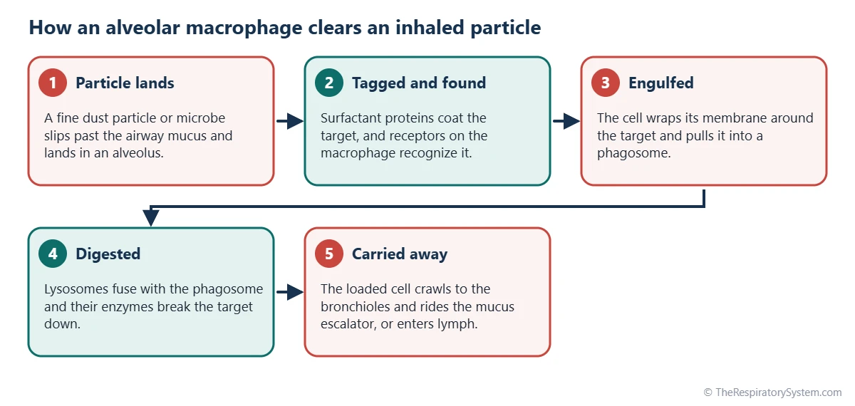
Alveolar macrophages work by phagocytosis: they wrap their cell membrane around a particle or microbe, pull it inside in a membrane bubble and then destroy it with digestive enzymes and toxic forms of oxygen. What cannot be broken down is carried out of the alveoli or stored inside the cell.
Recognizing a target
Macrophages carry receptors on their surface that recognize common features of bacteria and fungi, as well as foreign particles. Two surfactant proteins made by type II pneumocytes, SP-A and SP-D, help by sticking to microbes and marking them for capture, a process called opsonization.
Engulfing and digesting
Once a target is caught, the macrophage folds its membrane around it to form a phagosome. This bubble fuses with lysosomes, small sacs full of enzymes, to form a phagolysosome, where the contents are broken down. Many microbes are killed within this compartment, while mineral dust such as carbon cannot be digested and stays inside the cell.
Leaving the alveoli
Loaded macrophages leave the alveoli in two main ways. Many crawl up to the ciliated bronchioles, where the mucus escalator carries them to the throat to be swallowed or coughed out. Others enter the lung tissue and lymphatic vessels and travel to lymph nodes near the hilum of the lung. Clearing particles from the alveoli this way is slow and can take weeks to months, compared with hours in the larger airways.
| Feature | Description | Why it matters |
|---|---|---|
| Cell family | Tissue macrophage of the monocyte-macrophage line | Specialized for engulfing and digesting |
| Location | Free in the alveolar air spaces, on the surfactant layer | First contact with particles reaching the alveoli |
| Appearance | Large, irregular cells with many lysosomes and vacuoles, often with dark specks | Specks are engulfed dust or carbon |
| Origin | Mostly self-renewing cells seeded around birth; blood monocytes add more in infection | The lung can restock them locally |
| Growth signal | GM-CSF made by type II pneumocytes | Needed for normal maturity and surfactant clearance |
| Helpers | Surfactant proteins SP-A and SP-D tag microbes | Makes capture faster and more efficient |
| Main method | Phagocytosis into a phagolysosome | Destroys microbes with enzymes and oxidants |
| Exit routes | Up the mucociliary escalator, or into lymphatics | Carries debris out of the alveoli |
| Signals released | Cytokines that recruit neutrophils, and calming signals | Balances defense against lung damage |
| Source: Standard histology and physiology, our editors | ||
Where Do Alveolar Macrophages Come From?
Most alveolar macrophages come from cells that settle in the lungs around the time of birth and then renew themselves by dividing locally, while extra macrophages are made from blood monocytes when the lungs are infected or injured. Older textbooks describe them as coming only from blood monocytes, but research over the past few decades has shown that the resident population largely maintains itself.
Their growth depends on a signaling protein called GM-CSF (granulocyte-macrophage colony-stimulating factor), which is made mainly by type II pneumocytes. Without enough GM-CSF signaling, alveolar macrophages do not mature properly and cannot clear surfactant, which is why the two cell types are often studied together. The lung's general cell makeup is described on our respiratory system histology page.
Common Conditions That Affect Alveolar Macrophages
Conditions that affect alveolar macrophages include infections that survive inside them, dusts that damage them, smoking, and rare disorders in which they cannot clear surfactant. Our respiratory diseases section covers these conditions in more depth.
- Tuberculosis: the bacteria that cause tuberculosis are engulfed by alveolar macrophages but can resist being destroyed and multiply inside them.
- Pneumonia: when microbes arrive in numbers too large for the macrophages, they call in neutrophils, and the alveoli fill with fluid and inflammatory cells.
- Dust diseases: silica and asbestos fibers injure the macrophages that swallow them, and the signals they release drive scarring, as in silicosis and asbestosis; see pulmonary fibrosis.
- Smoking: smokers have more alveolar macrophages, filled with tar pigment, but they kill germs less well and release enzymes that can break down alveolar walls, a process linked to emphysema and COPD.
- Pulmonary alveolar proteinosis: a rare disorder, usually caused by antibodies that block GM-CSF, in which macrophages cannot clear surfactant and it fills the alveoli.
- Heart failure cells: when blood backs up in the lungs in left heart failure, red cells leak into the alveoli and macrophages fill with the iron pigment hemosiderin.
Anyone with a lasting cough or shortness of breath should see a doctor.
Key Facts
- Alveolar macrophages, or dust cells, are immune cells that live inside the alveoli and engulf inhaled particles and microbes.
- They are the main defense of the alveoli, which have no cilia or mucus of their own.
- They capture targets by phagocytosis and destroy them inside phagolysosomes with enzymes and toxic oxygen compounds.
- Surfactant proteins SP-A and SP-D made by type II pneumocytes help them recognize microbes.
- Loaded macrophages leave the alveoli by riding the mucociliary escalator or by entering the lymphatic vessels.
- Most alveolar macrophages renew themselves inside the lung, with extra cells made from blood monocytes during infection.
- Smoking increases their number but weakens their ability to kill germs.
Quick Quiz: Test What You Know About the Alveolar Macrophages
6 questions. Tap an answer to check it.
What is another name for alveolar macrophages?
They are called dust cells because engulfed dust and carbon are often visible inside them.
Where are alveolar macrophages found?
They live on the surfactant-coated inner surface of the alveoli.
By what process do alveolar macrophages engulf particles?
Phagocytosis means cell eating: the membrane wraps around a target and pulls it into the cell.
Why do the alveoli need macrophages instead of mucus and cilia?
A thick mucus layer would slow gas exchange, so the alveoli rely on wandering macrophages instead.
Which cells make the GM-CSF that alveolar macrophages need to mature?
Type II pneumocytes produce GM-CSF, which alveolar macrophages need for normal function.
How do many loaded macrophages leave the alveoli?
They crawl to the ciliated bronchioles, and the mucus escalator carries them to the throat.
Frequently Asked Questions
Are alveolar macrophages white blood cells?
Yes. Alveolar macrophages are a type of white blood cell, or leukocyte, belonging to the monocyte-macrophage family. Unlike white cells that circulate in the blood, they are tissue cells that live permanently in the lung's air spaces. They are part of the innate immune system, the fast, built-in defense that acts against many kinds of invaders without needing to have met them before.
What is the difference between alveolar macrophages and type II pneumocytes?
Type II pneumocytes are part of the alveolar wall: they are rounded epithelial cells that make surfactant and can divide to repair the lining. Alveolar macrophages are immune cells that are not part of the wall but move freely over its surface, engulfing particles and microbes. The two cooperate, since pneumocytes make surfactant proteins and GM-CSF that the macrophages rely on.
Can alveolar macrophages be coughed up?
Yes. Macrophages that carry their load up to the bronchioles are moved by the mucus escalator to the throat, where they are usually swallowed without notice. They can also be coughed up in sputum. Finding macrophages in a sputum sample tells laboratory staff that the sample came from deep in the lungs rather than from saliva in the mouth.
How long do alveolar macrophages live?
Alveolar macrophages are long-lived cells compared with many white blood cells, which survive only days. Studies suggest that in humans they can last for months or longer, and the population is topped up by cells dividing in the lung and by new cells from blood monocytes. Exact lifespans are hard to measure in people, so figures vary between studies.
What happens to dust that macrophages cannot digest?
Mineral dust such as carbon or silica cannot be broken down by the enzymes in a macrophage. The cell either carries it out of the alveoli by the mucus escalator or lymphatics, or keeps it stored inside itself. Over many years, macrophages packed with carbon collect in the lung tissue and lymph nodes, giving the lungs of city dwellers and smokers their gray, speckled look.
Do alveolar macrophages fight viruses?
Yes. Alveolar macrophages engulf virus particles and infected cells, and they release signaling proteins, including interferons and other cytokines, that warn neighboring cells and recruit more immune cells. In some infections, such as influenza, an overly strong macrophage response can add to lung inflammation, which is why their calming role is as important as their fighting role.
How does air pollution affect alveolar macrophages?
Fine particles from traffic, industry and smoke are small enough to reach the alveoli, where macrophages engulf them. A heavy load of particles can weaken the macrophages' ability to kill bacteria and can make them release inflammatory signals. This is one reason why long exposure to polluted air is linked to more lung infections and chronic lung disease.
Keep Exploring


