At a Glance
A bronchopulmonary segment is the largest part of a lung lobe that has its own segmental (tertiary) bronchus and segmental artery, separated from neighboring segments by connective tissue and drained by veins along its borders. The right lung has 10 segments and the left lung usually 8 to 10, because some left segments share one bronchus.
- Location
- Inside each lung lobe, apex toward the hilum and base at the lung surface
- Number
- 10 in the right lung; 8 to 10 in the left lung
- Supplied by
- One segmental (tertiary) bronchus and one segmental branch of the pulmonary artery
- Drained by
- Intersegmental pulmonary veins running along the borders
- Function
- Self-contained units of airway, blood supply and gas exchange
- Part of
- Lung lobes; level 3 of the bronchial tree
On this page
- What Are Bronchopulmonary Segments?
- How Many Bronchopulmonary Segments Are There?
- What Are Bronchopulmonary Segments Supplied By?
- Right Lung Anatomy: Which Segments Are in Each Lobe?
- Left Lung Anatomy: Which Segments Are in Each Lobe?
- Why Are Bronchopulmonary Segments Important?
- Common Conditions That Affect the Bronchopulmonary Segments
- Key Facts
- Quiz
- FAQ
What Are Bronchopulmonary Segments?
Bronchopulmonary segments are the anatomically and functionally separate units into which each lobe of the lungs is divided, each served by a single segmental bronchus and a single segmental artery. They sit one level below the lung lobes in the organization of the lung and are part of the lower respiratory tract.
Each segment has a set of features that make it a self-contained unit:
- Shape: roughly a pyramid or cone, with its apex pointing toward the root of the lung and its base on the lung surface under the pleura.
- Airway: one segmental (tertiary) bronchus enters near the apex and branches only within that segment.
- Artery: one segmental branch of the pulmonary artery runs alongside the bronchus.
- Borders: thin connective tissue partitions separate it from its neighbors, and pulmonary veins run in these borders.
Explained simply
Think of a lung lobe as an orange and the segments as its wedges. Each wedge has its own straw bringing in air and its own pipe bringing in blood. Because no straw or pipe crosses into the next wedge, one wedge can be studied, blocked or removed without cutting off the others.
How Many Bronchopulmonary Segments Are There?
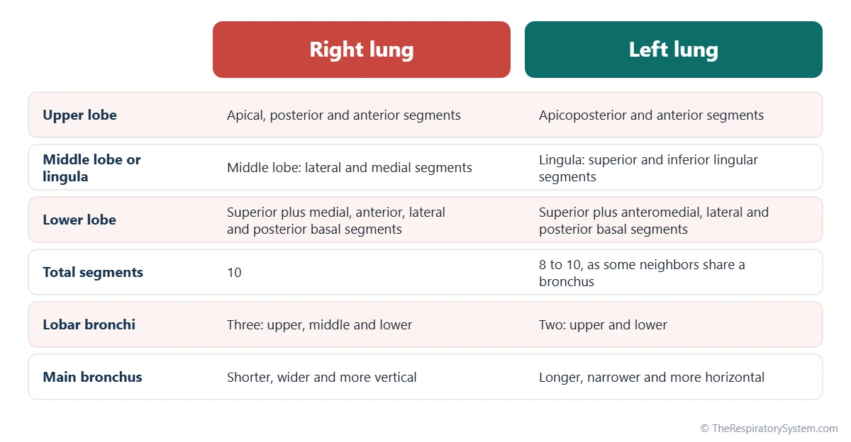
There are usually 10 bronchopulmonary segments in the right lung and 8 to 10 in the left lung, so most descriptions list 18 to 20 in total. The number on the left varies because some neighboring segments are served by one shared bronchus and are counted as a single combined segment.
The right lung's three lobes hold 3, 2 and 5 segments: upper, middle and lower. The left lung has only two lobes, and its upper lobe includes the lingula, a tongue-shaped part next to the cardiac notch that matches the right middle lobe. On the left, the apical and posterior segments usually share a bronchus and form one apicoposterior segment, and the anterior basal and medial basal segments often form one anteromedial basal segment.
How the segments are numbered
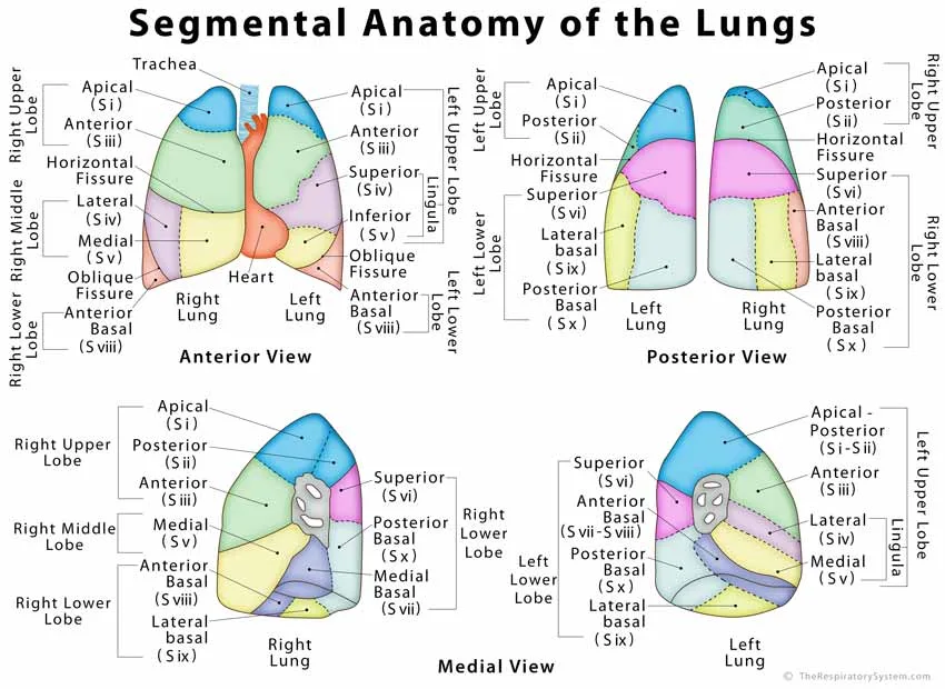
Segments are numbered from top to bottom with the letter S: S1 to S3 in the upper lobe, S4 and S5 in the right middle lobe or the left lingula, and S6 to S10 in the lower lobe. Their bronchi carry the same numbers with the letter B, so B6 is the bronchus to the superior segment of the lower lobe. The table below lists every segment in both lungs.
| Segment | Right lung | Left lung | Lobe |
|---|---|---|---|
| S1 | Apical | Apicoposterior (S1 and S2 combined) | Upper lobe |
| S2 | Posterior | Apicoposterior (shared with S1) | Upper lobe |
| S3 | Anterior | Anterior | Upper lobe |
| S4 | Lateral | Superior lingular | Right middle lobe; left lingula |
| S5 | Medial | Inferior lingular | Right middle lobe; left lingula |
| S6 | Superior | Superior | Lower lobe |
| S7 | Medial basal | Anteromedial basal (often joined with S8) | Lower lobe |
| S8 | Anterior basal | Anteromedial basal (shared with S7) | Lower lobe |
| S9 | Lateral basal | Lateral basal | Lower lobe |
| S10 | Posterior basal | Posterior basal | Lower lobe |
| Source: Standard segment names and numbers, our editors | |||
What Are Bronchopulmonary Segments Supplied By?
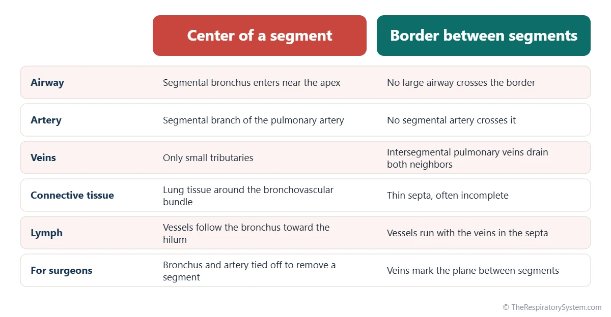
Each bronchopulmonary segment is supplied by its own segmental (tertiary) bronchus and its own segmental branch of the pulmonary artery, which travel together through the center of the segment as a bronchovascular bundle. The segment is drained by pulmonary veins that run in the borders between segments.
Airway supply
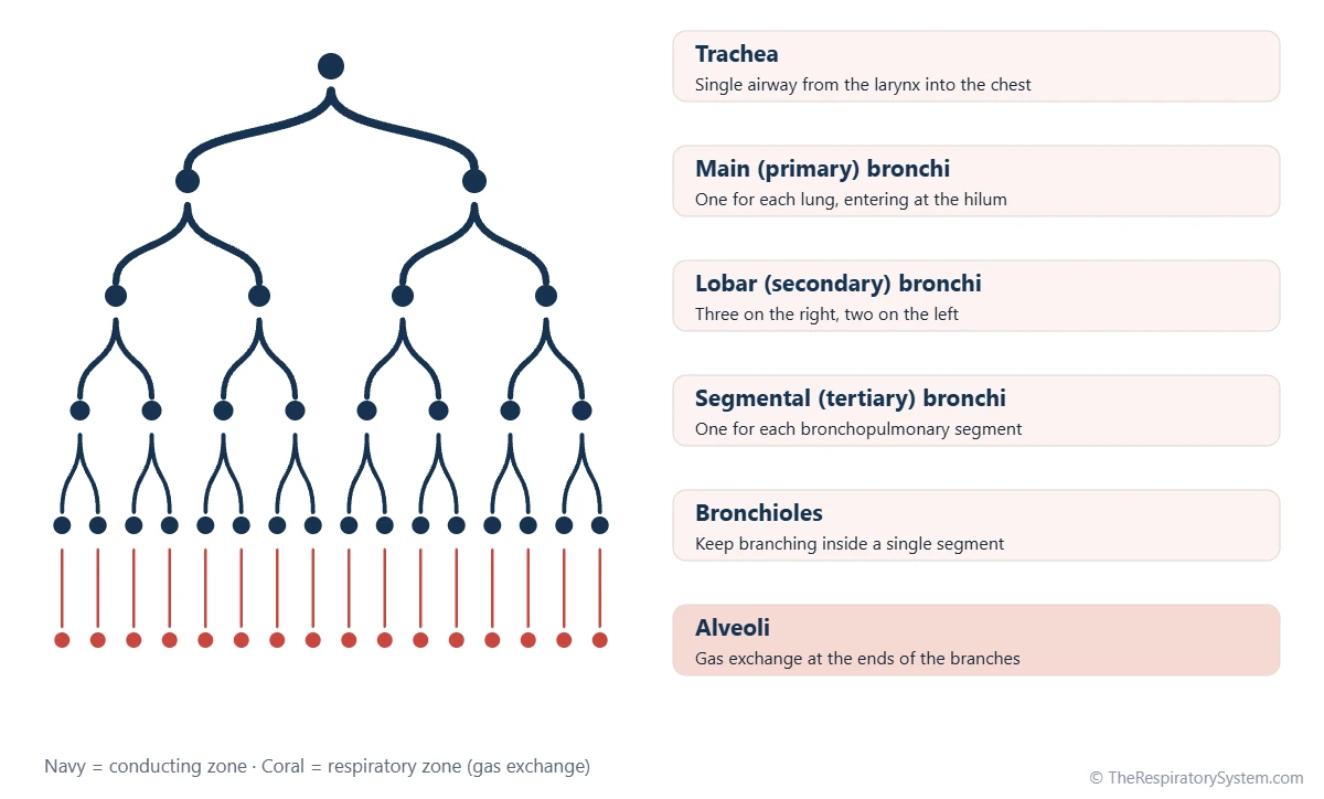
The trachea divides into the main bronchi, each main bronchus into lobar bronchi and each lobar bronchus into segmental bronchi, one for every segment. Inside its segment, the segmental bronchus keeps dividing into smaller bronchi and then bronchioles, ending in the alveoli; the whole branching pattern is described on our bronchial tree page.
Blood and lymph
The pulmonary arteries bring oxygen-poor blood from the heart and branch exactly with the airways, so each segmental artery matches one segmental bronchus. The pulmonary veins behave differently: they run between segments and collect blood from two neighbors at once, which makes them useful landmarks for the borders. The small bronchial arteries, part of the systemic circulation, feed the walls of the airways, and lymph vessels drain toward the nodes at the hilum. Our pulmonary circulation page follows the blood through this loop.
How segments develop
The segmental bronchi are among the first branches to form as the lung buds grow, appearing within the first two months of embryonic life. Each one grows into the surrounding tissue with its own artery, which is why the segment pattern is already set long before birth; see development of the respiratory system.
Right Lung Anatomy: Which Segments Are in Each Lobe?
The right lung has 10 bronchopulmonary segments: three in the upper lobe, two in the middle lobe and five in the lower lobe. They are named after their position in the lobe.
- Upper lobe: apical (S1) at the top, reaching into the root of the neck; posterior (S2) toward the back; anterior (S3) toward the front.
- Middle lobe: lateral (S4) on the outer side and medial (S5) next to the heart.
- Lower lobe: superior (S6) at the top of the lobe toward the back, then four basal segments resting on the diaphragm: medial basal (S7), anterior basal (S8), lateral basal (S9) and posterior basal (S10).
The superior segment of the lower lobe reaches surprisingly high, up to about the level of the shoulder blade, so on the back of the chest much of the area heard with a stethoscope belongs to the lower lobe.
Left Lung Anatomy: Which Segments Are in Each Lobe?
The left lung usually has 8 to 10 bronchopulmonary segments in two lobes, because some of its segments share a bronchus and are counted together. Its segments follow the same plan as the right lung, adjusted for the space taken by the heart.
- Upper lobe: apicoposterior (S1+2), a merged apical and posterior segment, and anterior (S3).
- Lingula of the upper lobe: superior lingular (S4) and inferior lingular (S5), the left-side match of the right middle lobe.
- Lower lobe: superior (S6), anteromedial basal (S7+8), lateral basal (S9) and posterior basal (S10).
When every merged segment is counted separately, the left lung reaches 10 segments, the same as the right. When both merged pairs are counted as one, it has 8.
Why Are Bronchopulmonary Segments Important?
Bronchopulmonary segments are important because each one is a separate unit with its own air and blood supply, so disease can be pinpointed to a segment and a single segment can be removed while the rest of the lobe keeps working. They turn the lung from one large organ into a precise map.
- Locating disease: doctors describe findings on CT scans and during bronchoscopy by segment, for example a shadow in the posterior segment of the right upper lobe.
- Surgery: because no major bronchus or artery crosses a segment border, surgeons can take out one segment, an operation called a segmentectomy, and spare more healthy lung than removing a whole lobe.
- Patterns of spread: infection or inhaled material often stays within one segment at first, producing segment-shaped shadows on imaging.
- Gravity and position: which segments lie lowest depends on body position, so the segments most often reached by inhaled material change between standing and lying down.
For gas exchange itself, each segment works like a miniature lung, with its airways ending in pulmonary lobules and alveoli.
Common Conditions That Affect the Bronchopulmonary Segments
Several lung conditions tend to involve particular bronchopulmonary segments, which is why segment anatomy appears in so many imaging and pathology reports. Our respiratory diseases section describes each condition, and anyone with symptoms such as a lasting cough or breathlessness should see a doctor.
- Aspiration pneumonia: infection after food, liquid or stomach contents are inhaled, most often in the lowest-lying segments of the right lung.
- Segmental collapse (atelectasis): when a segmental bronchus is blocked, by mucus or a tumor, the air beyond it is absorbed and that segment shrinks.
- Tuberculosis: reactivated disease in adults classically affects the apical and posterior segments of the upper lobes and the superior segments of the lower lobes.
- Bronchiectasis: permanently widened airways that may be limited to one segment or lobe.
- Pulmonary embolism: a clot lodged in a segmental artery cuts off blood to that segment and can leave a wedge-shaped area of damage.
- Lung cancer: tumors are located and staged by lobe and segment.
Key Facts
- A bronchopulmonary segment is the part of a lung lobe served by one segmental (tertiary) bronchus and one segmental branch of the pulmonary artery.
- The right lung has 10 segments: 3 in the upper lobe, 2 in the middle lobe and 5 in the lower lobe.
- The left lung has 8 to 10 segments, because its apicoposterior and anteromedial basal segments are often counted as one each.
- Segments are pyramid-shaped, with the apex toward the hilum and the base on the lung surface.
- Pulmonary veins run in the borders between segments, while the bronchus and artery run through the center.
- The lingula of the left upper lobe matches the right middle lobe and holds segments S4 and S5.
- Because each segment is self-contained, a surgeon can remove one segment and spare the rest of the lobe.
Quick Quiz: Test What You Know About the Bronchopulmonary Segments
6 questions. Tap an answer to check it.
How many bronchopulmonary segments does the right lung have?
The right lung has 10 segments: 3 in the upper lobe, 2 in the middle lobe and 5 in the lower lobe.
Which airway supplies a single bronchopulmonary segment?
Each segment is aerated by one segmental, or tertiary, bronchus.
Where do the pulmonary veins run in relation to the segments?
Pulmonary veins are intersegmental: they run between segments and drain neighboring segments.
Which part of the left lung matches the right middle lobe?
The lingula of the left upper lobe, with its superior and inferior lingular segments, matches the right middle lobe.
Which segment is labeled S6 in both lungs?
S6 is the superior segment of the lower lobe, the first segment of the lower lobe in both lungs.
Why can a single bronchopulmonary segment be removed surgically?
Because each segment has its own bronchus and artery and no major airway or artery crosses its borders, it can be separated from its neighbors.
Frequently Asked Questions
What is the difference between a lung lobe and a bronchopulmonary segment?
A lobe is a large division of the lung separated by fissures that are visible on the surface, served by a lobar (secondary) bronchus. A bronchopulmonary segment is a smaller division within a lobe, served by a segmental (tertiary) bronchus. Segments have no visible fissures between them; their borders are thin internal partitions marked by veins.
What is the difference between a bronchopulmonary segment and a pulmonary lobule?
A bronchopulmonary segment is a large unit served by one segmental bronchus and contains many smaller units. A pulmonary lobule is a much smaller unit, only about 1 to 2 cm across, served by a small bronchiole and surrounded by thin connective tissue. Many lobules pack together to build each segment, and each lobule contains several acini where gas exchange happens.
Why does the left lung have fewer segments than the right?
The left lung is smaller because the heart bulges toward the left side of the chest, and it has only two lobes. Its apical and posterior segments usually share one bronchus, and its anterior basal and medial basal segments often do too, so these pairs are counted as single segments. That gives the left lung 8 to 10 segments instead of 10.
Which segments are most often affected when something is inhaled?
Inhaled material more often enters the right lung, because the right main bronchus is wider, shorter and more vertical than the left. In a person who is upright, it tends to reach the posterior basal segment of the right lower lobe. In a person lying on their back, it tends to reach the superior segment of the lower lobe and the posterior segment of the upper lobe.
Can you see bronchopulmonary segments on an X-ray or CT scan?
The borders between segments are not normally visible on an X-ray or CT scan, because the partitions between them are thin and often incomplete. Radiologists identify segments by following the segmental bronchi and arteries, which are visible on CT. A segment becomes easy to see when it is filled with fluid or collapses, because it then shows up as a dense wedge.
Are bronchopulmonary segments the same in everyone?
The basic plan of 10 segments in the right lung and 8 to 10 in the left is the same in nearly everyone, but the exact branching of the segmental bronchi and arteries varies from person to person. Extra small subsegments, shared bronchi or unusual origins are common. Surgeons therefore study each patient's own scans before operating on a segment.
Do bronchopulmonary segments exchange air with each other?
Segments are separated mainly by thin connective tissue, and the partitions are often incomplete, so a little air can pass between neighboring segments through small connections between alveoli. This collateral flow is usually limited. That is why a segment whose bronchus is blocked often collapses as the trapped air is absorbed into the blood.


