At a Glance
Respiratory epithelium is a ciliated pseudostratified columnar epithelium containing goblet cells. It lines the nasal cavity, paranasal sinuses, nasopharynx, most of the larynx, the trachea and the bronchi, where it traps inhaled particles in mucus and moves them toward the pharynx with beating cilia. In the bronchioles it thins to simple columnar and then cuboidal cells.
- Type
- Ciliated pseudostratified columnar epithelium with goblet cells
- Location
- Nasal cavity, sinuses, nasopharynx, larynx, trachea and bronchi
- Main cells
- Ciliated columnar cells, goblet cells and basal cells
- Function
- Traps particles in mucus and sweeps them toward the throat
- Part of
- The respiratory mucosa of the conducting zone
- Changes to
- Simple columnar, then cuboidal cells in the bronchioles
On this page
- What Is Respiratory Epithelium?
- Where Is Respiratory Epithelium Located?
- What Cells Make Up the Respiratory Epithelium?
- What Is the Function of the Respiratory Epithelium?
- What Is the Respiratory Mucosa?
- How Does the Airway Lining Change Toward the Alveoli?
- Common Conditions That Affect the Respiratory Epithelium
- Key Facts
- Quiz
- FAQ
What Is Respiratory Epithelium?
Respiratory epithelium is the specialized lining tissue of the conducting airways, a ciliated pseudostratified columnar epithelium with goblet cells. It forms the inner surface of the airway tubes and is the first tissue that inhaled air touches after the nostrils.
What type of epithelium is respiratory epithelium?
Each word in its name describes a feature. Columnar means most cells are taller than they are wide. Ciliated means many cells carry hair-like cilia on their free surface. Pseudostratified means "falsely layered": every cell rests on the basement membrane, but only some reach the airway surface, so the nuclei sit at different heights and the tissue looks like several layers when it is really one.
Explained simply
Imagine a carpet of tiny brushes covered by a thin sheet of sticky syrup. Dust and germs land on the syrup and stick, and the brushes all sweep in the same direction, carrying the dirty syrup up and out of your airways like a slow conveyor belt.
Where Is Respiratory Epithelium Located?
Respiratory epithelium lines most of the conducting zone: the nasal cavity beyond the vestibule, the paranasal sinuses, the nasopharynx, most of the larynx, the trachea and the bronchi. It also lines the auditory tubes that connect the nasopharynx to the middle ears.
It is replaced by other linings wherever the surface faces friction or has another job. The skin-like lining of the nasal vestibule, the food passages of the oropharynx and laryngopharynx, and the vocal folds are covered by tougher stratified squamous epithelium. A small patch in the roof of the nasal cavity carries olfactory epithelium for the sense of smell. Deeper in the lungs, the lining becomes thinner and simpler in the bronchioles until it turns into the flat cells of the alveoli.
| Region | Epithelium | Why it fits |
|---|---|---|
| Nasal vestibule | Stratified squamous, keratinized near the nostrils | Skin-like surface with hairs that catch large particles |
| Nasal cavity (respiratory region) | Respiratory epithelium | Traps dust and moistens the air |
| Roof of the nasal cavity | Olfactory epithelium | Holds the smell receptor neurons |
| Paranasal sinuses | Respiratory epithelium, thinner | Mucus drains into the nasal cavity |
| Nasopharynx | Respiratory epithelium | Air-only passage behind the nose |
| Oropharynx and laryngopharynx | Stratified squamous, nonkeratinized | Resists rubbing by swallowed food |
| Vocal folds | Stratified squamous, nonkeratinized | Withstands constant vibration |
| Rest of the larynx, trachea and bronchi | Respiratory epithelium | Main zone of mucociliary clearance |
| Bronchioles | Simple ciliated columnar to cuboidal, with club cells | Thinner wall for narrow airways |
| Alveoli | Simple squamous (type I) with type II cells | Very thin barrier for gas exchange |
| Source: Standard histology of the adult airway, our editors | ||
What Cells Make Up the Respiratory Epithelium?
Respiratory epithelium contains five main cell types: ciliated columnar cells, goblet cells, basal cells, brush cells and small granule (neuroendocrine) cells. The first two do most of the visible work, while the others renew, sense and signal.
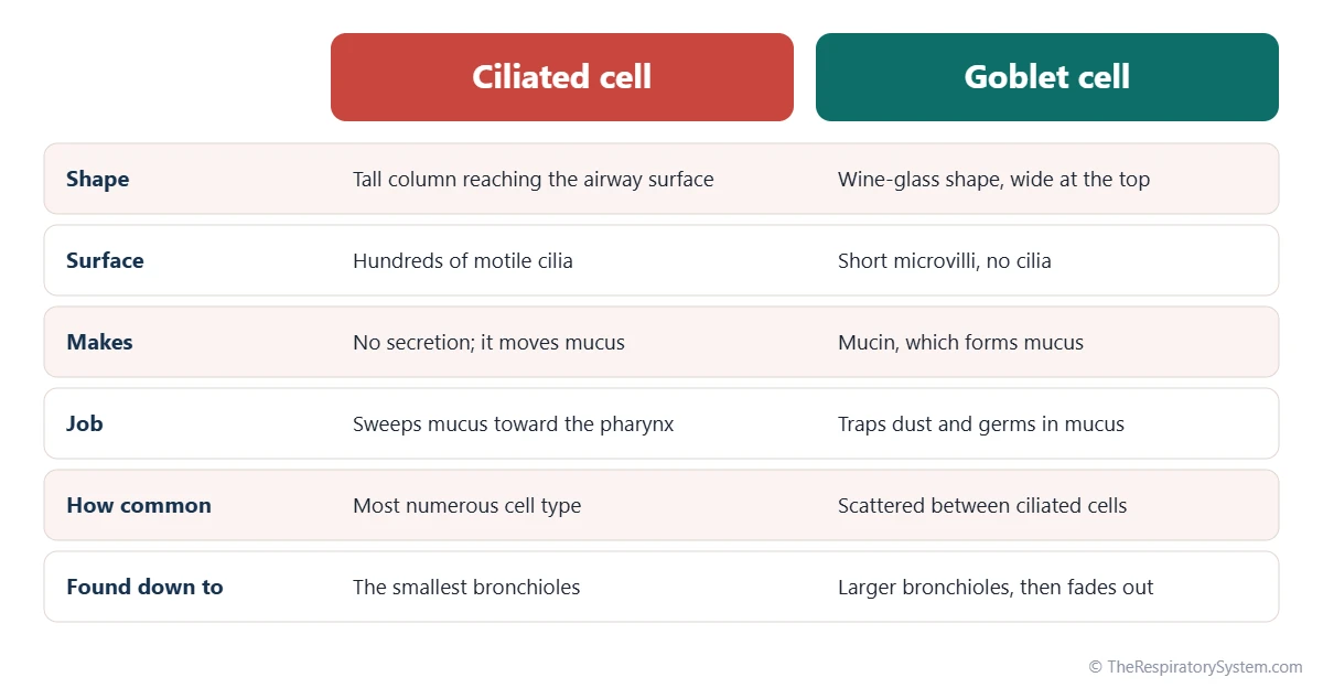
- Ciliated columnar cells are the most numerous. Each carries hundreds of motile cilia that beat in coordinated waves.
- Goblet cells are shaped like a wine glass, with a cup of mucus granules at the top. They release mucin, the protein that forms mucus.
- Basal cells are small cells on the basement membrane that act as stem cells, replacing ciliated and goblet cells.
- Brush cells carry short microvilli and are thought to sense chemicals in the airway.
- Small granule cells release signaling substances that act on nearby nerves, muscle and blood vessels.
For how these cells look under the microscope next to the other airway tissues, see respiratory system histology.
| Cell | Where it sits | Main job |
|---|---|---|
| Ciliated columnar cell | Reaches the airway surface; most numerous | Beats cilia to move mucus toward the pharynx |
| Goblet cell | Scattered between ciliated cells | Releases mucin, which forms mucus |
| Basal cell | On the basement membrane; does not reach the surface | Stem cell that replaces surface cells |
| Brush cell | Reaches the surface; short microvilli | Thought to sense chemicals in the airway |
| Small granule cell | Near the basement membrane | Releases signals that act on nerves and muscle |
| Club cell | Bronchioles, where goblet cells fade out | Secretes protective proteins and helps repair |
| Source: Standard histology of the adult airway, our editors | ||
What Is the Function of the Respiratory Epithelium?
The main function of the respiratory epithelium is to protect the lungs by trapping inhaled particles and germs in mucus and moving them out of the airways, while also moistening the air and acting as a living barrier.
- Mucociliary clearance: mucus traps particles, and the cilia carry it upward to the pharynx, where it is swallowed or coughed out. This conveyor is called the mucociliary escalator and is explained step by step on our cilia and mucus page.
- Humidifying and warming: the moist surface adds water vapor to the air, and blood vessels just beneath it give off heat.
- Chemical defense: the cells and glands release antibodies and enzymes such as lysozyme that weaken bacteria.
- Barrier: tight junctions between neighboring cells keep germs and particles from slipping between them.
- Repair: basal cells divide to replace damaged surface cells.
Goblet cells function
Goblet cells release mucin, which swells with water to form the sticky gel on top of the epithelium. They work with seromucous glands deeper in the wall, which add both mucus and watery fluid.
What Is the Respiratory Mucosa?
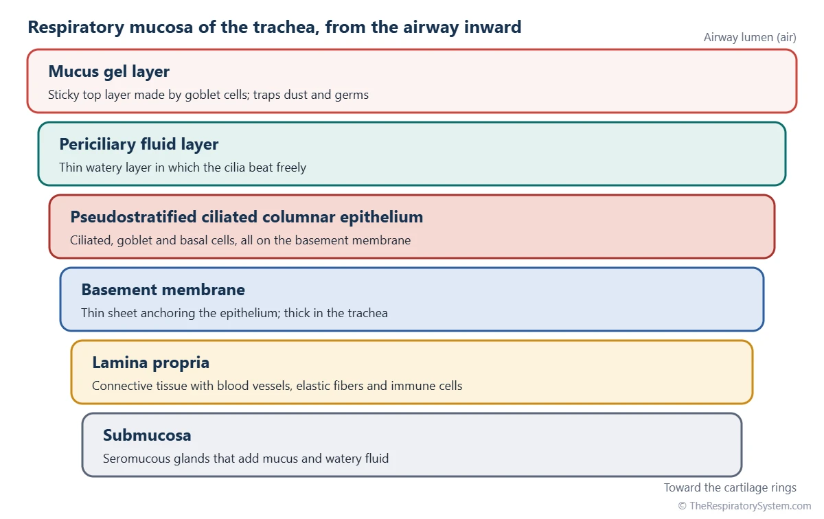
The respiratory mucosa is the full moist lining of the airway: the respiratory epithelium plus its basement membrane and the layer of connective tissue beneath it, called the lamina propria. The epithelium is the surface layer; the mucosa is the whole membrane.
On top of the epithelium sit two layers of fluid: a thin watery periciliary layer in which the cilia beat, and a thicker mucus gel that floats on the cilia tips. The basement membrane is especially thick in the trachea. The lamina propria holds blood vessels, elastic fibers and immune cells, and in the trachea and bronchi a deeper submucosa contains seromucous glands. In the nasal cavity the lamina propria is rich in wide veins that can swell, which warms the air and drives the nasal cycle.
How Does the Airway Lining Change Toward the Alveoli?
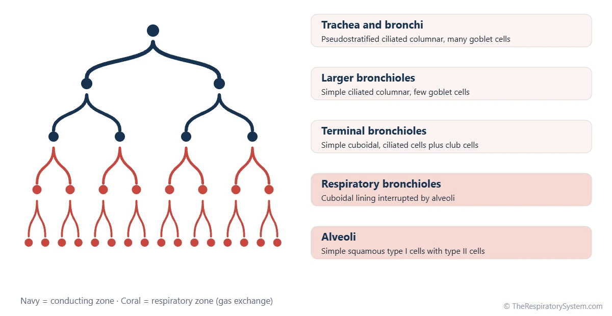
The airway lining becomes shorter and simpler as the airways narrow: pseudostratified in the trachea and bronchi, simple ciliated columnar in the larger bronchioles, simple cuboidal in the terminal bronchioles, and simple squamous in the alveoli.
Goblet cells thin out in the bronchioles and are normally absent by the terminal bronchioles. Their place is taken by club cells, dome-shaped cells that secrete protective proteins, help break down inhaled toxins and can divide to repair the lining. In the respiratory bronchioles the cuboidal lining is interrupted by the first alveoli, described on our terminal and respiratory bronchioles page. The alveoli themselves are lined by very flat type I and rounded type II pneumocytes, with no cilia or mucus, so they rely on alveolar macrophages for cleaning. This shift marks the border on the conducting zone vs respiratory zone page.
Common Conditions That Affect the Respiratory Epithelium
Conditions that affect the respiratory epithelium usually disturb mucus or cilia, which slows clearance and lets mucus and germs collect in the airways. Our respiratory diseases section explains them further, and anyone with breathing symptoms should see a doctor.
- Viral infections: colds and flu damage ciliated cells, which is one reason a cough can linger while the lining regrows.
- Cystic fibrosis: a faulty salt channel makes airway mucus thick and dry, so cilia cannot move it.
- Primary ciliary dyskinesia: an inherited defect leaves cilia unable to beat normally.
- Chronic bronchitis and asthma: goblet cells and glands increase, producing excess mucus.
- Squamous metaplasia: long exposure to tobacco smoke can replace ciliated cells with flat, tougher cells that cannot clear mucus.
Key Facts
- Respiratory epithelium is a ciliated pseudostratified columnar epithelium with goblet cells.
- It lines the nasal cavity, sinuses, nasopharynx, most of the larynx, the trachea and the bronchi.
- All of its cells rest on the basement membrane, but only some reach the surface, which is why it is called pseudostratified.
- Goblet cells make mucus, and ciliated cells sweep it toward the throat in the mucociliary escalator described on our cilia and mucus page.
- Basal cells are stem cells that replace damaged ciliated and goblet cells.
- The vocal folds and the oropharynx are lined by stratified squamous epithelium instead, because they face friction.
- In the bronchioles the lining thins to simple columnar and cuboidal cells, and goblet cells give way to club cells.
Quick Quiz: Test What You Know About the Respiratory Epithelium
6 questions. Tap an answer to check it.
What type of epithelium is respiratory epithelium?
Respiratory epithelium is ciliated pseudostratified columnar epithelium with goblet cells.
Which cells in the respiratory epithelium produce mucus?
Goblet cells release mucin, which forms mucus; seromucous glands in the wall add more.
Why is respiratory epithelium called pseudostratified?
It is a single layer of cells that only looks stratified because the nuclei are at different levels.
Which part of the airway is NOT lined by respiratory epithelium?
The vocal folds are covered by stratified squamous nonkeratinized epithelium, which withstands constant vibration.
Which cells act as stem cells in the respiratory epithelium?
Basal cells sit on the basement membrane and divide to replace ciliated and goblet cells.
What lines the terminal bronchioles?
By the terminal bronchioles the lining is simple cuboidal, and club cells replace goblet cells.
Frequently Asked Questions
Is respiratory epithelium the same as respiratory mucosa?
No. Respiratory epithelium is only the surface layer of cells. The respiratory mucosa is the whole moist lining, made of the epithelium, its basement membrane and the connective tissue beneath it, called the lamina propria. In everyday use the two terms are often mixed up, but in histology the epithelium is one part of the mucosa.
What do goblet cells do in the respiratory tract?
Goblet cells make and release mucin, a large protein that absorbs water and becomes the sticky mucus gel on the airway surface. This mucus catches dust, pollen, smoke particles and germs before they reach the lungs. Goblet cells are most common in the trachea and bronchi, become fewer in the bronchioles and are normally absent from the smallest airways.
Why are the vocal folds not covered by respiratory epithelium?
The vocal folds vibrate hundreds of times a second during speech, and their edges rub together. Delicate ciliated cells would be damaged by this, so the folds are covered by stratified squamous nonkeratinized epithelium, a tougher, many-layered lining similar to the inside of the mouth. Respiratory epithelium resumes just below the vocal folds.
How does respiratory epithelium look under a microscope?
On a standard stained slide it shows tall columnar cells with a fringe of cilia along the top edge and oval nuclei scattered at several different heights. Pale, rounded goblet cells sit between the ciliated cells, and small basal cells line the bottom. In the trachea a thick pink basement membrane is easy to see just below the epithelium.
What is the difference between respiratory and olfactory epithelium?
Both are pseudostratified columnar epithelia in the nasal cavity, but they do different jobs. Respiratory epithelium has goblet cells and motile cilia and cleans the air. Olfactory epithelium, found in a small area in the roof of the nasal cavity, has no goblet cells and contains smell receptor neurons whose non-moving cilia carry the receptors that detect odors.
Can respiratory epithelium repair itself?
Yes. When surface cells are damaged, for example by a viral infection, basal cells divide and develop into new ciliated and goblet cells. In the bronchioles, club cells also take part in repair. Recovery of the cilia after an infection usually takes days to weeks, which is one reason a cough can last after other symptoms have cleared.
How does smoking affect the respiratory epithelium?
Tobacco smoke slows and damages the cilia, so mucus is cleared less well, while goblet cells and mucus glands increase. Over time the ciliated lining can be replaced in patches by flat, stratified squamous cells, a change called squamous metaplasia. These cells resist irritation but cannot move mucus, so mucus builds up and a chronic cough develops.
Does respiratory epithelium line the alveoli?
No. Respiratory epithelium stops in the bronchioles. The alveoli are lined by a very thin simple squamous epithelium made mostly of type I pneumocytes, with scattered type II pneumocytes that make surfactant. A thick, ciliated lining would slow the diffusion of oxygen and carbon dioxide, so the alveoli use macrophages instead of cilia to stay clean.
Keep Exploring


