At a Glance
These respiratory system worksheets cover five skills: labeling the eight main stops on the path of air (nostrils to alveoli), filling in the blanks, matching each part to its job, putting the steps of breathing in into order, and answering short review questions. Every worksheet has an answer key in a table directly below it, and the whole page prints cleanly from any browser.
- Worksheets
- Five: labeling, fill in the blanks, matching, sequencing and sorting, short answer
- Level
- Middle school and high school biology; worksheet 1 also suits upper elementary
- Answer key
- In a table directly after each worksheet
- Format
- Print the page or save it as a PDF from your browser's print menu
- Covers
- Upper and lower tract parts, their functions and the mechanics of breathing
- Pairs with
- The labeled diagram page, the quiz page and the balloon lung model
On this page
- What Is in These Respiratory System Worksheets?
- Worksheet 1: Respiratory System Labeling (Name Each Stop)
- Worksheet 2: Fill in the Blanks
- Worksheet 3: Match Each Part to Its Job
- Worksheet 4: Order a Breath In and Sort Upper vs Lower Tract
- Worksheet 5: Short-Answer Review Questions
- Can These Diagrams Be Used as a Respiratory System Coloring Page?
- How Can Teachers Use Respiratory System Worksheets in Class?
- Key Facts
- FAQ
What Is in These Respiratory System Worksheets?
This page holds five short respiratory system worksheets that build from naming the parts to explaining how they work, and each one ends with an answer key in a table. They follow the order most biology courses teach the topic: structure first, then function, then the mechanics of breathing.
- Worksheet 1, Labeling: name the eight main stops on the path of air from written clues.
- Worksheet 2, Fill in the blanks: complete eleven sentences using a word bank.
- Worksheet 3, Matching: match nine parts of the system to their jobs.
- Worksheet 4, Sequencing and sorting: put the events of breathing in into order, then sort the parts into the upper and lower tract.
- Worksheet 5, Short answer: explain eight "why" questions in one or two sentences each.
To make a printable copy, open your browser's print menu and choose your printer or "Save as PDF". For a quick quiz version, print only the worksheet and fold or cut away the answer key underneath it.
The worksheets practice skills rather than introduce the topic, so students should first study the labeled respiratory system diagram and the parts of the upper respiratory tract and lower respiratory tract.
Worksheet 1: Respiratory System Labeling (Name Each Stop)
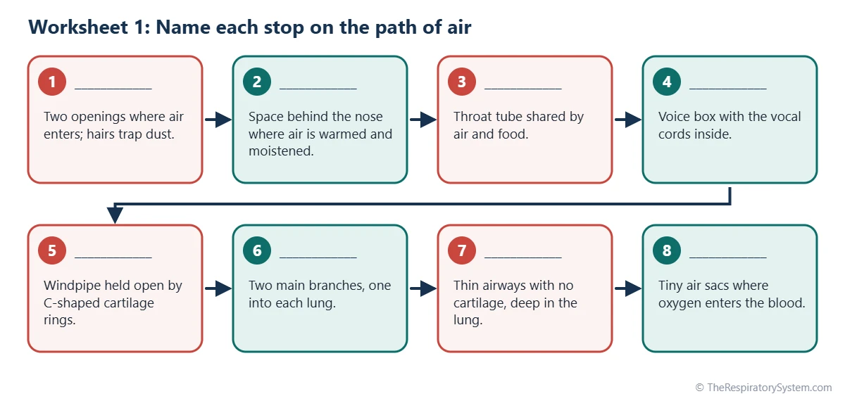
Worksheet 1 is a labeling exercise: the chart at the top of this page shows the eight main stops on the path of a breath, in order, with a blank line and a clue for each one, and students write the correct name on each line. It works as an unlabeled respiratory system diagram in text form, so it suits students who are still learning to read anatomical drawings.
Instructions: Read each clue and write the name of the part on the blank line. The stops are numbered in the order air passes through them on its way in.
Word bank: alveoli, bronchioles, larynx, nasal cavity, nostrils, pharynx, primary bronchi, trachea.
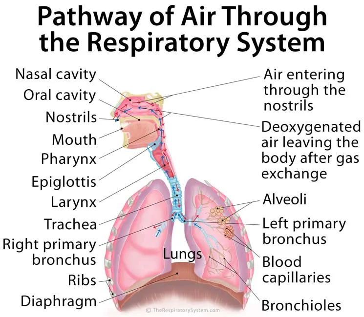
Extension: For a picture-based labeling quiz, cover the labels of the diagram below with sticky notes, number each note, and ask students to write the names on a separate sheet. The same trick turns any labeled image on our site into a blank diagram, including those on the nasal cavity and lungs pages.
| Stop | Answer | Clue it matches |
|---|---|---|
| 1 | Nostrils | Two openings where air enters; hairs trap dust |
| 2 | Nasal cavity | Space behind the nose where air is warmed and moistened |
| 3 | Pharynx | Throat tube shared by air and food |
| 4 | Larynx | Voice box with the vocal cords inside |
| 5 | Trachea | Windpipe held open by C-shaped cartilage rings |
| 6 | Primary bronchi | Two main branches, one into each lung |
| 7 | Bronchioles | Thin airways with no cartilage, deep in the lung |
| 8 | Alveoli | Tiny air sacs where oxygen enters the blood |
| Source: Answer key, our editors | ||
Worksheet 2: Fill in the Blanks
Worksheet 2 checks vocabulary by asking students to complete sentences about each part of the respiratory system with one word or short phrase from the word bank.
Word bank: alveoli, bronchi, cartilage, diaphragm, diffusion, epiglottis, larynx, nasal conchae, nostrils, pharynx, three.
- Air enters the body through the __________ and then passes into the nasal cavity.
- Curled shelves of bone called the __________ swirl the air inside the nasal cavity.
- The __________ is a muscular tube in the throat that carries both air and food.
- During swallowing, a flap called the __________ covers the entrance to the larynx.
- The vocal cords are found inside the __________, also called the voice box.
- The trachea is held open by C-shaped rings of __________.
- At its lower end, the trachea divides into the right and left primary __________.
- Gas exchange takes place in tiny air sacs called __________.
- The right lung has __________ lobes, while the left lung has two.
- When the __________ contracts, it flattens and air flows into the lungs.
- Oxygen moves from the alveoli into the blood by __________.
Students who finish early can write one more sentence of their own for any two words in the bank, using a fact from the trachea or alveoli pages.
| Sentence | Answer | Why |
|---|---|---|
| 1 | nostrils | The two openings of the nose are the entrance for air |
| 2 | nasal conchae | Also called turbinates; they create swirling airflow |
| 3 | pharynx | The throat is a shared passage for air and food |
| 4 | epiglottis | It tips back over the larynx when you swallow |
| 5 | larynx | The vocal cords stretch across the inside of the voice box |
| 6 | cartilage | Hyaline cartilage rings stop the trachea from collapsing |
| 7 | bronchi | One primary bronchus enters each lung |
| 8 | alveoli | Their thin walls touch a dense net of capillaries |
| 9 | three | Superior, middle and inferior lobes; the left lung has two |
| 10 | diaphragm | Its contraction enlarges the chest cavity |
| 11 | diffusion | Oxygen moves from higher to lower partial pressure |
| Source: Answer key, our editors | ||
Worksheet 3: Match Each Part to Its Job
Worksheet 3 is a matching exercise that links each part of the respiratory system to its main function, which is the skill most tests ask for after labeling.
Instructions: Write the number of the correct job next to each letter. Each job is used once.
Parts
- A. Nasal cavity
- B. Pharynx
- C. Epiglottis
- D. Larynx
- E. Trachea
- F. Bronchioles
- G. Alveoli
- H. Diaphragm
- I. Cilia
Jobs
- Exchange oxygen and carbon dioxide with the blood.
- Produces sound as air passes the vocal cords.
- Main muscle that pulls air into the lungs.
- Filters, warms and moistens incoming air.
- Stops food and drink from entering the airway.
- Carries both air and food.
- Small airways deep in the lung that can narrow or widen.
- Stiff tube that carries air from the neck into the chest.
- Sweep mucus and trapped dust up toward the throat.
A good follow-up question is to ask which two parts protect the airway (the epiglottis and the cilia) and how. Our pages on the epiglottis and on cilia and mucus explain both defenses.
| Part | Job number | Job |
|---|---|---|
| A. Nasal cavity | 4 | Filters, warms and moistens incoming air |
| B. Pharynx | 6 | Carries both air and food |
| C. Epiglottis | 5 | Stops food and drink from entering the airway |
| D. Larynx | 2 | Produces sound as air passes the vocal cords |
| E. Trachea | 8 | Stiff tube that carries air from the neck into the chest |
| F. Bronchioles | 7 | Small airways deep in the lung that can narrow or widen |
| G. Alveoli | 1 | Exchange oxygen and carbon dioxide with the blood |
| H. Diaphragm | 3 | Main muscle that pulls air into the lungs |
| I. Cilia | 9 | Sweep mucus and trapped dust up toward the throat |
| Source: Answer key, our editors | ||
Worksheet 4: Order a Breath In and Sort Upper vs Lower Tract
Worksheet 4 has two parts: students first number the events of one breath in, then sort a list of parts into the upper and lower respiratory tract.
Part A: Put the events of breathing in into order (1 to 7)
- ___ Air flows in through the airways until the pressures are equal.
- ___ The diaphragm contracts and flattens.
- ___ The brainstem sends signals along the nerves to the breathing muscles.
- ___ Air pressure inside the lungs falls below the air pressure outside.
- ___ The external intercostal muscles lift the ribs up and out.
- ___ The lungs expand along with the chest wall.
- ___ The chest cavity becomes larger.
Part B: Sort the parts
Write U for upper respiratory tract or L for lower respiratory tract next to each part: trachea, nasal cavity, alveoli, pharynx, bronchi, paranasal sinuses, bronchioles, nostrils.
Part B answers: upper tract = nasal cavity, pharynx, paranasal sinuses, nostrils; lower tract = trachea, alveoli, bronchi, bronchioles. The larynx was left off the list on purpose, because textbooks disagree on which tract it belongs to, as our upper vs lower respiratory tract page explains.
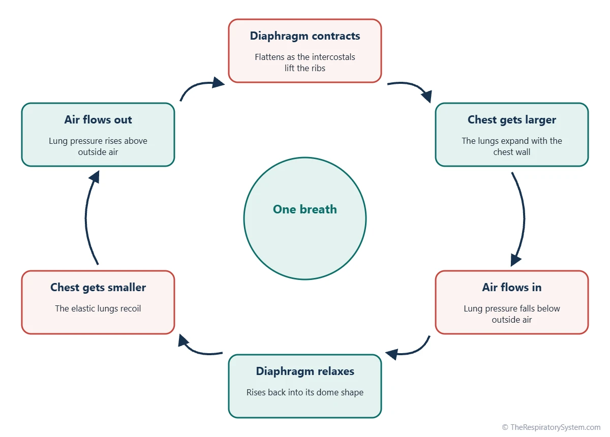
The cycle diagram below works as a picture answer key for Part A and adds the second half of the cycle, breathing out, for students who finish early. For the full explanation, see inhalation and exhalation and the diaphragm.
| Order | Event | Note |
|---|---|---|
| 1 | The brainstem sends signals to the breathing muscles | The phrenic nerve carries them to the diaphragm |
| 2 | The diaphragm contracts and flattens | Steps 2 and 3 happen at the same time |
| 3 | The external intercostal muscles lift the ribs up and out | Either order of 2 and 3 is correct |
| 4 | The chest cavity becomes larger | It grows from top to bottom and front to back |
| 5 | The lungs expand along with the chest wall | Pleural fluid holds the lungs against the chest wall |
| 6 | Air pressure inside the lungs falls below outside pressure | Larger volume means lower pressure |
| 7 | Air flows in until the pressures are equal | Then breathing out begins |
| Source: Answer key, our editors | ||
Worksheet 5: Short-Answer Review Questions
Worksheet 5 asks students to explain, in one or two sentences, why the respiratory system is built the way it is, which tests understanding rather than memory.
- Why is breathing through the nose better for the airway than breathing through the mouth at rest?
- Why are the cartilage rings of the trachea shaped like a C instead of a full circle?
- Why does the left lung have only two lobes?
- How are the alveoli suited to gas exchange? Give two features.
- What happens to the diaphragm during a quiet breath out?
- What are cilia, and why does the airway need them?
- Which two gases are exchanged in the lungs, and in which direction does each move?
- What is the difference between breathing and respiration?
Model answers are in the key below. Award full marks for any answer that gives the same idea in the student's own words, and use the linked pages, such as gas exchange and respiratory system functions, for follow-up reading.
| Question | Model answer |
|---|---|
| 1. Nose vs mouth | The nose filters, warms and moistens air before it reaches the lungs; the mouth does this far less well |
| 2. C-shaped rings | The rings keep the trachea open, while the soft open back lets the esophagus behind it bulge when food is swallowed |
| 3. Two lobes on the left | The heart sits slightly to the left, so the left lung is smaller and has a notch for it |
| 4. Alveoli and gas exchange | There are hundreds of millions of them, with very thin, moist walls wrapped in capillaries |
| 5. Quiet breath out | The diaphragm relaxes and rises back into a dome, so the chest gets smaller and air is pushed out |
| 6. Cilia | Tiny hair-like projections on airway cells that sweep mucus, dust and germs up toward the throat |
| 7. Gases exchanged | Oxygen moves from the alveoli into the blood; carbon dioxide moves from the blood into the alveoli |
| 8. Breathing vs respiration | Breathing moves air in and out; respiration also includes gas exchange and the use of oxygen by cells |
| Source: Answer key, our editors | |
Can These Diagrams Be Used as a Respiratory System Coloring Page?
Yes, any of the labeled diagrams on our site can be printed in black and white and colored in, and coloring each part a fixed color is a simple way for younger students to remember which part is which. Coloring works best when the class agrees on one color key and adds a small key box in the corner of the page.
- Nose, nasal cavity and sinuses: light pink, for the moist lining that warms the air.
- Pharynx and larynx: darker pink or red, for the muscular throat and voice box.
- Trachea and bronchi: light blue, with the cartilage rings outlined in white or gray.
- Lungs: pale pink or purple, with the lobes outlined in a darker pencil.
- Alveoli: orange or yellow, so the gas exchange sites stand out.
- Diaphragm: brown or dark red, for muscle.
After coloring, ask students to add one arrow showing where air enters and one showing where oxygen enters the blood. Our page on the respiratory system for kids has a simpler set of labels for younger classes.
How Can Teachers Use Respiratory System Worksheets in Class?
Respiratory system worksheets work best as short, focused practice after a lesson, used one or two at a time rather than all at once, with the answer key used for self-checking or peer marking. Most students finish a single worksheet in a short part of one class period.
- Warm-up: start the next lesson with Worksheet 1 to recall the parts.
- Homework: set Worksheets 2 and 3 together for vocabulary and function.
- Hands-on lesson: pair Worksheet 4 with a balloon lung model so students watch the diaphragm change the pressure.
- Revision: use Worksheet 5 before a test, then the online respiratory system quiz as a final check.
- Differentiation: give younger or struggling students the word banks; ask advanced students to complete Worksheet 1 without one and to add the epiglottis and diaphragm.
For students who want to go further, the pathway of air and organs of the respiratory system pages follow the same order as these worksheets.
Key Facts
- Labeling the parts of the airway in the order air flows, from the nostrils to the alveoli, makes them easier to remember.
- The eight main stops on the path of air are the nostrils, nasal cavity, pharynx, larynx, trachea, primary bronchi, bronchioles and alveoli.
- The right lung has three lobes and the left lung has two, a favorite labeling question on tests.
- During breathing in, the diaphragm contracts and flattens, the chest enlarges, lung pressure falls and air flows in.
- Quiet breathing out is passive: the diaphragm relaxes and the elastic lungs recoil.
- Any labeled diagram becomes a blank diagram for a labeling quiz when its labels are covered with numbered sticky notes.
- Textbooks disagree on whether the larynx belongs to the upper or lower tract, so follow your own course's rule.
Frequently Asked Questions
Where can I find a respiratory system blank diagram?
You can make one from any labeled diagram on our site. Print the labeled respiratory system diagram, cover each label with a numbered sticky note or strip of paper, and photocopy the page. The copy is an unlabeled respiratory system diagram, and the original serves as the answer key. Worksheet 1 on this page is a text-based version that needs no drawing at all.
Can I print these worksheets or save them as a PDF?
Yes. Open your browser's print menu, choose a printer or the option to save as PDF, and print the whole page or only the pages you need. The worksheets are free for personal and classroom use. To print a worksheet without its answers, print the page range for that worksheet only, or fold the answer key under before photocopying.
What should a respiratory system labeling quiz include?
A good labeling quiz asks for the main structures in airflow order: nostrils, nasal cavity, pharynx, epiglottis, larynx, trachea, primary bronchi, bronchioles, alveoli, the lobes of each lung and the diaphragm. Harder versions add the paranasal sinuses, the nasal conchae, the cartilage rings of the trachea and the intercostal muscles. One function question per part checks real understanding.
What grade level are these respiratory system worksheets for?
They are written for middle school and high school life science and biology, roughly grades 6 to 12. Worksheet 1 with its word bank also suits upper elementary students who have studied the parts. For anatomy and physiology courses, add the harder labels and ask for full-sentence answers to Worksheet 5.
Where can I find a nasal cavity diagram unlabeled?
Our nasal cavity page includes a labeled diagram of the nasal cavity and its bones. Print it and cover the labels to create an unlabeled version for practice. The key structures to label are the nostrils, nasal septum, the superior, middle and inferior nasal conchae, the meatuses beneath them, the hard and soft palate and the opening into the nasopharynx.
What does a thoracic cavity diagram show?
A thoracic cavity diagram shows the chest space enclosed by the ribs, sternum and spine, with the diaphragm as its floor. It contains the two lungs, each in its own pleural cavity, and the mediastinum in the middle, which holds the heart, the great blood vessels, the esophagus and the lower trachea. Unlabeled versions ask students to name these regions and their borders.
How long does each worksheet take?
Most students complete one worksheet in about ten to twenty minutes, depending on age and whether they use the word bank. Worksheets 1 to 3 are quickest because they test recall. Worksheets 4 and 5 take longer because they ask students to reason about how breathing works, so they suit homework or the second half of a lesson.
Keep Exploring


