At a Glance
This page holds 60 respiratory system questions with answers: 54 practice questions in seven topic sets (upper tract, lower tract and lungs, thoracic cavity, histology, breathing mechanics, gas exchange and control, lung volumes), each followed by an answer table, and a 6-question interactive quiz. Cover the answer column, write your answers, then check them.
- Questions
- 60 in total: 54 practice questions with answers and a 6-question interactive quiz
- Topics
- Anatomy, thoracic cavity, histology, breathing mechanics, gas exchange, control and lung volumes
- Level
- High school biology to first-year anatomy and physiology
- Format
- Short-answer sets with answer tables, plus tap-to-check multiple choice
- Values used
- Typical textbook values for a healthy young adult at rest
- Pairs with
- The labeled diagram and the printable worksheets
On this page
- How Does This Respiratory System Quiz Work?
- Respiratory System Anatomy Quiz: Upper Respiratory Tract
- Lower Respiratory Tract and Lung Anatomy Questions
- Thoracic Cavity Anatomy Quiz: Pleura, Mediastinum and Diaphragm
- Respiratory System Histology Quiz
- Breathing Mechanics Questions: How Does Air Move In and Out?
- Gas Exchange, Transport and Control of Breathing Questions
- Lung Volumes and Capacities Quiz
- How Can You Score Higher on a Respiratory System Test?
- Key Facts
- Quiz
- FAQ
How Does This Respiratory System Quiz Work?
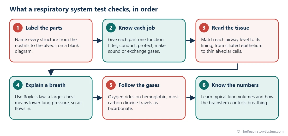
The quiz is split into seven practice sets that follow the order most courses teach the respiratory system, from naming the parts to explaining how breathing is controlled, and every set ends with a table of answers. A six-question multiple-choice quiz near the bottom of the page lets you tap an answer and check it straight away.
- Read the short note at the start of each set to see what it tests.
- Cover the answer column of the table with a sheet of paper.
- Write or say your answer to each question, then uncover it and mark yourself.
- For every question you miss, open the linked topic page and reread that part.
- Come back a day or two later and retry only the questions you missed.
If you are just starting the topic, study the labeled respiratory system diagram first, or use the printable respiratory system worksheets, which give more support with word banks. Numbers in the answers are typical textbook values for a healthy young adult at rest; real values vary with body size, age and sex.
Respiratory System Anatomy Quiz: Upper Respiratory Tract
This set tests the anatomy of the upper respiratory tract: the nose, nasal cavity, paranasal sinuses, pharynx and larynx, including the pharynx anatomy questions that often appear on tests. The key to this set is knowing each part's position relative to its neighbors, not just its name.
Before you start, picture a side view of the head. The nasal cavity sits above the roof of the mouth, the pharynx runs down behind both, and the larynx sits at the front of the neck where the airway separates from the food passage. Most wrong answers in this set come from mixing up the three parts of the pharynx or the cartilages of the larynx.
| Question | Answer |
|---|---|
| 1. What divides the nasal cavity into left and right halves? | The nasal septum, made of cartilage at the front and bone at the back |
| 2. Which nasal concha is a separate bone of its own? | The inferior nasal concha; the superior and middle conchae are parts of the ethmoid bone |
| 3. Name the four paired paranasal sinuses. | Frontal, maxillary, ethmoid and sphenoid sinuses |
| 4. What are the openings from the nasal cavity into the nasopharynx called? | The choanae, or internal nares |
| 5. Name the three parts of the pharynx from top to bottom. | Nasopharynx, oropharynx and laryngopharynx |
| 6. Which part of the pharynx carries air only, not food? | The nasopharynx, which the soft palate seals off during swallowing |
| 7. What is the largest cartilage of the larynx? | The thyroid cartilage, which forms the laryngeal prominence or Adam's apple |
| 8. Which laryngeal cartilage forms a complete ring? | The cricoid cartilage, just below the thyroid cartilage |
| Source: Standard human anatomy, our editors | |
Lower Respiratory Tract and Lung Anatomy Questions
This set covers the lower respiratory tract: the trachea, the branching bronchial tree and the anatomy of the lungs themselves, including lobes, fissures and the hilum. The questions most students miss are the ones about differences between the right and left sides.
Remember three right-left contrasts. The right main bronchus is wider, shorter and more vertical; the right lung has three lobes and two fissures while the left has two lobes and one fissure; and only the left lung has a cardiac notch and a lingula. For the full anatomy, see our pages on the trachea, the bronchi and the lungs.
| Question | Answer |
|---|---|
| 1. What holds the trachea open? | About 16 to 20 C-shaped rings of hyaline cartilage |
| 2. What is the ridge where the trachea divides called? | The carina, roughly level with the sternal angle |
| 3. Which main bronchus is wider, shorter and more vertical? | The right main bronchus, so inhaled objects lodge there more often |
| 4. How many lobes does each lung have? | Three in the right lung, two in the left lung |
| 5. Which fissures does the right lung have? | The oblique and horizontal fissures; the left lung has only an oblique fissure |
| 6. What and where is the lingula? | A tongue-shaped part of the superior lobe of the left lung, below the cardiac notch |
| 7. What is the hilum of the lung? | The area on the inner surface where the bronchus, blood vessels and nerves enter and leave |
| 8. Which is the first airway to have alveoli in its walls? | The respiratory bronchiole, the start of the respiratory zone |
| Source: Standard human anatomy, our editors | |
Thoracic Cavity Anatomy Quiz: Pleura, Mediastinum and Diaphragm
This thoracic cavity anatomy quiz checks how the lungs are packaged inside the chest: the pleural layers, the mediastinum between the lungs and the muscles that form the walls and floor. These structures are not airways, but questions about them appear on almost every respiratory system test.
Think of each lung as sitting inside a closed, double-layered bag. The inner layer covers the lung, the outer layer lines the chest wall, and a thin film of fluid between them lets the lung slide as it moves. The diaphragm closes the cavity from below, and the intercostal muscles fill the spaces between the ribs.
| Question | Answer |
|---|---|
| 1. What forms the floor of the thoracic cavity? | The diaphragm |
| 2. Name the two layers of the pleura. | Visceral pleura on the lung surface and parietal pleura lining the chest wall |
| 3. What fills the pleural cavity? | A thin film of serous pleural fluid that reduces friction |
| 4. What is the mediastinum? | The central compartment between the lungs, holding the heart, great vessels, esophagus and trachea |
| 5. Which nerve supplies the diaphragm? | The phrenic nerve, from spinal nerves C3 to C5 |
| 6. Which three major structures pass through the diaphragm? | The inferior vena cava, the esophagus and the aorta |
| 7. Which muscles lie between the ribs? | The external, internal and innermost intercostal muscles |
| Source: Standard human anatomy, our editors | |
Respiratory System Histology Quiz
This respiratory system histology quiz tests the tissues that line each level of the airway, a topic that trips up many students because the lining changes gradually from the nose to the alveoli. The general rule is that the epithelium becomes thinner and simpler the deeper you go, until it is thin enough for gas exchange.
In the trachea and bronchi the lining is tall, ciliated and full of mucus-making goblet cells, which trap and clear dust. In the bronchioles the cartilage disappears and the cells become shorter. In the alveoli, flat type I cells form most of the wall, while rounder type II cells make surfactant. Our page on cilia and mucus explains how the lining keeps the airway clean.
| Question | Answer |
|---|---|
| 1. What epithelium lines the trachea and bronchi? | Pseudostratified ciliated columnar epithelium, often called respiratory epithelium |
| 2. Which cells in that lining secrete mucus? | Goblet cells, helped by submucosal glands |
| 3. At which airway level does cartilage disappear from the wall? | The bronchioles |
| 4. Which cells form most of the alveolar surface? | Type I pneumocytes, very thin flat cells |
| 5. Which cells make pulmonary surfactant? | Type II pneumocytes |
| 6. Which cells clear dust and microbes from the alveoli? | Alveolar macrophages, also called dust cells |
| 7. What epithelium covers the vocal folds? | Stratified squamous epithelium, which resists wear from vibration |
| Source: Standard human histology, our editors | |
Breathing Mechanics Questions: How Does Air Move In and Out?
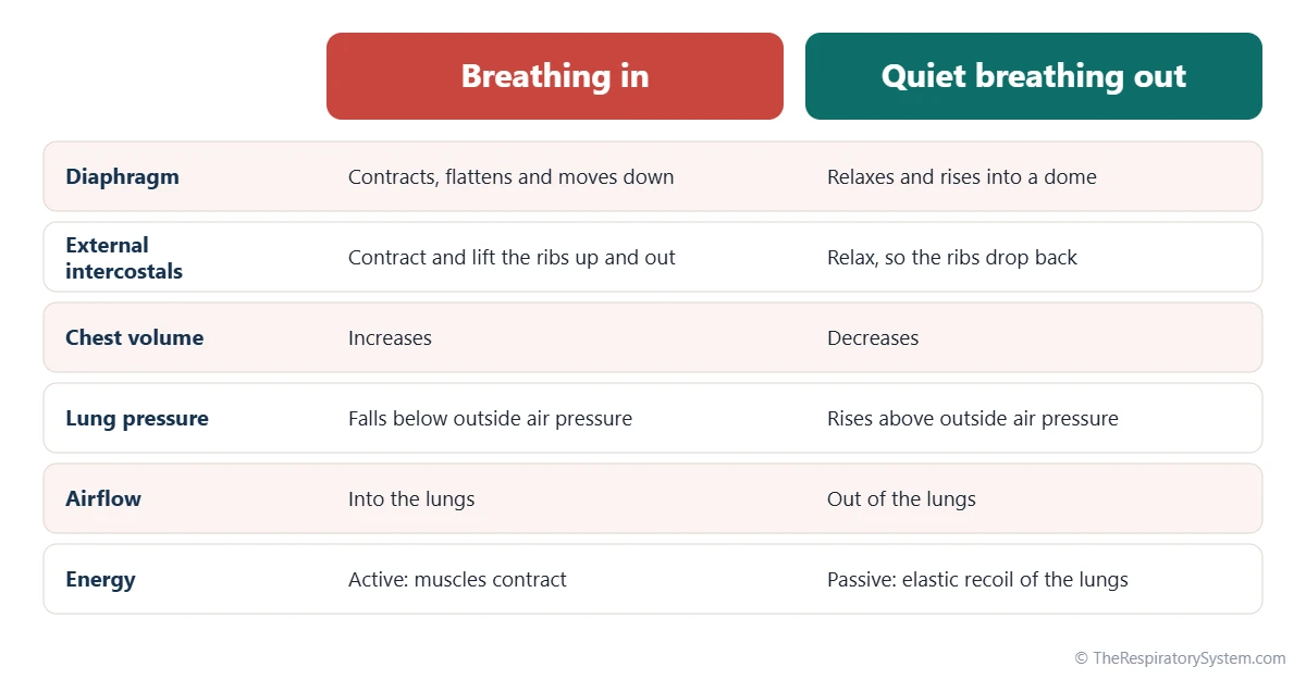
This set tests pulmonary ventilation, the physical process that moves air into and out of the lungs, and almost every answer comes back to one rule: when the volume of the chest increases, the pressure inside the lungs falls and air flows in. That rule is Boyle's law, which states that at a constant temperature the pressure of a gas falls as its volume rises.
Know which muscles act in each phase. Quiet breathing in uses the diaphragm and the external intercostals; quiet breathing out is passive recoil; forced breathing out adds the internal intercostals and the abdominal muscles. The comparison chart below summarizes the two phases, and our respiratory system functions page places breathing within everything else the system does.
| Question | Answer |
|---|---|
| 1. Which gas law explains how air moves into the lungs? | Boyle's law: as chest volume rises, lung pressure falls |
| 2. What is the main muscle of quiet breathing in? | The diaphragm |
| 3. Is quiet breathing out active or passive? | Passive: the muscles relax and the elastic lungs recoil |
| 4. Which muscles help with forced breathing out? | The internal intercostals and the abdominal muscles |
| 5. Name two accessory muscles of breathing in. | Any two of the sternocleidomastoid, scalenes and pectoralis minor |
| 6. What keeps the lungs pressed against the chest wall? | The pleural fluid and the slightly negative pressure in the pleural cavity |
| 7. What lowers surface tension inside the alveoli? | Pulmonary surfactant, made by type II pneumocytes |
| 8. What does lung compliance describe? | How easily the lungs stretch when pressure changes |
| Source: Standard human physiology, our editors | |
Gas Exchange, Transport and Control of Breathing Questions
This set tests what happens after air reaches the alveoli: how oxygen and carbon dioxide cross into and out of the blood, how the blood carries them, and how the brainstem adjusts breathing to keep their levels steady. It is the most physiology-heavy set and the one most often examined in anatomy and physiology courses.
Hold on to three ideas. Gases cross the respiratory membrane by simple diffusion, moving from higher to lower partial pressure, as explained on our gas exchange page. Oxygen travels mostly bound to hemoglobin, while carbon dioxide travels mostly as bicarbonate. And at rest, the level of carbon dioxide in the blood, sensed through its effect on acidity, is the main signal that sets how fast and deep you breathe.
| Question | Answer |
|---|---|
| 1. How do oxygen and carbon dioxide cross the respiratory membrane? | By simple diffusion, from higher to lower partial pressure |
| 2. What is the typical oxygen partial pressure in alveolar air? | About 100 mmHg, versus about 40 mmHg in blood arriving at the lungs |
| 3. How is most oxygen carried in the blood? | Bound to hemoglobin in red blood cells, about 98 to 99% |
| 4. How is most carbon dioxide carried in the blood? | As bicarbonate ions in the plasma, about 70% |
| 5. Which enzyme speeds the reaction of carbon dioxide with water? | Carbonic anhydrase, inside red blood cells |
| 6. What is the Bohr effect? | More carbon dioxide and acid lower hemoglobin's grip on oxygen, so tissues receive more |
| 7. Which part of the brain sets the basic breathing rhythm? | The medulla oblongata, fine-tuned by the pons |
| 8. What is the main chemical stimulus for breathing at rest? | A rise in carbon dioxide, sensed through the acidity it causes |
| 9. Where are the peripheral chemoreceptors? | In the carotid bodies and the aortic bodies |
| Source: Typical values for a healthy adult at rest at sea level, our editors | |
Lung Volumes and Capacities Quiz
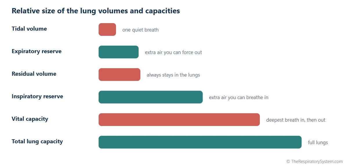
This lung volumes and capacities quiz checks the four basic lung volumes measured with a spirometer and the capacities made by adding them together. Learn the volumes first, because every capacity is simply a sum of two or more of them.
- Tidal volume: the air of one quiet breath.
- Inspiratory reserve volume: the extra air you can still breathe in after a normal breath in.
- Expiratory reserve volume: the extra air you can still force out after a normal breath out.
- Residual volume: the air that always stays in the lungs and cannot be breathed out.
The bar chart below shows their relative sizes without numbers; the exact typical values are in the answer table. Values in a textbook are averages for a healthy young adult, and real values are larger in taller people and smaller in shorter and older people.
| Question | Answer |
|---|---|
| 1. What is a typical resting tidal volume? | About 500 mL per breath |
| 2. What is a typical inspiratory reserve volume? | About 3,000 mL |
| 3. What is a typical expiratory reserve volume? | About 1,100 to 1,200 mL |
| 4. Which volume cannot be measured with simple spirometry? | Residual volume, about 1,200 mL, because it never leaves the lungs |
| 5. Vital capacity is the sum of which volumes? | Tidal volume + inspiratory reserve + expiratory reserve, about 4.5 to 5 liters |
| 6. What is a typical total lung capacity? | About 6 liters: vital capacity plus residual volume |
| 7. What is the normal resting breathing rate of an adult? | About 12 to 20 breaths per minute |
| Source: Typical values for a healthy young adult male at rest, our editors | |
How Can You Score Higher on a Respiratory System Test?
The fastest way to raise your score is to learn the parts in the order air flows through them, attach one function to each part, and then practice explaining processes out loud rather than rereading notes. Testing yourself, as on this page, works better than reading the same chapter again.
- Use the airflow order. Nose, pharynx, larynx, trachea, bronchi, bronchioles, alveoli: most labeling and sequencing questions follow it. Our pathway of air page walks through it.
- Watch the right-left traps. Diagrams show the body's right on your left, and the right lung has the extra lobe.
- Separate similar words. Pharynx and larynx, bronchi and bronchioles, ventilation and respiration, and visceral and parietal pleura are classic mix-ups.
- Learn mechanisms, not just facts. Be ready to explain why the chest pressure changes, why oxygen diffuses and why carbon dioxide drives breathing.
- Build a model. A balloon lung model makes the pressure changes of breathing easy to remember.
Key Facts
- The three parts of the pharynx from top to bottom are the nasopharynx, oropharynx and laryngopharynx.
- The right lung has three lobes and two fissures; the left lung has two lobes, one fissure, a cardiac notch and a lingula.
- Respiratory epithelium, the lining of the trachea and bronchi, is pseudostratified ciliated columnar epithelium with goblet cells.
- Quiet breathing in is active and driven mainly by the diaphragm; quiet breathing out is passive recoil.
- About 98 to 99% of the oxygen in the blood is carried by hemoglobin, while about 70% of the carbon dioxide travels as bicarbonate.
- At rest, a rise in carbon dioxide is the main signal that increases breathing, detected mainly by central chemoreceptors in the brainstem.
- A typical resting tidal volume is about 500 mL, and a typical total lung capacity is about 6 liters.
Quick Quiz: Test What You Know About the Respiratory System
6 questions. Tap an answer to check it.
Which structure covers the entrance to the larynx during swallowing?
The epiglottis tips back over the laryngeal inlet so food and drink pass into the esophagus instead of the airway.
Where does gas exchange take place in the lungs?
Oxygen and carbon dioxide diffuse across the thin walls of the alveoli and their capillaries. The trachea and bronchi only conduct air.
How many lobes does the left lung have?
The left lung has a superior and an inferior lobe. It is smaller than the right lung because the heart sits slightly to the left.
Which muscle does most of the work in quiet breathing in?
The diaphragm contracts and flattens, enlarging the chest cavity. The external intercostals help, and the sternocleidomastoid joins in only during forced breathing.
Which cells produce pulmonary surfactant?
Type II pneumocytes make surfactant, which lowers surface tension and keeps the alveoli from collapsing. Type I cells form the thin gas exchange surface.
Which part of the brain sets the basic rhythm of breathing?
Breathing centers in the medulla oblongata generate the basic rhythm, and centers in the pons help smooth it.
Frequently Asked Questions
What are the most common respiratory system quiz questions?
The questions that appear most often ask you to label the parts in airflow order, name the function of each part, state where gas exchange happens, explain how the diaphragm causes air to flow in, and compare the right and left lungs. Many tests also ask how oxygen and carbon dioxide are carried in the blood and what the main lung volumes are.
How should I study for a respiratory system test?
Start with a labeled diagram until you can name every part on a blank copy. Then attach one function to each part, practice explaining one full breath and the path of oxygen into the blood out loud, and finish with timed practice questions like the sets on this page. Spread the practice over several short sessions rather than one long one.
Is this respiratory system quiz suitable for medical students?
It suits first-year anatomy and physiology, nursing and allied health students as a warm-up and revision check. Medical school exams go deeper, into topics such as ventilation-perfusion matching, compliance curves, the oxygen-hemoglobin dissociation curve and acid-base balance, so medical students should use this quiz to check the foundations before moving on to clinical question banks.
What topics are on an anatomy and physiology respiratory system exam?
A typical anatomy and physiology exam covers the gross anatomy of the upper and lower tract, the pleura and mediastinum, the histology of the airway and alveoli, the mechanics of ventilation, gas exchange and partial pressures, oxygen and carbon dioxide transport, the control of breathing by the brainstem and chemoreceptors, and lung volumes and capacities measured by spirometry.
Why do lung volume values differ between textbooks?
Lung volumes depend on height, age, sex and fitness, so each textbook quotes averages from a slightly different reference group, often a healthy young adult male. The relationships stay the same in every source, though: vital capacity is the sum of tidal volume and the two reserve volumes, and total lung capacity adds the residual volume. Use the values your course teaches on an exam.
What is the difference between ventilation and respiration?
Ventilation, or breathing, is the movement of air into and out of the lungs by the breathing muscles. Respiration is a broader term covering external respiration, the gas exchange between alveoli and blood, internal respiration, the exchange between blood and body cells, and cellular respiration, the use of oxygen inside cells to release energy. Exams often test this distinction.
What should I do with the questions I get wrong?
Write each missed question on a card with the correct answer on the back, then read the linked topic page for that set, such as the trachea, alveoli or gas exchange page, to understand why the answer is right. Retest yourself on only the missed cards a day or two later, and keep a card in the pile until you answer it correctly twice in a row.
Keep Exploring


