At a Glance
The nasal septum is the partition that separates the left and right halves of the nasal cavity. Its front part is flexible septal cartilage made of hyaline cartilage, and its back part is bone: the perpendicular plate of the ethmoid bone above and the vomer below. Mucous membrane covers both sides, and its front carries the small blood vessel network where most nosebleeds start.
- Location
- Midline of the nasal cavity, from the columella between the nostrils back to the choanae
- Made of
- Septal cartilage in front; perpendicular plate of the ethmoid bone and vomer behind
- Covering
- Ciliated respiratory mucosa, with olfactory epithelium on its highest strip
- Function
- Divides airflow, supports the bridge and tip of the nose, helps warm and moisten air
- Blood supply
- Branches of the ophthalmic, maxillary and facial arteries, meeting at Kiesselbach's plexus
- Part of
- Nose and nasal cavity, upper respiratory tract
On this page
- What Is the Nasal Septum?
- Where Is the Nasal Septum Located?
- What Is the Nasal Septum Made Of?
- Nasal Septum vs Vomer: What Is the Difference?
- Blood Supply and Nerves of the Nasal Septum
- What Is the Function of the Nasal Septum?
- How Does the Nasal Septum Develop and Change With Age?
- Common Conditions That Affect the Nasal Septum
- Key Facts
- Quiz
- FAQ
What Is the Nasal Septum?
The nasal septum is the vertical wall in the middle of the nose that separates the right nasal passage from the left one. Each face of the septum forms the medial (inner) wall of one half of the nasal cavity, while the curled nasal conchae project from the opposite, lateral wall. The name comes from the Latin saeptum, meaning a fence or partition.
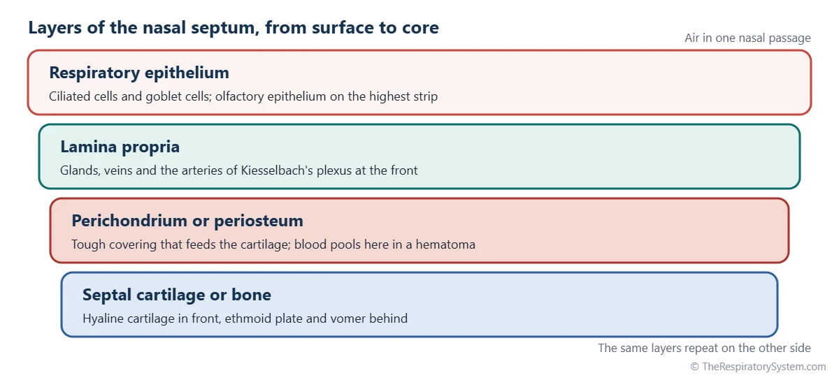
The septum is a composite structure. Its front portion is a flexible plate of hyaline cartilage, its back portion is bone, and a short strip of skin and fibrous tissue at the very front sits between the nostrils. A mucous membrane covers both of its faces, so in life it is a soft, moist surface with a firm core. It belongs to the nose and is one of the first structures inhaled air meets in the upper respiratory tract.
Explained simply: think of the inside of your nose as two narrow hallways side by side. The nasal septum is the wall between them. It is bendy at the front, where you can wiggle the tip of your nose, and hard bone further back.
Where Is the Nasal Septum Located?
The nasal septum lies in the midline of the face, running from the nostrils at the front to the choanae at the back, where the nasal cavity opens into the throat. You can feel its front end as the column of tissue between your two nostrils, called the columella.
Borders of the nasal septum
- Front: the columella and the tip of the nose, where the septum meets the alar cartilages that shape the nostrils.
- Roof: the nasal bones and the nasal spine of the frontal bone in front, then the thin cribriform plate of the ethmoid bone, which separates the nose from the brain.
- Floor: the hard palate, formed by the maxillae and palatine bones, which is also the roof of the mouth.
- Back: the free rear edge of the vomer, which forms the inner border of each choana and faces the nasopharynx.
Most of the septum is hidden behind the bridge of the nose. Only the columella and the area just inside the nasal vestibule can be seen without instruments.
What Is the Nasal Septum Made Of?
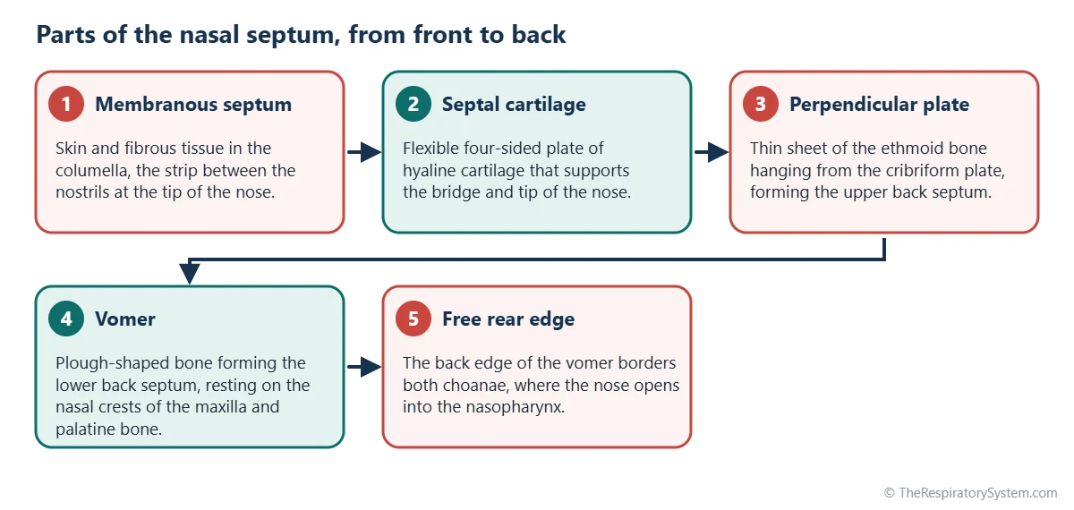
The nasal septum is made of cartilage in front and bone behind, joined by fibrous tissue and covered on both sides by mucous membrane. Anatomists describe three parts: membranous, cartilaginous and bony.
Membranous and cartilaginous septum
The membranous septum is the short, skin-covered segment in the columella, between the inner limbs of the two major alar cartilages. Behind it lies the septal cartilage, also called the quadrangular cartilage because of its roughly four-sided shape. It is hyaline cartilage, the same glassy type found in the rings of the trachea, and it fills the angle between the two bones behind it. Its lower edge sits in a groove on the nasal crest of the maxilla, and a narrow strip called the vomeronasal cartilage lies along its lower border.
Bony septum
The bony septum has two main pieces. The perpendicular plate of the ethmoid bone forms the upper back part and hangs down from the cribriform plate. The vomer, a thin bone shaped like a plough blade, forms the lower back part and rests on the nasal crests of the maxillae and palatine bones. Small contributions come from the nasal bones, the frontal bone and the crest of the sphenoid bone at the back of the roof.
| Part | Tissue | Position in the septum | Connects to |
|---|---|---|---|
| Membranous septum (columella) | Skin and dense fibrous tissue | Very front, between the nostrils | Inner limbs of the major alar cartilages |
| Septal (quadrangular) cartilage | Hyaline cartilage | Front and middle | Nasal bones, ethmoid plate, vomer and maxillary crest |
| Vomeronasal cartilage | Thin strip of cartilage | Lower edge of the septal cartilage | Septal cartilage and vomer |
| Perpendicular plate of the ethmoid | Thin bone | Upper back | Cribriform plate, septal cartilage and sphenoid crest |
| Vomer | Thin bone | Lower back, with a free rear edge at the choanae | Ethmoid plate, sphenoid, maxillary and palatine crests |
| Nasal crest of the maxilla | Low bony ridge | Floor, front part | Septal cartilage and vomer |
| Nasal crest of the palatine bone | Low bony ridge | Floor, back part | Vomer |
| Nasal spine of the frontal bone and nasal bones | Bone | Top of the roof, at the front | Perpendicular plate and septal cartilage |
| Sphenoidal crest (rostrum) | Bone | Back of the roof | Perpendicular plate and vomer |
| Source: Typical adult anatomy, our editors | |||
Nasal Septum vs Vomer: What Is the Difference?
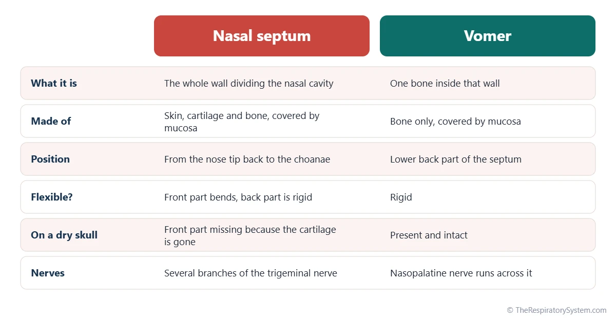
The nasal septum is the whole dividing wall, while the vomer is only one bone inside it, forming the lower back part of that wall. People often mix the two up because the vomer is a whole bone in its own right, while the perpendicular plate is only one part of the ethmoid bone, so on a skull the vomer is the piece most easily named as the septum.
The difference shows clearly on skull models. On a dry skull the cartilage has usually decayed, so the septum looks short and the pear-shaped opening at the front of the nose, the piriform aperture, appears empty in the middle. In a living person the septal cartilage fills that gap and carries the wall forward to the tip of the nose.
The septum should also not be confused with the nasal cavity itself. The nasal cavity is the air space; the septum is the wall that splits it into two halves, each with its own conchae and nasal meatuses on the outer side.
Blood Supply and Nerves of the Nasal Septum
The nasal septum receives blood from branches of both the internal and the external carotid arteries, which meet in a dense network at the front of the septum. This network, called Kiesselbach's plexus or Little's area, lies in the lower front part of the septum just inside the nostril, where the lining is thin and exposed to drying air.
Because the vessels lie so close to the surface there, most nosebleeds start in Kiesselbach's plexus. Fewer nosebleeds start further back, from branches of the sphenopalatine artery. The veins follow the arteries and drain toward the facial vein, the pterygoid venous plexus and the ophthalmic veins.
Nerves of the septum
Touch and pain from the septum travel in branches of the trigeminal nerve. The nasopalatine nerve, from the maxillary division, runs forward and down across the vomer to the incisive canal in the roof of the mouth. The anterior ethmoidal nerve, from the ophthalmic division, supplies the upper front part. At the very top of the septum, olfactory nerve fibers pass through the cribriform plate to a small patch of smell-sensing lining, described on the sense of smell page.
| Vessel or nerve | Comes from | Area of the septum it serves |
|---|---|---|
| Anterior ethmoidal artery | Ophthalmic artery (internal carotid system) | Upper front part |
| Posterior ethmoidal artery | Ophthalmic artery (internal carotid system) | Upper back part |
| Sphenopalatine artery (posterior septal branches) | Maxillary artery (external carotid system) | Back and middle, running forward along the vomer |
| Greater palatine artery | Maxillary artery, rising through the incisive canal | Lower front part |
| Septal branch of the superior labial artery | Facial artery (external carotid system) | Front, close to the nostril |
| Nasopalatine nerve | Maxillary division of the trigeminal nerve | Lower and back parts, along the vomer |
| Anterior ethmoidal nerve | Ophthalmic division of the trigeminal nerve | Upper front part |
| Olfactory nerves | Olfactory epithelium (cranial nerve I) | Highest strip, just below the cribriform plate |
| Source: Typical adult anatomy, our editors | ||
What Is the Function of the Nasal Septum?
The nasal septum divides inhaled air into two streams, holds up the middle of the nose and adds a large moist surface that conditions air before it reaches the lungs.
- Structural support. The septal cartilage props up the bridge and tip of the nose and keeps the nostrils open. Without it, the lower nose would sag.
- Dividing airflow. Splitting the nose into two narrow passages keeps air close to the mucosa on both sides, where it is warmed, moistened and filtered.
- Air conditioning and cleaning. The lining is respiratory epithelium with mucus-making goblet cells and cilia, part of the mucociliary escalator that sweeps trapped dust toward the throat.
- Smell. The highest strip of the septum carries olfactory epithelium, which helps detect odors.
- The nasal cycle. Vascular tissue on the conchae and a thickened area of the septum, the septal swell body, fill and empty in turn, shifting more airflow to one side and then the other every few hours.
These jobs are why breathing through the nose differs from breathing through the mouth, as explained in nose breathing vs mouth breathing. Air that travels along the septum and conchae reaches the pharynx warmer, wetter and cleaner.
How Does the Nasal Septum Develop and Change With Age?
The nasal septum forms in the embryo from tissue in the middle of the developing face, the frontonasal prominence, and grows downward to meet the palate. When the two palatal shelves fuse with each other and with the lower edge of the septum, the nasal cavity is sealed off from the mouth and split into right and left halves. This stage is outlined on the development of the respiratory system page.
In newborns the septum is mostly cartilage. The vomer and the perpendicular plate of the ethmoid turn to bone gradually through childhood, and the septal cartilage acts as a growth center that helps push the middle of the face forward and down. In adults the septum is rarely perfectly straight: small bends and bony spurs, often where cartilage meets bone, are common and usually cause no trouble.
Common Conditions That Affect the Nasal Septum
The septum can be bent, injured or affected through its lining. The main conditions are listed here for education only; broader overviews are on the respiratory diseases page.
- Deviated septum: a septum that bends noticeably to one side, present from birth or caused by injury. A large bend can narrow one nasal passage. See deviated septum.
- Nosebleeds (epistaxis): most begin in Kiesselbach's plexus on the front of the septum, often when the lining is dry or irritated.
- Septal hematoma: blood collecting between the cartilage and its covering after a blow to the nose. The cartilage gets its nutrients from that covering, so doctors treat it as urgent.
- Septal perforation: a hole through the septum, which can follow injury, surgery, infection or irritating substances, and may cause crusting or a whistling sound when breathing.
- Nasal fracture: a break of the nasal bones that often also bends or shifts the septal cartilage.
Swelling of the lining during colds and allergies affects the septum along with the rest of the nose, and inflammation can spread to the paranasal sinuses, as in sinusitis. Anyone with lasting blockage, repeated nosebleeds or a nose injury should see a doctor.
Key Facts
- The nasal septum divides the nasal cavity into right and left halves and forms the inner wall of each half.
- Its front part is the septal, or quadrangular, cartilage, made of hyaline cartilage; its back part is bone.
- The perpendicular plate of the ethmoid bone forms the upper back septum, and the vomer forms the lower back septum.
- Kiesselbach's plexus, a network of small arteries on the front of the septum, is where most nosebleeds begin.
- The nasopalatine and anterior ethmoidal nerves, both branches of the trigeminal nerve, carry touch and pain from the septum.
- A slight bend is normal in adults; a marked bend is called a deviated septum.
- The septal cartilage acts as a growth center that helps shape the middle of the face during childhood.
Quick Quiz: Test What You Know About the Nasal Septum
6 questions. Tap an answer to check it.
Which bone forms the lower back part of the nasal septum?
The vomer forms the lower back part of the septum. The perpendicular plate of the ethmoid forms the upper back part.
What type of tissue is the septal cartilage?
The septal cartilage is hyaline cartilage, the same glassy type that forms the rings of the trachea.
Where do most nosebleeds begin?
Most nosebleeds start in Kiesselbach's plexus, a network of small arteries in the lower front part of the septum.
Which part of the ethmoid bone helps form the nasal septum?
The perpendicular plate of the ethmoid hangs down from the cribriform plate and forms the upper back part of the septum.
What does the nasal septum separate?
The septum divides the nasal cavity into right and left passages. The hard palate is what separates the nose from the mouth.
Which nerve runs across the vomer toward the roof of the mouth?
The nasopalatine nerve, a branch of the maxillary division of the trigeminal nerve, runs down along the vomer to the incisive canal.
Frequently Asked Questions
Is the nasal septum bone or cartilage?
It is both. The front part, which you can bend by pressing the tip of your nose, is septal cartilage made of hyaline cartilage. The back part is bone: the perpendicular plate of the ethmoid bone above and the vomer below. A small skin-covered segment at the very front, in the columella between the nostrils, contains neither and is called the membranous septum.
Is it normal for the nasal septum to be crooked?
Yes, a slightly off-center septum is very common and usually causes no symptoms. Small bends and bony spurs often form where the cartilage meets the bone as the face grows. Only a marked bend that narrows one side of the nose is usually described as a deviated septum, and whether it matters depends on how much it affects airflow.
What is the columella of the nose?
The columella is the narrow bridge of tissue between the two nostrils at the base of the nose. It contains the membranous part of the nasal septum and the inner limbs of the two major alar cartilages, covered by skin. It forms the front end of the septum and helps support the tip of the nose.
What is the difference between the nasal cavity and the nasal septum?
The nasal cavity is the air-filled space inside the nose, running from the nostrils to the choanae. The nasal septum is a wall inside that space, dividing it into two halves. Each half of the cavity has the septum on its inner side and the conchae and meatuses on its outer side, so the septum is part of the walls of the cavity rather than a space itself.
How thick is the nasal septum?
The nasal septum is thin, only a few millimeters thick in most places, including its mucous membrane on both sides. It is usually thicker near the floor of the nose, where the cartilage and vomer sit on the nasal crests, and in the septal swell body higher up. Its thickness also changes a little as the blood vessels in its lining fill and empty.
Where is the nasal septum on a diagram?
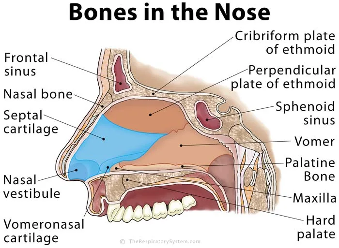
On a front view of the face, the septum is the vertical midline wall between the two nasal passages. On a side, or sagittal, view of the nose, the septum fills the whole picture: septal cartilage at the front, the perpendicular plate of the ethmoid at the top back, the vomer at the bottom back, and the hard palate as the floor.
What is the vomeronasal organ?
The vomeronasal organ is a tiny pouch near the base of the nasal septum. In many mammals it detects chemical signals from other animals of the same species. In humans it appears during fetal development, but in adults it is small or missing, and most researchers consider it to have no working sensory function.
Is the nasal septum part of the respiratory system?
Yes. The nasal septum belongs to the nose and nasal cavity, which form the first part of the upper respiratory tract. Air passes along both sides of the septum on its way to the pharynx, larynx and lungs, and its mucous lining helps warm, moisten and clean that air before it travels deeper into the airways.


