At a Glance
The sphenoid sinus is a pair of air spaces hollowed out of the body of the sphenoid bone, behind the nasal cavity and directly below the pituitary gland. Each side opens through a small ostium into the sphenoethmoidal recess at the top back of the nose. It is lined with ciliated respiratory mucosa, lightens the skull base, adds mucus to the nasal cavity and helps give the voice resonance.
- Location
- Body of the sphenoid bone, center of the skull base, behind the nasal cavity
- Type
- Paired paranasal sinus (left and right), split by a bony septum
- Drains into
- Sphenoethmoidal recess, above the superior nasal concha
- Lining
- Thin mucoperiosteum with ciliated respiratory epithelium
- Key neighbors
- Pituitary gland above, optic nerves and cavernous sinuses at the sides, nasopharynx below
- Part of
- Paranasal sinuses of the upper respiratory tract
On this page
- What Is the Sphenoid Sinus?
- Where Is the Sphenoid Sinus Located?
- Sphenoid Sinus Anatomy: Walls, Opening and Lining
- What Is the Function of the Sphenoid Sinus?
- How Does the Sphenoid Sinus Develop and Change With Age?
- How Is the Sphenoid Sinus Seen on CT and X-Ray?
- Common Conditions That Affect the Sphenoid Sinus
- Key Facts
- Quiz
- FAQ
What Is the Sphenoid Sinus?
The sphenoid sinus is the air-filled cavity inside the sphenoid bone, the butterfly-shaped bone that forms the middle of the skull base. It is one of four pairs of paranasal sinuses, the hollow spaces that surround the nose and open into it, and it belongs to the upper respiratory tract. Of the four pairs it lies farthest back, so it is sometimes called the deepest sinus.
Sphenoid sinus definition
In anatomy, the sphenoid sinus is defined as the pneumatized (air-containing) part of the body of the sphenoid bone, divided into left and right halves by an intersinus septum and opening into the nasal cavity through one ostium on each side. Like the ethmoid, frontal and maxillary sinuses, it is named after the bone that holds it.
Explained simply
Imagine a small hollow room in the very middle of your head, right behind the back of your nose and under the gland that controls your hormones. That room is filled with air, has a thin wet lining, and has a tiny door that lets mucus drip out into your nose. That room is the sphenoid sinus.
Where Is the Sphenoid Sinus Located?
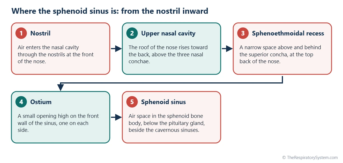
The sphenoid sinus is located in the body of the sphenoid bone, behind the upper back part of the nasal cavity and below the sella turcica, the bony seat of the pituitary gland. It sits roughly at the level of the eyes but much deeper, close to the center of the head. In front of it are the posterior ethmoid air cells and the roof of the nose, and below it is the roof of the nasopharynx.
Where is the left sphenoid sinus?
The left sphenoid sinus is the left half of the cavity, separated from the right half by a thin bony intersinus septum. This septum is rarely in the midline: it often bends to one side, so one sinus can be much larger than the other, and it may attach to the bone over the optic nerve or the internal carotid artery. Each half has its own opening and drains into the nose on its own side.
What surrounds it
The sphenoid sinus is unusual because so many important structures lie just beyond its thin walls:
- Above: the pituitary gland in the sella turcica, and the optic chiasm a little further forward.
- On each side: the cavernous sinus, a venous channel that carries the internal carotid artery and several cranial nerves that move the eye.
- Upper side wall: the optic nerve on its way from the eye to the brain.
- Below: the roof of the nasopharynx and the pterygoid canal.
- Behind: the clivus, the sloping bone in front of the brainstem.
Sphenoid Sinus Anatomy: Walls, Opening and Lining
Sphenoid sinus anatomy is defined by its six bony walls, a single opening on each side and a thin lining of respiratory mucosa. The walls are often thin, and in some people the bony ridges made by the optic nerve or the internal carotid artery bulge into the sinus.
The ostium and drainage
Each sphenoid sinus opens through an ostium high on its front wall. The opening leads into the sphenoethmoidal recess, a narrow space above and behind the superior nasal concha, which is why the sphenoid is the only sinus that does not drain into one of the main nasal meatuses. Because the ostium sits high on the wall, mucus must be carried upward by cilia rather than simply running out.
Lining and mucus
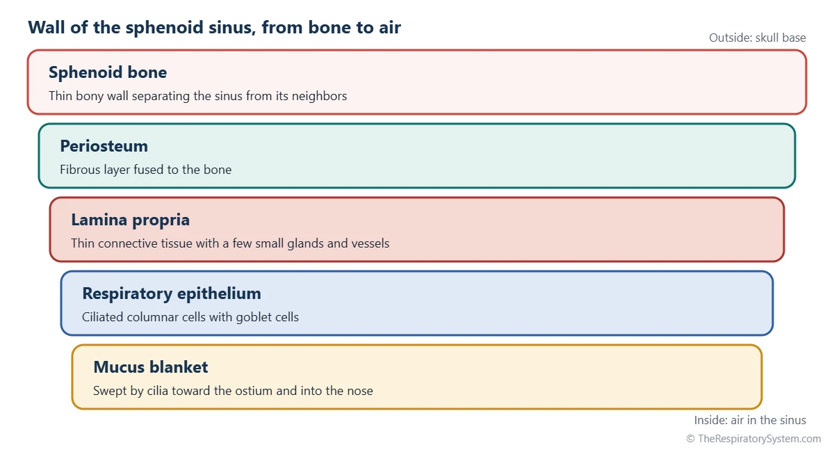
The inner surface is covered by a thin mucoperiosteum: respiratory epithelium of ciliated columnar cells and goblet cells, resting on a thin layer of connective tissue fused to the bone. The cilia beat in a set direction, sweeping a blanket of mucus toward the ostium, the same mucociliary clearance that cleans the rest of the airway.
Blood and nerve supply
Blood reaches the sinus mainly from the posterior ethmoidal arteries and branches of the sphenopalatine artery. Sensation comes from the posterior ethmoidal nerve and small orbital branches linked to the maxillary nerve, both parts of the trigeminal nerve. Lymph drains toward the retropharyngeal lymph nodes behind the throat.
Pneumatization patterns
The amount of bone the sinus hollows out varies from person to person. Anatomists describe three common patterns, based on how far back the air space reaches compared with the sella turcica:
- Conchal: a very small sinus that stays in front of the sella, typical of young children.
- Presellar: the sinus reaches the front wall of the sella but not under it.
- Sellar: the sinus extends under and behind the sella; the most common pattern in adults.
| Wall | What lies beyond it | Why it matters |
|---|---|---|
| Roof (superior wall) | Sella turcica with the pituitary gland; optic chiasm further forward | Explains why the pituitary gland can be reached through the nose |
| Lateral wall (each side) | Cavernous sinus with the internal carotid artery and nerves to the eye muscles | The artery may bulge into the sinus as a ridge |
| Upper lateral wall | Optic nerve in the optic canal | Bone over the nerve can be very thin |
| Lower lateral wall | Maxillary nerve near the foramen rotundum | Can leave a small ridge on the wall |
| Floor (inferior wall) | Roof of the nasopharynx and the pterygoid canal | The sinus sits directly over the back of the throat |
| Anterior wall | Sphenoethmoidal recess, nasal cavity and posterior ethmoid cells | Holds the ostium that drains the sinus |
| Posterior wall | Clivus, with the brainstem behind it | Thick bone in most people |
| Medial wall | Intersinus septum and the opposite sphenoid sinus | Often off center, so the two sides differ in size |
| Source: Typical adult anatomy, our editors | ||
What Is the Function of the Sphenoid Sinus?
The function of the sphenoid sinus is not fully settled, but it is thought to lighten the skull base, add mucus and moisture to the nasal cavity, and help give the voice resonance. These are the same roles proposed for all the paranasal sinuses, and they likely work together rather than alone.
- Lighter skull: replacing solid bone with air reduces the weight of the head at its center.
- Mucus supply: goblet cells and glands add mucus to the back of the nose, helping trap dust and germs.
- Voice resonance: air spaces in the skull affect how the voice sounds, which is one reason the voice changes when the sinuses are blocked; see how the voice is produced.
- Protective buffer: an air-filled space may help absorb force and insulate nearby structures.
Sphenoid sinus function in the respiratory system
Within the respiratory system, the sphenoid sinus works as a small mucus-producing side chamber of the nasal cavity. Inhaled air does not flow through it with each breath; instead, air exchanges slowly through the ostium, while the mucus it produces joins the stream of mucus moving back toward the pharynx, where it is swallowed. This keeps the lining of the nose moist and helps it filter the air that continues along the pathway of air to the lungs.
How Does the Sphenoid Sinus Develop and Change With Age?
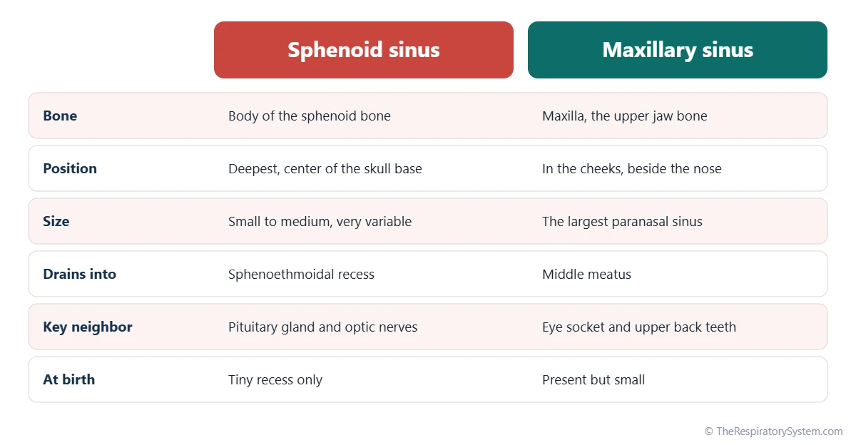
The sphenoid sinus is only a tiny recess at birth and grows into the sphenoid bone during childhood, reaching adult size around the teenage years. It is one of the later sinuses to form, together with the frontal sinus, while the maxillary and ethmoid sinuses are already present in newborns.
Pneumatization, the process of air spaces hollowing out bone, starts from the back of the nasal cavity in early childhood. The sinus spreads backward under the sella turcica during the school years, and its final shape depends on how far this process goes.
This growth pattern is covered for all four pairs on our respiratory system development page.
How Is the Sphenoid Sinus Seen on CT and X-Ray?
On a CT scan, a healthy sphenoid sinus looks black because it is full of air, outlined by thin white bone. CT is the standard imaging method for sinus anatomy because it shows the bony walls, the septum, the ostium and the bulges of the optic nerve and carotid artery clearly in slices.
Radiologists read the sphenoid sinus in three planes: coronal slices show the left and right halves and the septum, axial slices show the sinus between the cavernous sinuses, and sagittal slices show it below the pituitary gland. On a plain side X-ray of the skull, the sinus appears as a dark area just below the sella turcica. MRI is used more for soft tissue, such as the pituitary gland, than for the air space itself.
Common Conditions That Affect the Sphenoid Sinus
The sphenoid sinus can be affected by inflammation, blockage and, rarely, growths, although problems here are less common than in the maxillary and ethmoid sinuses. These are described for education only; see our respiratory diseases section for more.
- Sphenoid sinusitis: inflammation of the lining, usually together with other sinuses; when it happens alone it is often described with a deep headache behind the eyes or at the top of the head. See sinusitis.
- Mucocele: a slowly expanding sac of trapped mucus that forms when the ostium is blocked for a long time.
- Fungal ball: a clump of fungal material that can collect in one sinus without invading the tissue.
- Tumors: rare growths of the lining or of nearby structures such as the pituitary gland.
Because the optic nerves and cavernous sinuses lie so close, symptoms such as changes in vision with sinus pain need prompt medical assessment.
Key Facts
- The sphenoid sinus is the most posterior of the four pairs of paranasal sinuses, hollowed out of the body of the sphenoid bone.
- Its left and right halves are split by an intersinus septum that is usually off center, so the two sides are rarely equal.
- It drains through a small ostium into the sphenoethmoidal recess, above the superior nasal concha.
- The pituitary gland sits directly above it, and the optic nerves and cavernous sinuses run along its sides.
- Its lining is ciliated respiratory epithelium that sweeps mucus toward the nose.
- It is only a tiny recess at birth and grows into the sphenoid bone during childhood.
- On CT a healthy sphenoid sinus looks black because it is filled with air.
Quick Quiz: Test What You Know About the Sphenoid Sinus
6 questions. Tap an answer to check it.
Which bone contains the sphenoid sinus?
The sphenoid sinus is hollowed out of the body of the sphenoid bone, in the center of the skull base.
Where does the sphenoid sinus drain?
Its ostium opens into the sphenoethmoidal recess, above and behind the superior nasal concha.
Which gland lies directly above the sphenoid sinus?
The pituitary gland rests in the sella turcica, which forms the roof of the sphenoid sinus.
What separates the left and right sphenoid sinuses?
A thin bony intersinus septum divides the cavity into two halves, often unequally.
Which pneumatization pattern is most common in adults?
In most adults the sinus extends under the sella turcica, the sellar pattern.
What lines the sphenoid sinus?
Like the other sinuses, it is lined by ciliated columnar epithelium with goblet cells.
Frequently Asked Questions
Is the sphenoid sinus behind the eyes?
Yes, roughly. The sphenoid sinus sits deep in the skull base behind the nasal cavity, at about the level of the eyes but much further back, between the back ends of the two eye sockets. The optic nerves leave the eyes and run along its upper side walls, which is why pressure or inflammation in this sinus is often felt as a deep ache behind the eyes.
How many sphenoid sinuses do you have?
Most people have two sphenoid sinuses, a left and a right one, inside the same bone and separated by a thin bony septum. The septum is often off to one side, so one sinus may be much larger. Some people have extra partial septa that make the space look divided into several chambers on a CT scan, and in rare cases one side barely develops.
Why is the sphenoid sinus important in surgery?
The sphenoid sinus is a natural corridor to the center of the skull. The pituitary gland sits on its roof, so surgeons can reach pituitary tumors through the nose and the sinus. Its closeness to the optic nerves and internal carotid arteries also means that surgeons study its walls carefully on CT before any sinus operation.
What is the sphenoethmoidal recess?
The sphenoethmoidal recess is a small space at the top back of the nasal cavity, above and behind the superior nasal concha. It sits between the front wall of the sphenoid bone and the ethmoid bone. The sphenoid sinus opens into it, and from there mucus flows down and back toward the nasopharynx, where it is swallowed without being noticed.
Do babies have a sphenoid sinus?
Newborns have only a tiny recess where the sphenoid sinus will form. The air space begins to hollow out the sphenoid bone in early childhood, spreads backward under the pituitary gland during the school years, and usually reaches adult size around the teenage years. This is why sphenoid sinus problems are uncommon in very young children.
Can you feel the sphenoid sinus from outside?
No. Unlike the frontal and maxillary sinuses, which lie under the forehead and cheeks, the sphenoid sinus is buried in the middle of the skull. It cannot be pressed, tapped or seen from the outside, so doctors rely on endoscopes passed through the nose and on CT or MRI scans to look at it.
What is the difference between the sphenoid sinus and the cavernous sinus?
The sphenoid sinus is an air space in bone that belongs to the respiratory tract. The cavernous sinus is a channel of venous blood inside the dura, the tough covering of the brain, lying against each side of the sphenoid bone. It carries the internal carotid artery and nerves that move the eye. They share a name but have different jobs.
Is the sphenoid sinus the same as the sphenoid bone?
No. The sphenoid bone is the whole butterfly-shaped bone in the middle of the skull base, with a central body and wings that reach out to the sides. The sphenoid sinus is the air-filled hollow inside the body of that bone. A bone can exist without being hollowed out, but the sinus always lies within the bone.


