At a Glance
The larynx (voice box) is a hollow tube about 5 cm (2 inches) long in adults, built from 9 cartilages and sitting in the front of the neck between the third and sixth cervical vertebrae. It joins the pharynx above to the trachea below. Its three jobs are to keep the airway open for breathing, to close and protect the airway during swallowing, and to produce voice with the vocal folds.
- Location
- Front of the neck, below the hyoid bone and in front of the laryngopharynx, at the level of vertebrae C3 to C6
- Function
- Air passage, airway protection during swallowing, voice production, coughing
- Tissue
- 9 cartilages (hyaline and elastic) joined by ligaments, membranes and muscles, lined by mucous membrane
- Size
- About 5 cm (2 inches) long in adults, larger in men than in women
- Connects to
- Laryngopharynx above, trachea below; suspended from the hyoid bone
- Part of
- Upper respiratory tract
On this page
- What Is the Larynx?
- Where Is the Larynx Located?
- What Is the Larynx Made Of?
- Larynx Anatomy: Inside the Laryngeal Cavity and Vocal Cords
- Muscles, Nerves and Blood Supply of the Larynx
- What Does the Larynx Do?
- How Does the Larynx Produce Sound and Speech?
- Pharynx vs Larynx vs Trachea: What Is the Difference?
- How Does the Larynx Develop and Change With Age?
- Common Conditions That Affect the Larynx
- Key Facts
- Quiz
- FAQ
What Is the Larynx?
The larynx is the hollow, cartilage-walled organ at the top of the windpipe that works as the airway's gatekeeper and as the body's sound source. It is also known as the voice box, and the name is pronounced "LAIR-inks" (not "LAR-nix"). Air reaches it from the pharynx (throat) and leaves it through the trachea on the way to the lungs. Because the paths for air and for food cross just above it, the larynx belongs to the breathing system but works closely with the digestive system as well.
Most anatomy books place the larynx at the lower end of the upper respiratory tract, together with the nose, nasal cavity and pharynx. Some textbooks count it as the first part of the lower tract instead, because everything from the larynx down is normally kept free of food and germs.
What does the larynx look like?
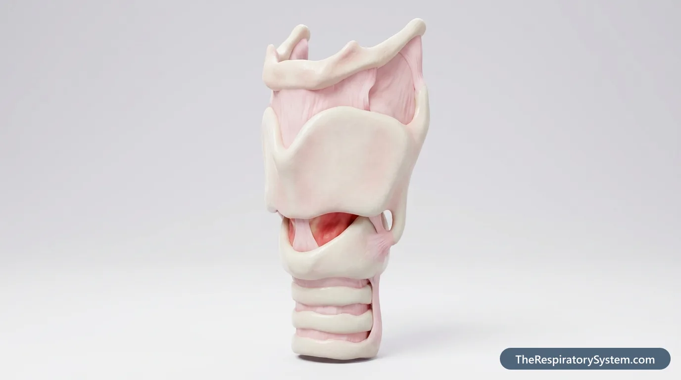
From the front, the larynx looks like a short, slightly flattened tube with a pointed ridge, the Adam's apple, in the middle. Inside, its passage narrows to a slit between two pale, shiny bands, the vocal folds, which open into a V shape when we breathe and close into a straight line when we speak. Above them sits the leaf-shaped epiglottis, which stands up behind the root of the tongue.
Explained simply
Think of the larynx as a valve with a built-in musical instrument. When you breathe, the valve stays open so air can flow freely. When you swallow, it snaps shut so food slides past into the food pipe. When you talk, it closes almost all the way, and air from your lungs makes the two edges of the valve buzz like the lips of a trumpet player.
Where Is the Larynx Located?
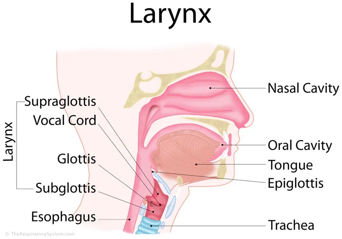
The larynx is located in the front of the neck, in the midline, below the hyoid bone and above the trachea. In adults it spans the third to sixth cervical vertebrae (C3 to C6), and it lies directly in front of the lowest part of the throat, the laryngopharynx, into which its upper opening faces.
Where is the larynx in relation to the pharynx and trachea?
- Above: the larynx hangs from the U-shaped hyoid bone by the thyrohyoid membrane, and its inlet opens backward into the laryngopharynx.
- Below: at the lower border of the cricoid cartilage, level with C6, it continues into the trachea.
- Behind: the laryngopharynx, which leads down into the esophagus, the food pipe.
- In front: skin, fat and the thin infrahyoid "strap" muscles of the neck.
- To the sides: the lobes of the thyroid gland, which wrap around the lower larynx and upper trachea, and the large blood vessels of the neck.
Where is the larynx located in the throat?
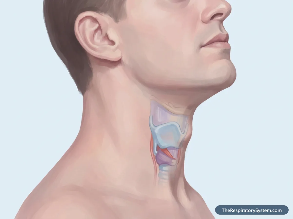
In everyday language the whole front of the neck is "the throat", but anatomically the larynx sits in front of the throat (pharynx), not inside it. You can feel it by placing a finger on the Adam's apple and swallowing: the whole larynx moves up and then down again. Just below the Adam's apple lies a soft gap, the cricothyroid membrane, and below that the firm ring of the cricoid cartilage.
The larynx sits higher in babies than in adults. In a newborn it lies roughly opposite the first to fourth cervical vertebrae, and it gradually descends through childhood, which changes both breathing and the sound of the voice.
What Is the Larynx Made Of?
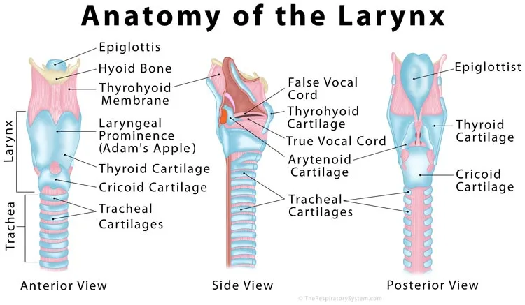
The larynx is made of 9 cartilages, 3 single and 3 paired, held together by joints, ligaments and membranes, moved by small muscles and lined with mucous membrane. The cartilages give it a stiff frame that cannot collapse when we breathe in, while the joints let some of them tilt and slide to change the shape of the airway.
The unpaired cartilages
The thyroid cartilage is the largest. It is formed by two flat plates (laminae) that meet in front at an angle, creating the laryngeal prominence, or Adam's apple. The angle is sharper, about 90 degrees, in men and wider, about 120 degrees, in women, which is why the prominence is easier to see in men. Its upper horns are tied to the hyoid bone, and its lower horns form small joints with the cricoid cartilage.
The cricoid cartilage lies below it and is shaped like a signet ring, narrow in front and broad at the back. It is the only complete ring of cartilage anywhere in the airway, so it keeps the bottom of the larynx open at all times. The epiglottis is a flexible, leaf-shaped plate of elastic cartilage attached to the back of the thyroid angle and rising behind the tongue.
The paired cartilages
The two arytenoid cartilages are small pyramids that sit on the upper back edge of the cricoid lamina. Each has a vocal process, to which the vocal ligament attaches, and a muscular process, where the muscles that move it pull. Because the arytenoids can glide and rotate, they open and close the vocal folds. The tiny corniculate cartilages perch on the tips of the arytenoids, and the rod-like cuneiform cartilages lie inside the aryepiglottic folds, where they stiffen the edges of the laryngeal inlet.
Membranes and ligaments
The thyrohyoid membrane slings the larynx from the hyoid bone, and the cricotracheal ligament joins it to the first ring of the trachea. Inside, two sheets of connective tissue shape the cavity: the quadrangular membrane, whose free lower edge is the vestibular ligament inside each false vocal cord, and the conus elasticus, whose thickened upper edge is the vocal ligament inside each true vocal cord. The front of the conus elasticus forms the cricothyroid ligament, the soft spot below the Adam's apple used for an emergency airway when the airway above is blocked.
| Cartilage | Number | Cartilage type | Where it is | What it does |
|---|---|---|---|---|
| Thyroid | 1 | Hyaline | Front and sides of the larynx; forms the Adam's apple | Shields the vocal folds and anchors their front ends |
| Cricoid | 1 | Hyaline | Below the thyroid cartilage, at the level of C6 | The only complete cartilage ring in the airway; keeps the lower larynx open |
| Epiglottis | 1 | Elastic | Behind the root of the tongue, attached to the thyroid angle | Tips over the laryngeal inlet during swallowing |
| Arytenoid | 2 | Mostly hyaline | On the upper back edge of the cricoid | Pivot and slide to open, close and tense the vocal folds |
| Corniculate | 2 | Elastic | On the tips of the arytenoids | Support the back of the aryepiglottic folds |
| Cuneiform | 2 | Elastic | Inside the aryepiglottic folds | Stiffen the edges of the laryngeal inlet |
| Source: Typical adult anatomy, our editors | ||||
Larynx Anatomy: Inside the Laryngeal Cavity and Vocal Cords
The inside of the larynx, the laryngeal cavity, runs from the laryngeal inlet at the top to the lower edge of the cricoid cartilage, and two pairs of folds divide it into three levels: the supraglottis, the glottis and the subglottis.
- Laryngeal inlet: the oval entrance, bordered by the epiglottis in front, the aryepiglottic folds at the sides and the notch between the arytenoids at the back.
- Supraglottis: everything above the true vocal folds, including the epiglottis, the vestibule, the vestibular folds (false vocal cords) and the laryngeal ventricles, two small side pockets between the false and true folds.
- Glottis: the true vocal folds and the gap between them, called the rima glottidis. In adults this is the narrowest part of the airway, and its width changes constantly with breathing, speaking and swallowing.
- Subglottis: the short space from below the vocal folds to the lower border of the cricoid cartilage, where the larynx becomes the trachea.
True and false vocal cords
The true vocal folds (vocal cords) are two smooth, pearly-white bands that stretch from the inside of the thyroid angle in front to the vocal processes of the arytenoids at the back. Each contains the vocal ligament and the vocalis muscle, the inner part of the thyroarytenoid. The false vocal folds lie just above them; they are thicker, pinker and contain only a little muscle, so they do not normally vibrate in speech. Their main job is to help seal the airway, for example when holding the breath to lift something heavy.
Larynx histology: the lining
Most of the larynx is lined by respiratory epithelium, a pseudostratified ciliated columnar layer with mucus-making goblet cells, the same lining found in the nose and trachea. The true vocal folds and the upper part of the epiglottis are different: they are covered by non-keratinized stratified squamous epithelium, a tougher, many-layered surface that withstands the constant rubbing of vibration. Under the vocal fold epithelium lies a loose, jelly-like layer called Reinke's space, which lets the surface ripple freely over the deeper ligament and muscle.
The thyroid, cricoid and most of the arytenoid cartilages are hyaline cartilage, while the epiglottis, corniculate and cuneiform cartilages and the tips of the vocal processes are elastic cartilage, which bends and springs back.
Muscles, Nerves and Blood Supply of the Larynx
The larynx is moved by two groups of muscles, controlled by two branches of the vagus nerve, and fed by two pairs of laryngeal arteries, with the vocal folds forming the dividing line for both its nerve supply and its lymph drainage.
Extrinsic muscles
Extrinsic muscles run from the larynx or the hyoid bone to structures outside it and move the whole larynx up or down. The suprahyoid group (digastric, geniohyoid, mylohyoid and stylohyoid) lifts the hyoid and larynx during swallowing. The infrahyoid group (omohyoid, sternohyoid, sternothyroid and thyrohyoid) pulls them back down afterward. You can feel this up-and-down movement every time you swallow.
Intrinsic muscles
The intrinsic muscles lie entirely within the larynx and change the position and tension of the vocal folds, which controls the size of the rima glottidis. They are the posterior cricoarytenoid, lateral cricoarytenoid, transverse arytenoid, oblique arytenoid, thyroarytenoid and cricothyroid muscles. All come in left and right pairs except the transverse arytenoid, which is a single muscle bridging the two arytenoids. The table below sums up what each one does.
Nerve supply
Both sensory and motor nerves of the larynx come from the vagus nerve (cranial nerve X) through two branches. The superior laryngeal nerve arises from the inferior ganglion of the vagus and splits in two: its internal branch carries sensation from the lining above the vocal folds, and its external branch supplies a single muscle, the cricothyroid. The recurrent laryngeal nerve supplies every other intrinsic muscle and carries sensation from below the vocal folds. On the left it loops under the arch of the aorta and on the right under the subclavian artery before climbing back up beside the trachea, so the left nerve is longer.
Arteries, veins and lymph
The superior laryngeal artery, a branch of the superior thyroid artery, pierces the thyrohyoid membrane with the internal laryngeal nerve to feed the glands, muscles and lining of the upper larynx. The inferior laryngeal artery comes from the inferior thyroid artery, a branch of the thyrocervical trunk, and enters near the junction of the trachea and esophagus alongside the recurrent laryngeal nerve. Matching veins follow the same paths: the superior laryngeal vein drains through the superior thyroid vein to the internal jugular vein, and the inferior laryngeal vein drains through the inferior thyroid veins to the left brachiocephalic vein.
Lymph from above the vocal folds follows the superior laryngeal artery to the deep cervical lymph nodes, while lymph from below the vocal folds follows the inferior thyroid artery to nodes in front of and beside the upper trachea. The true vocal folds themselves have very few lymph vessels.
| Muscle | Main action | Nerve |
|---|---|---|
| Posterior cricoarytenoid | Opens (abducts) the vocal folds; the only muscle that widens the glottis | Recurrent laryngeal nerve |
| Lateral cricoarytenoid | Closes (adducts) the vocal folds | Recurrent laryngeal nerve |
| Transverse arytenoid (single) | Pulls the arytenoids together to close the back of the glottis | Recurrent laryngeal nerve |
| Oblique arytenoid | Helps close the back of the glottis and narrow the laryngeal inlet | Recurrent laryngeal nerve |
| Thyroarytenoid (with vocalis) | Shortens and relaxes the vocal folds, lowering pitch; fine-tunes their tension | Recurrent laryngeal nerve |
| Cricothyroid | Tilts the thyroid cartilage forward, lengthening and tensing the vocal folds to raise pitch | External branch of the superior laryngeal nerve |
| Source: Typical adult anatomy, our editors | ||
What Does the Larynx Do?
The larynx does three main jobs: it keeps an open passage for air between the throat and the lungs, it closes to protect the airway when we swallow, and it produces the sound of the voice. It also powers coughing and lets us hold the breath against a closed airway.
Function of the larynx in the respiratory system
During breathing, air drawn in through the nose or mouth passes down the pharynx, through the larynx and into the trachea, which splits into the two bronchi leading to the lungs. The rigid cartilage frame keeps this passage from collapsing under the suction of a breath in, and the cilia and mucus of its lining help trap dust and sweep it upward. The larynx also adjusts airflow: the vocal folds open wider on a deep breath and narrow slightly as we breathe out, which helps control how fast the lungs empty.
Function of the larynx in the digestive system
The larynx is where the paths for air and food separate, so it must close every time we swallow. In a fraction of a second the larynx is pulled up and forward under the base of the tongue, the epiglottis tips backward over the inlet, the aryepiglottic and false vocal folds squeeze together, and the true vocal folds shut. Breathing stops briefly, food and drink slide around the sides of the inlet into the esophagus, and then everything reopens. If anything still slips into the larynx, its very sensitive lining triggers a strong cough to blast it back out.
Other jobs of the larynx
- Coughing: the vocal folds close, pressure builds in the chest, and the folds fly open to release a burst of air that clears the airway.
- Effort closure: shutting the glottis traps air in the chest and stiffens the trunk for lifting, pushing or straining.
- Voice: vibration of the true vocal folds makes the sound we shape into speech and song.
How Does the Larynx Produce Sound and Speech?
The larynx produces sound when air pushed up from the lungs makes the closed true vocal folds vibrate, chopping the airflow into rapid puffs that we hear as a buzzing tone. That tone is then shaped into words by the pharynx, the oral cavity, the tongue, the lips and the nasal passages.
- The lateral cricoarytenoid and arytenoid muscles bring the vocal folds together near the midline.
- Air pressure from the lungs builds up below the closed folds until it pushes them apart.
- The elastic folds spring back together, partly sucked inward by the fast-moving air, and the cycle repeats many times a second.
- Each opening releases a tiny puff of air; the stream of puffs is the raw sound of the voice.
What controls pitch and loudness?
Pitch depends on how fast the vocal folds vibrate, which depends on their length, thickness and tension. The cricothyroid muscle tilts the thyroid cartilage forward, stretching and thinning the folds to raise the pitch, while the thyroarytenoid shortens and relaxes them to lower it. In a typical speaking voice the folds vibrate roughly 100 to 150 times per second in adult men and around 200 times per second in adult women. Loudness depends mainly on how hard air is pushed from the lungs and how firmly the folds press together.
The larynx makes the sound, but it does not make words. Vowels and consonants are formed above it, in the resonating spaces of the throat, mouth and nose, which also give each person's voice its own color. That is why people who whisper can still be understood: whispering uses air passing through a partly open glottis without vibration.

Pharynx vs Larynx vs Trachea: What Is the Difference?
The pharynx, larynx and trachea are three linked sections of the airway: the pharynx is the shared muscular throat behind the nose and mouth, the larynx is the cartilage valve and voice box below it, and the trachea is the windpipe that carries air from the larynx down into the chest. Air passes through all three in that order, but only the pharynx also carries food.
The pharynx is divided into the nasopharynx, the oropharynx and the laryngopharynx. Its walls are mostly muscle, which squeezes food downward. The larynx opens off the front of the laryngopharynx, and at that point air turns forward into the larynx while food continues backward into the esophagus. Below the cricoid cartilage, the trachea takes over as a tube of 16 to 20 C-shaped cartilage rings, open at the back where it rests against the esophagus.
In short, the pharynx belongs to both the respiratory and digestive systems, the larynx guards the split between them and makes the voice, and the trachea belongs only to the respiratory system. The comparison table sums up the main differences.
For more detail on the first pair, see our larynx vs pharynx comparison.
| Feature | Pharynx | Larynx | Trachea | Esophagus |
|---|---|---|---|---|
| Common name | Throat | Voice box | Windpipe | Food pipe |
| Position | Behind the nose, mouth and larynx | Front of the neck, C3 to C6 | Front of the neck and chest, below the larynx | Behind the trachea |
| Carries | Air and food | Air only | Air only | Food and liquid only |
| Wall | Mostly skeletal muscle | 9 cartilages with ligaments and muscles | 16 to 20 C-shaped hyaline cartilage rings | Muscle, collapsed when empty |
| Body system | Respiratory and digestive | Respiratory | Respiratory | Digestive |
| Main job | Passage for air and food; swallowing | Airway protection and voice | Conducts air to the bronchi | Moves food to the stomach |
| Ends at | Larynx and esophagus | Lower border of the cricoid cartilage | Split into the two main bronchi | Stomach |
| Source: Typical adult anatomy, our editors | ||||
How Does the Larynx Develop and Change With Age?
The larynx begins forming in about the fourth week of embryonic life, sits high in the neck at birth, descends during childhood, grows quickly at puberty, especially in boys, and stiffens slowly in later life.
Before birth
The larynx and trachea develop from a groove in the floor of the embryonic foregut, the same bud that later forms the lungs. The laryngeal cartilages and muscles come from the fourth and sixth pharyngeal arches, which is why the larynx is supplied by the nerves of those arches: the superior laryngeal nerve (fourth arch) and the recurrent laryngeal nerve (sixth arch). The epiglottis develops from a separate swelling in the floor of the pharynx. For a while the inside of the larynx is temporarily sealed by its own lining and then opens up again; failure to reopen fully can leave a thin web across the glottis.
Infancy and childhood
A baby's larynx sits high, so the epiglottis can almost touch the soft palate, which helps an infant breathe through the nose while feeding. The infant epiglottis is relatively long, narrow and soft, and the subglottic region is small, so even mild swelling there can noticeably narrow the airway. Over the first years of life the larynx moves down the neck, creating a longer throat above it that adult speech depends on.
Puberty and older age
At puberty the larynx enlarges under the influence of sex hormones. In boys the thyroid cartilage grows forward to form a visible Adam's apple and the vocal folds become longer and thicker, so the voice drops by about an octave, often with a period of cracking. Girls' voices deepen too, but much less. From early adulthood onward the hyaline cartilages slowly begin to calcify and turn partly to bone. In older age the vocal folds can lose some muscle bulk and elasticity, a change called presbylarynx, which may make the voice thinner, breathier or weaker.
Common Conditions That Affect the Larynx
Most disorders of the larynx change the voice, so hoarseness, loss of voice, a sore throat, cough and trouble swallowing are the most common signs. The list below names the main conditions and the part of the larynx each one involves; our respiratory diseases guide covers the wider airway, and anyone with lasting hoarseness or breathing changes should see a doctor.
- Laryngitis: inflammation of the larynx, usually from a viral infection or voice overuse. It can bring a burning or painful throat, swollen glands in the neck, trouble swallowing, tiredness and a mild fever along with hoarseness.
- Croup: a childhood viral infection that swells the subglottis and upper trachea, causing a barking cough and noisy breathing in.
- Vocal fold nodules, polyps, cysts and granulomas: benign growths on the vocal folds that keep them from closing evenly, leading to a rough or breathy voice.
- Laryngeal cancer: most often squamous cell carcinoma of the lining, strongly linked to smoking and heavy alcohol use. It may cause lasting hoarseness, a persistent cough or sore throat, painful swallowing, a lump in the neck and ear pain.
- Vocal fold paralysis: loss of movement of one or both vocal folds when a laryngeal nerve, most often the recurrent laryngeal nerve, is damaged.
- Laryngopharyngeal reflux (LPR): stomach contents travel up the esophagus and reach the larynx. It differs from typical heartburn and can be harder to recognize; adults may notice hoarseness, throat clearing or a bitter taste, while infants may have a chronic barking cough, pauses in breathing or feeding trouble.
- Laryngospasm: a sudden, involuntary closure of the vocal folds, often during a breath in, that briefly stops breathing and speech. Though frightening, it is usually short-lived.
- Irritable larynx syndrome: an oversensitive larynx that reacts with cough, throat tightness, a lump feeling, swallowing difficulty or episodes of laryngospasm, sometimes linked to chronic reflux or nerve changes.
- Laryngeal fracture or dislocation: injury from a hard blow to the neck, for example in a sports collision or car crash, which can threaten the airway.
- Laryngomalacia: the most common cause of noisy breathing (stridor) in newborns; soft, floppy tissue above the glottis falls inward on each breath in. Most cases improve on their own by about 18 to 24 months of age.
- Laryngeal cleft: a rare birth defect in which a gap between the larynx and esophagus lets food and liquid enter the airway, causing feeding and breathing problems.
Doctors examine the larynx by looking at it with a mirror or a thin camera (laryngoscopy), and may use imaging such as CT scans, ultrasound or X-rays, or take a small tissue sample (biopsy).
Key Facts
- The larynx, or voice box, is about 5 cm (2 inches) long in adults and lies in the front of the neck between vertebrae C3 and C6.
- It is built from 9 cartilages: the thyroid, cricoid and epiglottis, plus paired arytenoid, corniculate and cuneiform cartilages.
- The cricoid cartilage is the only complete ring of cartilage in the entire airway; the trachea below it has C-shaped rings.
- The posterior cricoarytenoid is the only muscle that opens the vocal folds, so it keeps the airway open with every breath.
- All intrinsic laryngeal muscles are supplied by the recurrent laryngeal nerve except the cricothyroid, which is supplied by the superior laryngeal nerve.
- The larynx separates the paths for air and food: it rises and closes during every swallow so food goes into the esophagus, not the lungs.
- At puberty the male larynx grows and the vocal folds lengthen and thicken, deepening the voice by about an octave.
Quick Quiz: Test What You Know About the Larynx
6 questions. Tap an answer to check it.
Which cartilage forms the Adam's apple?
The two plates of the thyroid cartilage meet in front to form the laryngeal prominence, known as the Adam's apple.
Which is the only complete ring of cartilage in the airway?
The cricoid cartilage is a full signet-ring shape; the tracheal cartilages are open at the back.
Which muscle is the only one that opens the vocal folds?
The posterior cricoarytenoid rotates the arytenoids outward, widening the glottis; all the others close or tense the folds.
Which nerve supplies the cricothyroid muscle?
The cricothyroid is the one intrinsic muscle supplied by the external branch of the superior laryngeal nerve.
Where are the false vocal folds found?
The false (vestibular) folds lie above the laryngeal ventricles, in the supraglottis, just above the true vocal folds.
What happens to the larynx when you swallow?
The larynx is pulled up and forward, the epiglottis tips over the inlet and the vocal folds shut, so food passes into the esophagus.
Frequently Asked Questions
What is the larynx also known as?
The larynx is commonly known as the voice box, because it contains the vocal folds that produce the sound of the voice. Anything belonging to it is described with the adjective "laryngeal", as in laryngeal cartilages or laryngeal nerves, and the name itself comes from the ancient Greek word for the upper windpipe. The bulge it makes in the front of the neck is called the Adam's apple or laryngeal prominence.
Is the larynx part of the upper or lower respiratory tract?
Most anatomy courses place the larynx in the upper respiratory tract, as its lowest part, with the nose, nasal cavity, sinuses and pharynx above it. Some textbooks and clinical guides instead count it as the first part of the lower respiratory tract. Either way, the dividing line falls at the larynx: at its upper opening in some books, and at the lower edge of the cricoid cartilage, where the trachea begins, in others.
What is the difference between the larynx and the vocal cords?
The larynx is the whole organ, a framework of cartilages, ligaments and muscles about 5 cm long. The vocal cords, or vocal folds, are two small bands of tissue inside it that stretch from front to back across the airway. They are only one part of the larynx, but they are the part that vibrates to make sound and closes to protect the airway.
Why do men have a bigger Adam's apple than women?
During puberty, testosterone makes the male larynx grow larger, and the two plates of the thyroid cartilage meet at a sharper angle, about 90 degrees compared with about 120 degrees in women. This makes the front ridge stick out more. The same growth lengthens and thickens the vocal folds, which is why male voices drop more than female voices at puberty.
What does the larynx feel like?
From the outside, the larynx feels like a firm, slightly movable ridge of cartilage in the middle of the front of the neck. The upper notch of the thyroid cartilage is easy to find, and below it there is a small soft dip before the hard ring of the cricoid cartilage. When you swallow, you can feel the whole larynx rise and drop under your fingers.
Can a person live without a larynx?
Yes. When the entire larynx is removed, an operation called a total laryngectomy, the trachea is brought to an opening in the front of the neck called a stoma, and the person breathes through it for life. The mouth and nose are no longer connected to the lungs. People can learn to speak again with a voice prosthesis, an electrolarynx or esophageal speech, and a few larynx transplants have been performed successfully.
Why does your voice get hoarse when you have a cold?
A cold virus often spreads down from the nose and throat to the larynx, making its lining red and swollen. Swollen vocal folds are heavier and cannot meet evenly or vibrate smoothly, so the voice sounds rough, deeper or breathy, and it may disappear for a while. Mucus dripping onto the folds and frequent coughing add to the irritation. The voice usually returns as the swelling settles.
What is the function of the larynx in a pig?
In a fetal pig, which is often studied in biology class, the larynx has the same basic jobs as in humans: it is the passage between the pharynx and the trachea, it closes with the help of the epiglottis during swallowing, and it holds vocal folds used for grunting and squealing. Its cartilages are similar, though the shape and the position in the neck differ somewhat from the human larynx.
How do doctors look at the larynx?
Because the larynx is hidden behind the tongue and throat, doctors view it with laryngoscopy. In indirect laryngoscopy a small angled mirror is held at the back of the mouth. More often a thin flexible camera is passed through the nose, or a rigid scope through the mouth, and a strobe light can be used to show the vocal folds vibrating in slow motion. CT scans show the cartilages and surrounding tissues.
Keep Exploring


