At a Glance
Tonsils are collections of lymphoid tissue in the throat lining that act as immune lookout posts at the entrance of the airway and digestive tract. There are four kinds, forming Waldeyer's ring: two palatine tonsils on the side walls of the oropharynx, one lingual tonsil on the back of the tongue, two tubal tonsils near the Eustachian tube openings and one pharyngeal tonsil (the adenoids) in the nasopharynx. They are largest in childhood and shrink after puberty.
- Location
- Ring around the upper pharynx: nasopharynx, oropharynx and base of the tongue
- Type of tissue
- Mucosa-associated lymphoid tissue with lymphoid follicles
- Function
- Sample germs in inhaled air and food and start immune responses
- Number
- Four kinds: palatine (2), lingual (1), tubal (2), pharyngeal (1)
- Most active
- Childhood; they shrink after puberty
- Part of
- Waldeyer's ring, the immune system and the upper airway
On this page
What Are the Tonsils?
The tonsils are clusters of lymphoid tissue, the same type of immune tissue found in lymph nodes, set into the lining of the throat where air and food enter the body. Unlike lymph nodes, they have no incoming lymph vessels; instead they sample material directly from the surface of the throat. They belong to the immune system but sit along the upper respiratory tract, so they are often studied with it.
When people say "the tonsils" they usually mean the palatine tonsils, the two oval lumps visible at the back of the mouth on either side. Anatomists use the word for all four kinds of tonsil in the throat.
Explained simply
Your tonsils are like guards standing at the gate where air and food come in. They look at the germs that pass by, learn what they look like and send messages to the rest of your immune system. That way your body can fight those germs faster next time.
What Is Waldeyer's Ring?
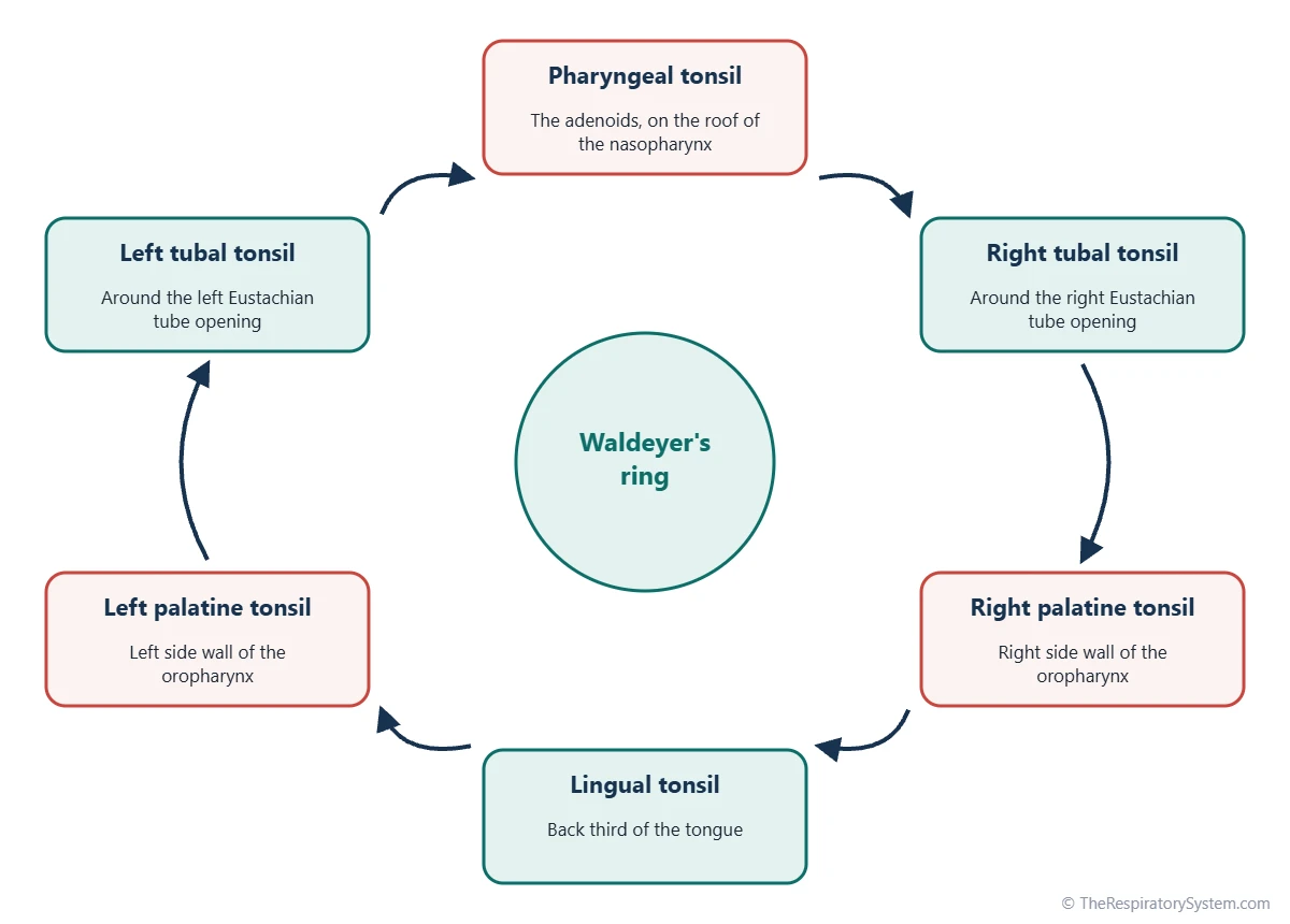
Waldeyer's ring is the ring of tonsil tissue that circles the upper throat where the nasal cavity and the mouth open into the pharynx. It is named after the 19th century German anatomist Heinrich Wilhelm von Waldeyer. The ring has four parts:
- Pharyngeal tonsil (adenoids): one mass on the roof and back wall of the nasopharynx, behind the nose.
- Tubal tonsils: two small patches around the openings of the Eustachian tubes, on the side walls of the nasopharynx.
- Palatine tonsils: two oval masses on the side walls of the oropharynx, between the arches at the back of the mouth.
- Lingual tonsil: lymphoid tissue spread over the back third of the tongue, at its base.
Because the ring surrounds both the air entrance from the nasal cavity and the food entrance from the oral cavity, almost everything breathed in or swallowed passes close to tonsil tissue.
Where Are the Tonsils Located?
The tonsils are located in the upper throat: the palatine tonsils at the sides of the back of the mouth, the lingual tonsil at the base of the tongue, and the adenoids and tubal tonsils higher up behind the nose. Only the palatine tonsils can normally be seen by looking into the mouth.
Palatine tonsils location
Each palatine tonsil sits in a shallow hollow called the tonsillar fossa, on the side wall of the oropharynx. The fossa is framed by two folds of mucosa over muscle: the palatoglossal arch in front and the palatopharyngeal arch behind, often called the anterior and posterior pillars. The uvula hangs in the middle between the two tonsils, and the superior constrictor muscle of the pharynx forms the floor of the fossa.
Lingual tonsils location
The lingual tonsil lies on the back third of the tongue, behind the V-shaped groove called the sulcus terminalis. It looks like a bumpy surface of small mounds rather than one lump, and it faces the oropharynx just in front of the epiglottis.
What is the nasopharyngeal tonsil?
The nasopharyngeal tonsil is another name for the pharyngeal tonsil, better known as the adenoids. It lies high on the back wall of the nasopharynx, behind the choanae, and cannot be seen through the mouth. It has its own page: adenoids (pharyngeal tonsil).
Tonsils Anatomy: Crypts, Follicles and Capsule
Tonsils anatomy is built for sampling: each tonsil is a mass of lymphoid follicles covered by an epithelium that folds inward to trap material from the throat. The palatine tonsil shows these features most clearly.
Surface and crypts
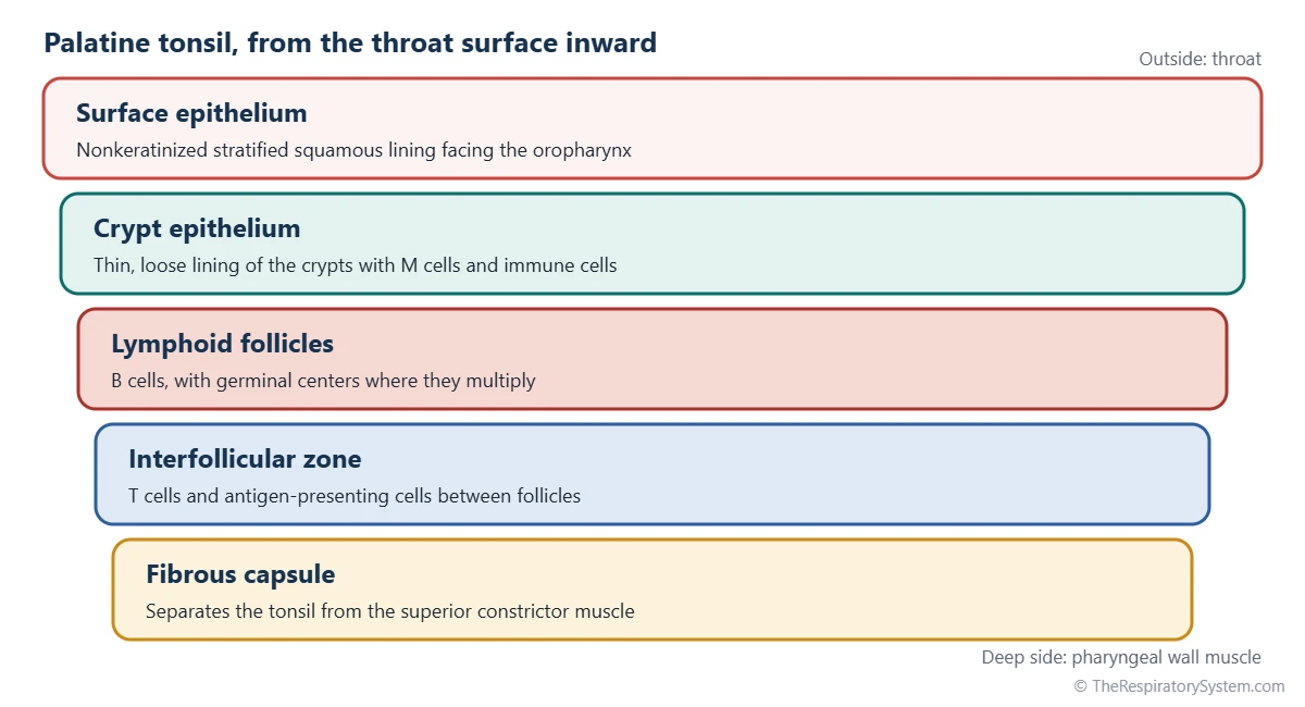
The palatine and lingual tonsils are covered by nonkeratinized stratified squamous epithelium, the same lining as the rest of the oropharynx. In the palatine tonsil this surface dips inward to form many deep, branching pits called tonsillar crypts, which greatly increase the area in contact with the throat. The lining of the crypts is thin and loose, with spaces for immune cells and specialized M cells that pass particles to the tissue below.
Follicles and germinal centers
Under the surface lie rounded lymphoid follicles packed with B lymphocytes. Active follicles have pale germinal centers where B cells multiply and mature after meeting an antigen. T lymphocytes fill the spaces between follicles.
Capsule, blood and nerves
On its deep side, the palatine tonsil is wrapped by a fibrous capsule that separates it from the muscle of the pharyngeal wall. Its main blood supply is the tonsillar branch of the facial artery, with smaller branches from nearby arteries, and sensation comes mainly from the glossopharyngeal nerve. This shared nerve supply with the ear is why a sore tonsil can make the ear ache. Lymph drains to the jugulodigastric node, just below the angle of the jaw.
| Feature | Pharyngeal tonsil (adenoids) | Tubal tonsils | Palatine tonsils | Lingual tonsil |
|---|---|---|---|---|
| How many | One | Two | Two | One |
| Where | Roof and back wall of the nasopharynx | Around the Eustachian tube openings | Side walls of the oropharynx, between the arches | Back third of the tongue |
| Surface lining | Mostly ciliated respiratory epithelium | Ciliated respiratory epithelium | Nonkeratinized stratified squamous | Nonkeratinized stratified squamous |
| Surface shape | Folds and shallow pits | Small diffuse patch | Deep branching crypts | Many shallow single crypts |
| Capsule | Thin, incomplete | None | Fibrous capsule on the deep side | Thin, incomplete |
| Seen through the mouth | No | No | Yes | Only partly, with a mirror |
| Samples mainly | Inhaled air | Air near the tube openings | Air and food | Food and drink |
| Largest | Early childhood | Childhood | Childhood | Childhood; can stay prominent in adults |
| Source: Typical anatomy, our editors | ||||
What Is the Function of the Tonsils?
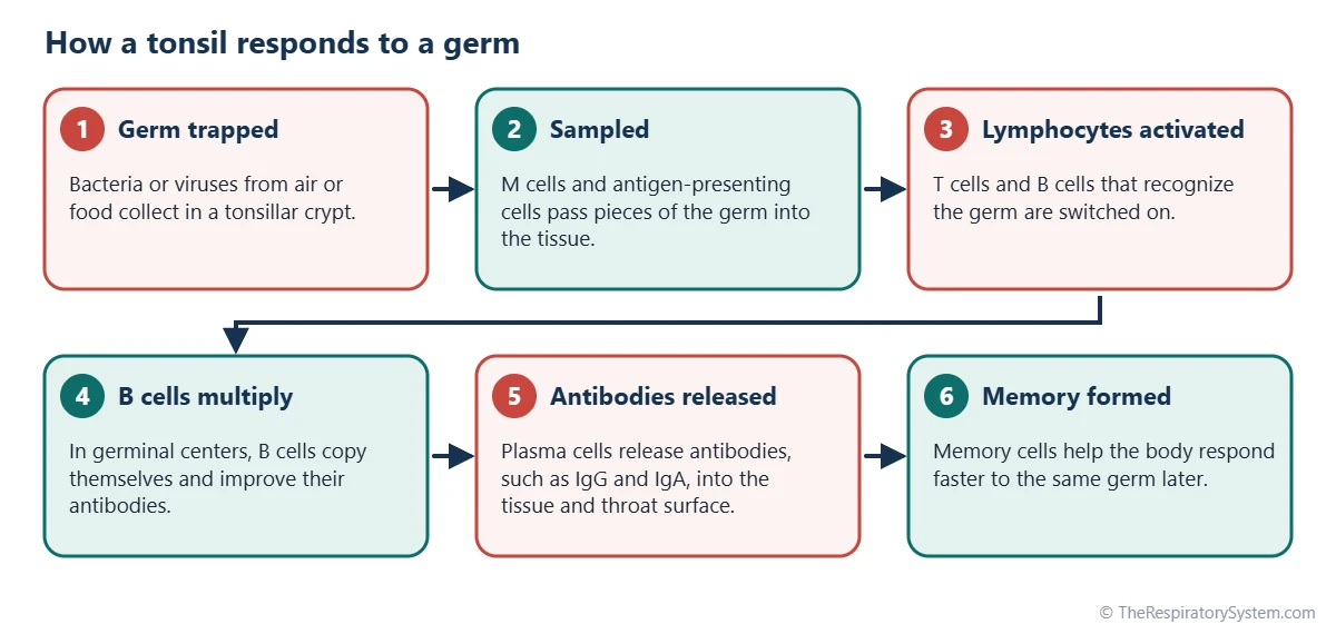
The function of the tonsils is to sample germs and particles that enter through the nose and mouth and to start immune responses against them. They are an early warning system rather than a wall: their job is to recognize invaders and train the immune system, not to block air or food.
- Sampling: crypts and M cells take in small amounts of bacteria, viruses and food proteins from the surface.
- Activating lymphocytes: antigen-presenting cells show these samples to T cells and B cells in the follicles.
- Making antibodies: activated B cells become plasma cells that produce antibodies, mainly IgG together with IgA, some of which reach the moist surfaces of the throat.
- Building memory: memory cells formed in the tonsils help the body respond faster to the same germ later.
Palatine tonsil function in the respiratory system
Within the respiratory system, the palatine tonsils guard the point where air from the mouth and nose meets in the oropharynx on its way down the pathway of air. They work alongside the mucus and cilia of the airway, described on our cilia and mucus page, which trap particles before they reach the lungs.
Lingual tonsils function in the digestive system
The lingual tonsil sits at the start of the digestive tract, at the base of the tongue, and monitors food and drink as they are swallowed. It does not digest food. Its crypts are flushed by small mucous glands, which helps keep them clean.
How Do the Tonsils Change With Age?
The tonsils grow during early childhood, are most active through the school years and gradually shrink after puberty. This pattern matches the period when a child meets many new germs for the first time and the immune system is building its memory.
The palatine tonsils develop from the second pharyngeal pouch of the embryo, and lymphocytes move into them before birth. They enlarge in the first years of life, and in young children they can look large compared with the size of the throat without being abnormal. In adults the tonsils become smaller and more fibrous, and in older people only a little active lymphoid tissue may remain. The adenoids follow a similar but earlier pattern; see respiratory system development for growth of the airway as a whole.
Common Conditions That Affect the Tonsils
The tonsils can become inflamed, enlarged or collect debris in their crypts. These conditions are described here for education only, with more in our respiratory diseases section.
- Tonsillitis: inflammation of the palatine tonsils, usually caused by viruses and sometimes by bacteria such as group A Streptococcus; see tonsillitis.
- Peritonsillar abscess: a collection of pus between the tonsil capsule and the throat wall, usually on one side.
- Tonsil stones (tonsilloliths): small hard deposits of debris and minerals that form in the crypts.
- Enlarged tonsils: large tonsils in children, which can narrow the throat during sleep and are linked to snoring and sleep apnea.
A severe sore throat with trouble swallowing or breathing should be checked by a doctor.
Key Facts
- The tonsils are masses of lymphoid tissue in the throat lining that sample germs from inhaled air and swallowed food.
- Four kinds of tonsils form Waldeyer's ring: the pharyngeal tonsil (adenoids), two tubal tonsils, two palatine tonsils and the lingual tonsil.
- The palatine tonsils sit in the tonsillar fossae on the side walls of the oropharynx, between the palatoglossal and palatopharyngeal arches.
- Deep branching crypts in the palatine tonsils greatly increase the surface that is in contact with the throat.
- Plasma cells in the tonsils produce antibodies, mainly IgG together with IgA, that help protect the throat.
- The tonsils are largest and most active in childhood and gradually shrink after puberty.
- Only the palatine tonsils are normally visible when looking into the open mouth.
Quick Quiz: Test What You Know About the Tonsils
6 questions. Tap an answer to check it.
Which tonsils can normally be seen through the open mouth?
The palatine tonsils sit on the side walls of the oropharynx and are visible at the back of the mouth.
What is another name for the pharyngeal tonsil?
The pharyngeal tonsil in the nasopharynx is commonly called the adenoids.
Where is the lingual tonsil found?
The lingual tonsil covers the back third of the tongue, behind the sulcus terminalis.
What are tonsillar crypts?
Crypts are branching pits of the surface epithelium that trap material for immune sampling.
Where do B cells multiply inside an active tonsil?
Active lymphoid follicles have pale germinal centers, where B cells multiply and mature after meeting an antigen.
What is the ring of tonsils around the upper throat called?
Waldeyer's ring is the circle of pharyngeal, tubal, palatine and lingual tonsils.
Frequently Asked Questions
How many tonsils do you have?
Most people have six tonsil masses in four kinds: one pharyngeal tonsil (the adenoids), two tubal tonsils, two palatine tonsils and one lingual tonsil. In everyday speech "tonsils" means only the two palatine tonsils at the back of the mouth. The lingual tonsil is sometimes described as a pair or as many small units because it is spread over the base of the tongue.
What is the difference between tonsils and adenoids?
The adenoids are themselves a tonsil, the pharyngeal tonsil, so the difference is mainly position. The palatine tonsils sit at the sides of the back of the mouth and are covered by stratified squamous epithelium with deep crypts. The adenoids sit higher, on the back wall of the nasopharynx behind the nose, are covered mostly by ciliated respiratory epithelium and cannot be seen through the mouth.
Are tonsils part of the lymphatic system?
Yes. The tonsils are secondary lymphoid organs, part of the mucosa-associated lymphoid tissue that guards the moist linings of the body. They contain the same B cells, T cells and follicles as lymph nodes. Their lymph drains to the deep cervical lymph nodes in the neck, which is why those nodes can become tender when the throat is infected.
Why are tonsils bigger in children?
Children meet many germs for the first time, and their tonsils work hard to sample them and build immune memory. This activity makes the lymphoid follicles grow, so the tonsils are relatively large from early childhood through the school years. After puberty the immune system has seen most common germs, and the tonsils slowly shrink and become more fibrous.
What are the white spots on tonsils?
White spots on the palatine tonsils can be several things. Small, hard, whitish lumps that come and go are often tonsil stones, deposits that form in the crypts. Spots or patches during a sore throat can be pus from an infection. Because different causes look alike, the spots alone do not show which one is present.
Do tonsils grow back?
When the palatine tonsils are removed, most of the tissue is taken out, but small amounts of lymphoid tissue can remain at the edges of the tonsillar fossa. In some people this leftover tissue enlarges over time, which can look like regrowth. The rest of Waldeyer's ring, including the lingual tonsil and adenoids, also remains and keeps working.
Why does a sore tonsil make the ear hurt?
The palatine tonsil receives most of its sensation from the glossopharyngeal nerve, which also carries sensation from part of the middle ear. When the tonsil is inflamed, the brain can interpret pain signals from this shared nerve as coming from the ear. This is called referred pain, and it is common with throat infections even when the ear itself is healthy.
Can you live without tonsils?
Yes. People whose palatine tonsils have been removed still have the rest of Waldeyer's ring, the lymph nodes of the neck and other immune tissues in the gut and airways. These continue to sample germs and make antibodies, so the immune system keeps working. The tonsils are one part of a large network rather than an organ the body cannot do without.


