At a Glance
The adenoids are the pharyngeal tonsil, one mass of lymphoid tissue on the upper back wall of the nasopharynx, behind the nasal cavity and above the soft palate. They sample germs carried in by inhaled air and help start immune responses. They cannot be seen through the mouth, grow during early childhood and usually shrink during adolescence, often becoming very small by adulthood.
- Location
- Roof and back wall of the nasopharynx, behind the choanae
- Also called
- Pharyngeal tonsil, nasopharyngeal tonsil
- Type of tissue
- Mucosa-associated lymphoid tissue covered mostly by ciliated respiratory epithelium
- Function
- Sample inhaled germs and help start immune responses
- Size over life
- Largest in early childhood, shrinking in adolescence
- Part of
- Waldeyer's ring of tonsils in the upper pharynx
On this page
What Are Adenoids?
Adenoids are a patch of lymphoid tissue high in the throat, behind the nose, that helps the immune system recognize germs breathed in through the nose. The correct anatomical name is the pharyngeal tonsil, because it is one of the tonsils of the pharynx. Although the word is plural, there is only one adenoid mass, in the midline.
Adenoids definition
In anatomy, the adenoids are defined as the unpaired collection of mucosa-associated lymphoid tissue in the mucosa of the roof and posterior wall of the nasopharynx. They form the upper part of Waldeyer's ring, the circle of lymphoid tissue that also includes the tubal, palatine and lingual tonsils. They sit along the airway of the upper respiratory tract but belong to the immune system.
Explained simply
Adenoids are a small soft cushion of germ-fighting tissue hidden behind your nose. When you breathe in, the air passes right over them, so they can check it for germs. They are bigger when you are little and get much smaller as you grow up.
Where Are the Adenoids Located?
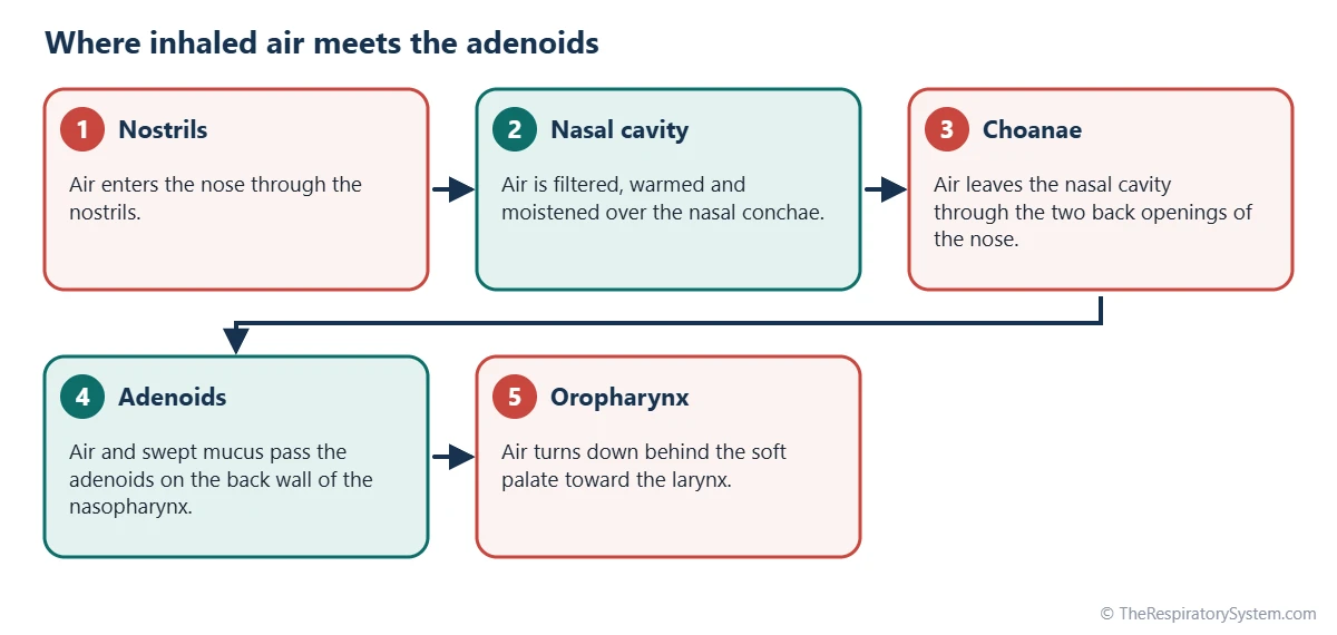
The adenoids are located on the roof and back wall of the nasopharynx, the space behind the nasal cavity and above the soft palate. They lie directly behind the choanae, the two back openings of the nose, so air from the nostrils flows straight toward them.
Adenoids location in the nasopharynx
The adenoid mass rests against the underside of the skull base, over the body of the sphenoid bone and the basilar part of the occipital bone. Its neighbors include:
- In front: the choanae and the back edge of the nasal septum.
- On each side: the openings of the Eustachian tubes, ringed by the small tubal tonsils.
- Above: the skull base, with the sphenoid sinus inside the bone above it.
- Below: the upper surface of the soft palate and the opening into the oropharynx.
Why you cannot see them
Looking into the open mouth, you can see the palatine tonsils and the uvula, but the adenoids are hidden above and behind the soft palate. Doctors view them with a thin endoscope passed through the nose or with a small mirror at the back of the mouth. A side X-ray of the neck also shows them as a soft tissue shadow on the back wall of the airway.
Adenoids Anatomy: Shape, Lining and Supply
Adenoids anatomy is that of a soft, pyramid-shaped pad of lymphoid tissue with its surface thrown into vertical folds, covered by respiratory lining. Its base is attached to the roof and back wall of the nasopharynx, and its tip points toward the nasal septum.
Surface and lining
The surface of the adenoids is covered mainly by pseudostratified ciliated columnar epithelium, the respiratory epithelium that also lines the nose, with patches of stratified squamous epithelium. Instead of the deep branching crypts of the palatine tonsils, the adenoids have folds and shallow pits. A thin, incomplete layer of connective tissue separates them from the tissue underneath.
Inside the adenoids
Beneath the lining are lymphoid follicles packed with B lymphocytes, many with germinal centers, and zones of T lymphocytes between them. This is the same basic design as the other tonsils: a sampling surface on top and immune cells ready to respond below.
Blood, nerve and lymph supply
Blood reaches the adenoids from several small arteries, including the ascending pharyngeal artery, the ascending palatine artery and the pharyngeal branch of the maxillary artery. Sensation is carried mainly by branches of the glossopharyngeal and vagus nerves. Lymph drains to the retropharyngeal lymph nodes and then to the deep cervical nodes of the neck.
| Feature | Adenoids | Why it matters |
|---|---|---|
| Number | One midline mass | Plural name, single structure |
| Position | Roof and back wall of the nasopharynx | Lies in the path of air from the nose |
| Shape | Pyramid-shaped pad with vertical folds | Folds increase the sampling surface |
| Surface lining | Mostly ciliated respiratory epithelium | Matches the lining of the nose |
| Capsule | Thin and incomplete | Unlike the palatine tonsils, no full capsule |
| Arterial supply | Ascending pharyngeal, ascending palatine and pharyngeal arteries | Several small vessels from the external carotid system |
| Nerve supply | Glossopharyngeal and vagus nerve branches | Shared with the rest of the pharynx |
| Lymph drainage | Retropharyngeal then deep cervical nodes | Neck nodes may swell with infection |
| Neighbors | Choanae in front, Eustachian tube openings at the sides | Explains effects on nose breathing and ears |
| Size over life | Grows in early childhood, shrinks in adolescence | Often very small in adults |
| Source: Typical anatomy, our editors | ||
What Is the Function of the Pharyngeal Tonsils?
The function of the pharyngeal tonsils, or adenoids, is to sample germs in air breathed in through the nose and to help start immune responses against them, especially in young children. Their position behind the choanae puts them in the path of almost every breath taken through the nose.
- Sampling: surface cells take in small amounts of bacteria, viruses and particles carried in by inhaled air and mucus.
- Activating lymphocytes: these samples are presented to T cells and B cells in the follicles.
- Making antibodies: activated B cells become plasma cells that release antibodies, including IgG and IgA, some of which reach the lining of the nose and throat.
- Training the immune system: in early childhood, the adenoids help build immune memory against common airborne germs.
The adenoids also sit on the route of mucus cleared from the nose. Cilia in the nasal lining sweep mucus back through the choanae, over the adenoids and down into the throat to be swallowed, the process described on our cilia and mucus page. Air then continues down the pathway of air to the larynx and lungs.
Adenoids vs Tonsils: What Is the Difference?
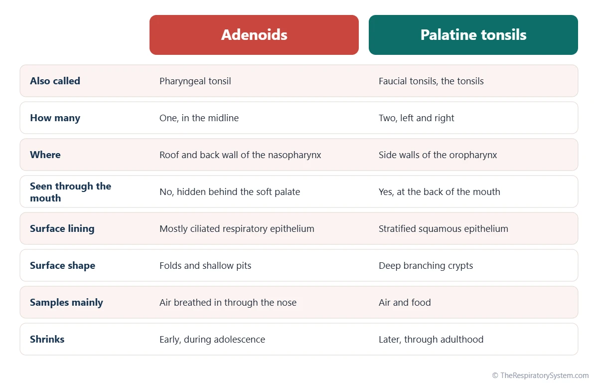
The adenoids are one of the tonsils, so the real difference is between the adenoids and the palatine tonsils: the adenoids sit behind the nose and sample inhaled air, while the palatine tonsils sit at the back of the mouth and sample both air and food. Both belong to Waldeyer's ring and share the same immune design.
The two also differ in lining and surface: the adenoids have mostly ciliated respiratory epithelium with folds, while the palatine tonsils have stratified squamous epithelium with deep crypts. The adenoids are hidden from view and tend to shrink earlier, during adolescence. The full set of four tonsil types is compared on our tonsils page, and the phrase "tonsils and adenoids" in everyday speech usually means the palatine tonsils plus the pharyngeal tonsil.
How Do the Adenoids Change With Age?
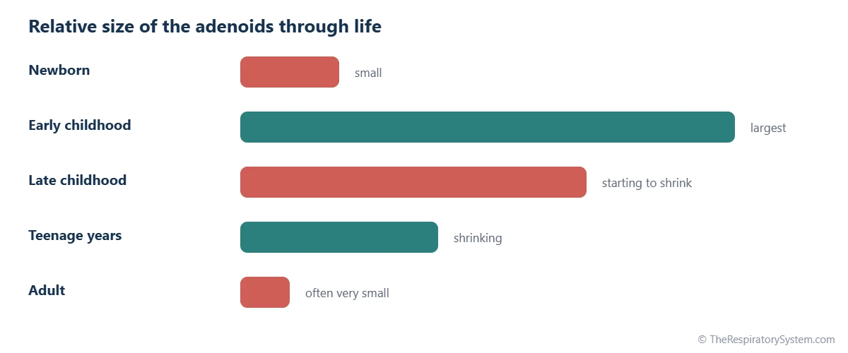
The adenoids are small at birth, grow during early childhood, are usually largest roughly between the ages of 3 and 7, and then shrink during adolescence. In many adults only a thin layer of lymphoid tissue remains on the back wall of the nasopharynx.
Their growth reflects the immune work of early childhood, when a child is exposed to many new germs. Because the nasopharynx of a young child is small, adenoids that are large for the space can narrow the airway behind the nose more easily than in an older child, whose nasopharynx has grown. Growth of the airway as a whole is described on our respiratory system development page.
Common Conditions That Affect the Adenoids
The adenoids can become enlarged or inflamed, and because of their position this can affect breathing through the nose and the Eustachian tubes. These conditions are described for education only; see our respiratory diseases section for more.
- Enlarged adenoids (adenoid hypertrophy): adenoids large enough to narrow the airway behind the nose, linked in children with blocked nose, mouth breathing and snoring; see nose vs mouth breathing.
- Adenoiditis: inflammation of the adenoids during an infection, which can cause a stuffy nose and thick nasal mucus.
- Eustachian tube blockage: large adenoids near the tube openings are associated with fluid in the middle ear and repeated ear infections in children.
- Sleep-disordered breathing: together with large palatine tonsils, enlarged adenoids are a common feature of sleep apnea in children.
Ongoing snoring with pauses in breathing, or a constantly blocked nose, is a reason to see a doctor.
Key Facts
- The adenoids are the pharyngeal tonsil, a single mass of lymphoid tissue on the roof and back wall of the nasopharynx.
- They form the top of Waldeyer's ring, together with the tubal, palatine and lingual tonsils.
- They sit directly behind the choanae, so air breathed in through the nose flows straight over them.
- Their surface is covered mainly by ciliated respiratory epithelium, unlike the stratified squamous lining of the palatine tonsils.
- The adenoids cannot be seen through the open mouth; they are viewed with an endoscope, a mirror or a side X-ray of the neck.
- They are usually largest in early childhood and shrink during adolescence.
- The openings of the Eustachian tubes lie on either side of them, which links the adenoids to the middle ear.
Quick Quiz: Test What You Know About the Adenoids
6 questions. Tap an answer to check it.
What is the anatomical name for the adenoids?
The adenoids are the pharyngeal tonsil, the top part of Waldeyer's ring.
Where are the adenoids located?
The adenoids sit on the roof and back wall of the nasopharynx, behind the choanae.
What mainly covers the surface of the adenoids?
Unlike the palatine tonsils, the adenoids are covered mostly by pseudostratified ciliated columnar epithelium.
Which openings lie on either side of the adenoids?
The Eustachian tubes open into the side walls of the nasopharynx, next to the adenoids.
When are the adenoids usually largest?
The adenoids grow in early childhood and then shrink during adolescence.
Why can the adenoids not be seen through the open mouth?
The adenoids lie above and behind the soft palate in the nasopharynx.
Frequently Asked Questions
Do adults have adenoids?
Most adults have only a small remnant of adenoid tissue. The adenoids shrink during adolescence, and by adulthood they are often reduced to a thin layer on the back wall of the nasopharynx. In some adults a little more tissue remains or enlarges again, but large adenoids are much less common than in children.
Are adenoids and tonsils the same thing?
Not exactly. The adenoids are one kind of tonsil, the pharyngeal tonsil, so they belong to the same family of lymphoid tissue. In everyday speech, though, "tonsils" means the two palatine tonsils at the back of the mouth, and "adenoids" means the single mass behind the nose. The two differ in position, lining and surface shape.
Why are they called adenoids?
The name comes from the Greek word for gland, with an ending meaning like, so adenoid means gland-like. Early anatomists thought the tissue was a gland because of its soft, lumpy look. It is actually lymphoid tissue, so its proper anatomical name is the pharyngeal tonsil, which places it with the other tonsils of the throat.
What are tonsils and adenoids made of?
Both are made of lymphoid tissue: lymphoid follicles full of B lymphocytes, many with germinal centers, and areas of T lymphocytes and antigen-presenting cells between them. The tissue is covered by the lining of the throat, ciliated respiratory epithelium over the adenoids and stratified squamous epithelium over the palatine tonsils, and supported by fine connective tissue.
How do doctors see the adenoids?
Because the adenoids are hidden behind the soft palate, doctors cannot see them through the open mouth. They can look at them with a thin flexible or rigid endoscope passed through the nose, with a small angled mirror placed at the back of the mouth, or on a side X-ray of the neck, where they appear as a soft tissue bulge on the airway's back wall.
Are the adenoids connected to the ears?
Not directly, but they are close neighbors. The Eustachian tubes, which link the middle ears to the throat and balance air pressure, open into the side walls of the nasopharynx right beside the adenoids. When the adenoids are large or inflamed, they can affect how well these tubes open and drain, which is why adenoids and ear problems are often discussed together.
Do adenoids help you breathe?
No, the adenoids do not move air or take part in gas exchange. They are immune tissue that lines the airway behind the nose and checks the air for germs. Air simply flows past them on its way from the nose to the throat. When they are very large, they can partly narrow this passage, which is the opposite of helping breathing.
Can adenoids grow back?
Adenoid tissue is not wrapped in a full capsule, so when the adenoids are removed, small amounts of lymphoid tissue can be left on the back wall of the nasopharynx. In some children this remaining tissue grows again, which is called regrowth. This is uncommon.


