At a Glance
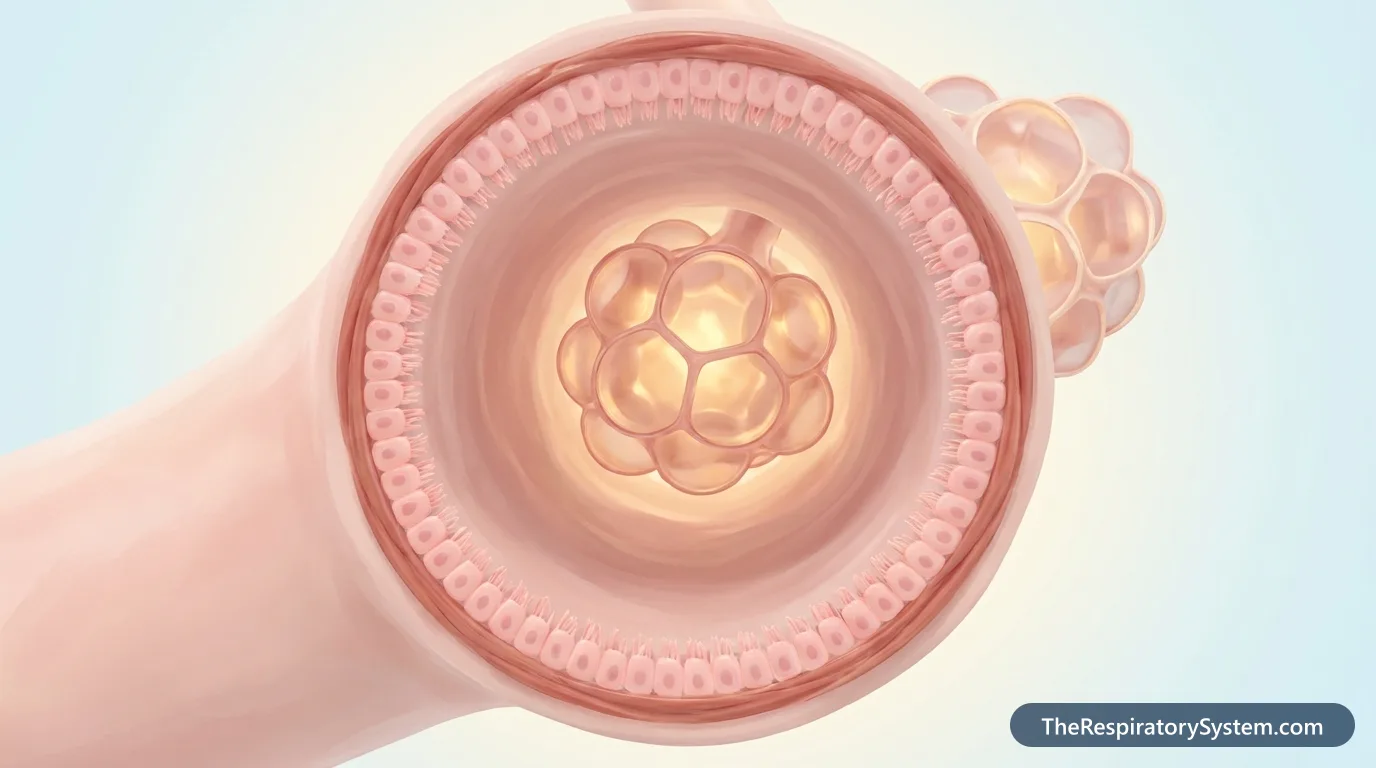
Bronchioles are the smallest airways of the lungs, about 1 mm wide or less, with no cartilage in their walls. They branch from the bronchi and end in terminal bronchioles (about 0.5 mm wide), which lead to respiratory bronchioles, alveolar ducts and the alveoli. The two lungs together have tens of thousands of terminal bronchioles.
- Location
- Inside the lungs, between the smallest bronchi and the alveolar ducts, within every lung lobule
- Function
- Conduct air to the alveoli and control airflow with smooth muscle; respiratory bronchioles also exchange some gas
- Tissue
- Ciliated columnar to cuboidal epithelium with club cells, smooth muscle, elastic fibers; no cartilage or glands
- Size
- About 1 mm wide or less; terminal bronchioles about 0.5 mm
- Connects to
- Bronchi above; alveolar ducts and alveoli below
- Part of
- Bronchial tree: conducting zone ending in the respiratory zone
On this page
- What Are the Bronchioles?
- Where Are the Bronchioles Located?
- Bronchioles Anatomy: Lobular, Terminal and Respiratory Bronchioles
- What Are the Bronchioles Made Of?
- What Is the Difference Between the Bronchi and the Bronchioles?
- What Do the Bronchioles Do in the Respiratory System?
- How Do the Bronchioles Work During Breathing?
- How Do the Bronchioles Develop and Change With Age?
- Common Conditions That Affect the Bronchioles
- Key Facts
- Quiz
- FAQ
What Are the Bronchioles?
The bronchioles are the narrow, cartilage-free airways inside the lungs that link the bronchi to the alveoli, the air sacs where gas exchange takes place. After the trachea divides into the right and left main bronchi, these keep branching into smaller and smaller bronchi; once a tube is about 1 mm wide and its cartilage has disappeared, it is called a bronchiole. The trachea, bronchi, bronchioles and alveoli together form the lower respiratory tract.
What does the word bronchiole mean?
Bronchiole means "little bronchus". It is pronounced BRONG-kee-ole, and the plural, bronchioles, is BRONG-kee-oles. The term covers three kinds of airway, from largest to smallest: lobular (preterminal) bronchioles, terminal bronchioles and respiratory bronchioles.
What do the bronchioles look like?
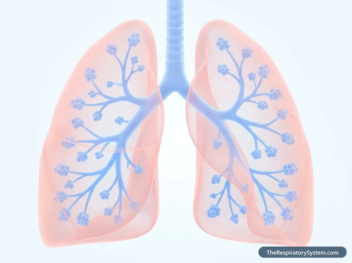
Bronchioles look like very fine, thin-walled branching tubes, many of them narrower than a pencil lead. Each one divides into two smaller branches again and again, like the twigs at the end of a tree branch. The last ones are dotted with tiny bulges, the first alveoli, and end in grape-like clusters of air sacs wrapped in a net of capillaries.
How many bronchioles are there?
The bronchioles are by far the most numerous airways. The two lungs together have tens of thousands of terminal bronchioles, and every one of them leads to its own small cluster of gas-exchanging tissue. Counting the larger bronchioles above them and the respiratory bronchioles below, the total number of bronchioles runs into the hundreds of thousands.
Explained simply
If the airways of a lung are a tree, the bronchioles are its thinnest twigs. They are too small to need stiff cartilage, so instead they are wrapped in muscle that can loosen or tighten, a bit like adjusting a garden hose nozzle. At the very tip of every twig hangs a bunch of tiny balloons, the alveoli, where oxygen moves into the blood.
Where Are the Bronchioles Located?
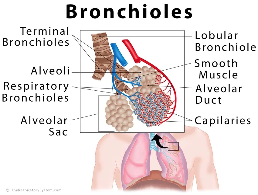
The bronchioles are located deep inside both lungs, in every lobe and segment, where they form the last few millimeters of the airway between the smallest bronchi and the alveolar ducts. None of them lie outside the lung tissue.
Bronchioles inside the lung lobules
The lung tissue is divided into small pyramid-shaped units called secondary pulmonary lobules, each about 1 to 2.5 cm across and outlined by thin walls of connective tissue. A lobular bronchiole enters each lobule at its center, together with a branch of the pulmonary artery, and divides into several terminal bronchioles inside it. Each terminal bronchiole supplies a pulmonary acinus, the cluster of respiratory bronchioles, alveolar ducts and alveoli beyond it.
Where are the bronchioles and alveoli located in the respiratory system?
Both lie at the far end of the airway. Air enters through the nose or mouth, passes through the pharynx, larynx, trachea and bronchi, then flows through the bronchioles into the alveoli. The bronchioles sit in every part of each lung lobe, from the apex near the collarbone to the base resting on the diaphragm.
Bronchioles Anatomy: Lobular, Terminal and Respiratory Bronchioles
There are three types of bronchioles: lobular bronchioles, which enter the lung lobules; terminal bronchioles, the last purely air-conducting tubes; and respiratory bronchioles, the first airways with alveoli in their walls. Their walls become thinner and their lining flatter at each step.
Lobular (preterminal) bronchioles
Lobular bronchioles, also called preterminal bronchioles, are the first bronchioles after the last small bronchi, about 1 mm wide. Each one passes into a pulmonary lobule and there divides into several terminal bronchioles. Their lining is ciliated columnar epithelium with a few goblet cells, and a well-developed layer of smooth muscle surrounds them.
Terminal bronchioles
Terminal bronchioles are about 0.5 mm wide and mark the end of the conducting zone: they are the last airways that only carry air and take no part in gas exchange. They are lined with ciliated cuboidal epithelium that has no goblet cells, mixed with dome-shaped club cells. Each terminal bronchiole divides into two or more respiratory bronchioles.
Respiratory bronchioles
Respiratory bronchioles are the start of the respiratory zone. Their walls are interrupted by scattered alveoli that bulge out of them, so a little gas exchange already happens here, and the alveoli become more numerous along each branch. They are lined with low cuboidal epithelium, with few cilia and many club cells, and wrapped in thin bands of smooth muscle and elastic fibers. Each respiratory bronchiole ends in about 2 to 11 alveolar ducts.
Alveolar ducts and sacs
Alveolar ducts are thin passages whose walls are made almost entirely of alveolar openings, with rings of elastin, collagen and smooth muscle around the openings. They lead into alveolar sacs, the dead-end clusters of alveoli that are each wrapped in pulmonary capillaries.
| Airway | Approximate width | Lining | Role |
|---|---|---|---|
| Small bronchi | More than 1 mm | Pseudostratified ciliated columnar with goblet cells | Conduct air; supported by cartilage plates |
| Lobular (preterminal) bronchioles | About 1 mm | Ciliated simple columnar with a few goblet cells | Enter a lung lobule and divide into terminal bronchioles |
| Terminal bronchioles | About 0.5 mm | Ciliated cuboidal with club cells; no goblet cells | Last purely conducting airway |
| Respiratory bronchioles | Smaller than terminal bronchioles | Low cuboidal with club cells and few cilia | Conduct air and begin gas exchange through scattered alveoli |
| Alveolar ducts | Very small | Walls made almost entirely of alveolar openings | Lead air into the alveolar sacs; 2 to 11 per respiratory bronchiole |
| Alveolar sacs and alveoli | Each alveolus about 0.2 mm | Type I and type II pneumocytes | Main site of gas exchange |
| Source: Typical adult anatomy, our editors | |||
What Are the Bronchioles Made Of?
Bronchioles are made of a thin lining epithelium, a thin layer of connective tissue rich in elastic fibers, and a relatively thick layer of smooth muscle, with no cartilage, no submucosal glands and, in the smaller ones, no goblet cells. Their structure is a simplified version of the bronchial wall.
Epithelium
The lining changes gradually along the bronchioles. The largest ones are lined with ciliated simple columnar epithelium that still has a few goblet cells; in smaller ones the cells become cuboidal and the goblet cells disappear. In the respiratory bronchioles the lining is low cuboidal with only scattered cilia, and it flattens into the very thin alveolar lining where the alveoli begin.
Club cells
Club cells, formerly called Clara cells, are dome-shaped cells without cilia that become the main secretory cell in the terminal and respiratory bronchioles. They release protective proteins and a thin surfactant-like fluid that lines the small airways, help break down inhaled toxins with their enzymes, and can divide to replace damaged lining cells. In this way they partly take over the protective job that goblet cells and glands do in the larger airways.
Smooth muscle and elastic fibers
Compared with the size of the tube, bronchioles have more smooth muscle than any other airway, arranged in spiral bundles. Contraction of this muscle narrows the bronchiole. Elastic fibers in the wall and in the surrounding alveolar walls attach to the outside of each bronchiole and pull it open from all sides, so the tube stays open even though it has no cartilage.
Blood supply and nerves
The walls of the bronchioles are nourished by the bronchial arteries down to about the level of the respiratory bronchioles, while a branch of the pulmonary artery runs right beside every bronchiole on its way to the alveoli. Vagus nerve fibers narrow the bronchioles, and sympathetic nerve signals and the hormone epinephrine (adrenaline) widen them by acting on beta-2 receptors in the smooth muscle.
What Is the Difference Between the Bronchi and the Bronchioles?
The bronchi are larger airways, more than about 1 mm wide, that are supported by cartilage and contain mucus glands, while the bronchioles are smaller airways, about 1 mm wide or less, that have no cartilage or glands and rely on smooth muscle and elastic fibers instead.
Under the microscope the difference is easy to see. A cross section of a bronchus shows plates of hyaline cartilage, seromucous glands and a tall pseudostratified lining with many goblet cells. A bronchiole shows a thin, often wavy lining of columnar or cuboidal cells, a prominent ring of smooth muscle, and no cartilage at all. The table below compares the bronchus, the bronchiole and the alveolus.
Our side-by-side guide to bronchi vs bronchioles compares the two airways feature by feature.
| Feature | Bronchi | Bronchioles | Alveoli |
|---|---|---|---|
| Width | More than about 1 mm | About 1 mm or less | About 0.2 mm across |
| Cartilage | Rings in main bronchi, plates inside the lungs | None | None |
| Glands | Seromucous glands in the submucosa | None | None |
| Lining | Pseudostratified ciliated columnar | Simple columnar to cuboidal | Very thin type I pneumocytes plus type II pneumocytes |
| Goblet cells | Many | Few in larger bronchioles, none in terminal ones | None |
| Smooth muscle | Present, in a spiral layer | Prominent for the size of the tube | Only around the openings of alveolar ducts |
| Kept open by | Cartilage | Elastic pull of the surrounding lung tissue | Surfactant and elastic fibers |
| Gas exchange | No | Only in respiratory bronchioles | Yes, the main site |
| Zone | Conducting | Conducting, then the start of the respiratory zone | Respiratory |
| Source: Typical adult histology, our editors | |||
What Do the Bronchioles Do in the Respiratory System?
The main function of the bronchioles is to carry air from the bronchi to every alveolus and to control how much air reaches each part of the lung by widening and narrowing. The respiratory bronchioles also take part in gas exchange through the alveoli in their walls. Our overview of respiratory system functions shows how this fits with the rest of the system.
Main functions
- Delivering air to the alveoli: the bronchioles spread incoming air through tens of thousands of fine branches, so that every alveolus receives fresh air with each breath.
- Controlling airflow: their smooth muscle adjusts the width of each tube, matching the air sent to an area of lung with the blood flowing through it.
- Gas exchange: the respiratory bronchioles exchange a small amount of oxygen and carbon dioxide through the alveoli that bud from their walls.
- Cleaning: in the larger bronchioles, goblet cells and cilia trap dust and germs in mucus and sweep them up toward the bronchi, trachea and throat, where they are coughed out or swallowed.
- Protection: club cells secrete protective proteins, help neutralize inhaled toxins and repair the lining after injury.
Function of the terminal bronchioles
The terminal bronchioles are the last conducting airways. They carry air into the respiratory zone and take no part in gas exchange themselves. Together with all the larger airways above them, they form the anatomical dead space, about 150 mL of air in an adult that is breathed in and out without reaching the alveoli.
Function of the respiratory bronchioles
The respiratory bronchioles are a transition zone. They still conduct air onward to the alveolar ducts, but the alveoli scattered along their walls already exchange gases with the blood. They are the point where the airways change from moving air to exchanging it.
How Do the Bronchioles Work During Breathing?
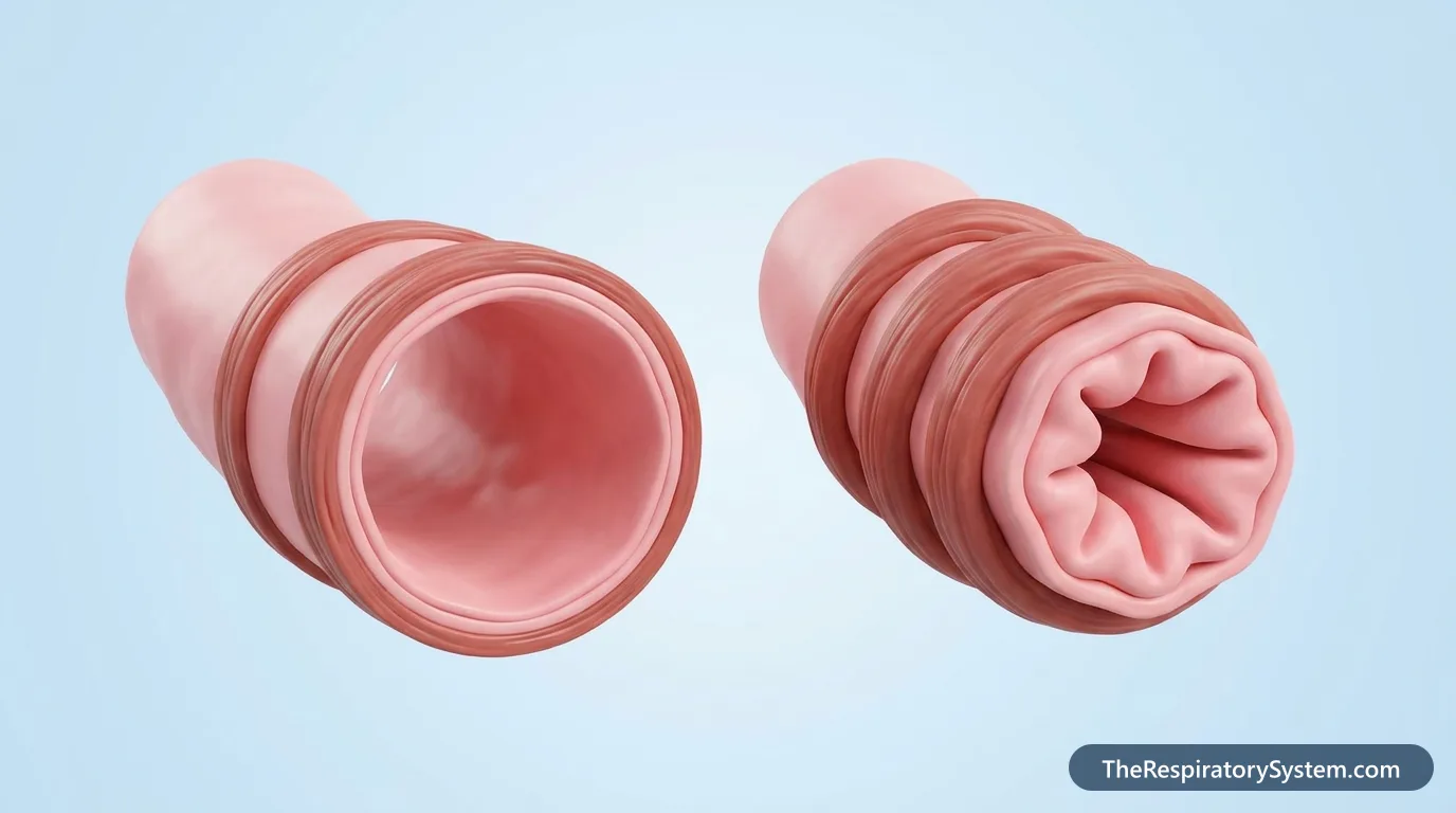
During a breath in the bronchioles are stretched open by the expanding lung tissue around them, and during a breath out they narrow again as the lung recoils, so their width changes with every breath.
Breathing in and out
When the diaphragm and the intercostal muscles enlarge the chest, the elastic alveolar walls attached to each bronchiole pull outward on it and widen it. On a quiet breath out the lung recoils and the bronchioles return to their resting width. During a forced breath out, pressure around the small airways rises, and without cartilage they rely entirely on that elastic tethering to stay open.
Why airflow slows down
Although each bronchiole is tiny, there are so many of them that their combined cross-section is far larger than that of the trachea. As a result, air slows down sharply as it moves into the bronchioles, and in the respiratory zone it moves mainly by diffusion rather than by flowing. For the same reason, the small airways together cause only a small part of the total resistance to airflow in healthy lungs; most of it comes from the medium-sized bronchi.
Bronchoconstriction and bronchodilation
Bronchoconstriction is the narrowing of the bronchioles by their smooth muscle, triggered by the vagus nerve and by substances such as histamine released during allergic reactions. Bronchodilation is the widening of the bronchioles, driven by the sympathetic nervous system and epinephrine during exercise or stress. Because the bronchioles are so narrow, even a small amount of narrowing has a large effect on airflow through them.
How Do the Bronchioles Develop and Change With Age?
The bronchioles form before birth: the terminal bronchioles appear by about the sixteenth week of pregnancy, and the respiratory bronchioles develop during the following canalicular stage, from about the sixteenth to the twenty-sixth week.
Before birth
The branching of the lung buds builds all the conducting airways, down to the terminal bronchioles, during the pseudoglandular stage of lung development. In the canalicular stage, each terminal bronchiole divides into respiratory bronchioles, the airways widen, and capillaries grow close to the lining, preparing the first sites where gas exchange will be possible.
Babies and children
A baby's bronchioles are extremely narrow, so swelling of the lining or a little mucus can block them easily. This is why viral infections of the small airways cause much more trouble in infants than in older children and adults. The bronchioles lengthen and widen as the lungs grow through childhood.
Older adults
With age, the lung tissue loses some of its elastic recoil, so it pulls less on the outside of the bronchioles. The small airways at the bottom of the lungs then tend to close earlier during a breath out, trapping a little more air in the lungs, which is one reason the residual volume rises in older age.
Common Conditions That Affect the Bronchioles
Conditions of the bronchioles include infection, scarring, spasm of their smooth muscle and loss of their elastic support, and the list below names the main problem in each. Our respiratory diseases section covers them in more depth, and anyone with breathing symptoms should see a doctor.
- Bronchiolitis: a common viral infection of the bronchioles, most often caused by respiratory syncytial virus (RSV). It mainly affects babies and children under 2 years old, whose small airways swell and fill with mucus, causing coughing, congestion, wheezing and difficulty breathing.
- Bronchospasm: a sudden tightening of the smooth muscle in the walls of the bronchi and bronchioles. It causes wheezing, shortness of breath, coughing and chest tightness, and is most often linked to asthma and allergic reactions.
- Asthma: long-term inflammation of the airways in which the bronchioles and small bronchi narrow too easily, with swelling of their lining and extra mucus.
- Emphysema: a form of chronic obstructive pulmonary disease (COPD) in which the walls of the alveoli are destroyed. The bronchioles lose the elastic tissue that holds them open, so they collapse during a breath out and trap air in the lungs. Smoking is the main cause.
- Bronchiolitis obliterans: scarring that narrows or blocks the bronchioles. It can follow severe infections, the breathing in of toxic fumes, or a lung transplant; one form linked to the flavoring chemical diacetyl is known as popcorn lung.
- Bronchiectasis: permanent widening and damage of the bronchi and bronchioles, which can follow severe infections or occur with genetic disorders, immune system problems and allergic lung conditions. It causes a chronic cough, phlegm, wheezing, shortness of breath, chest pain and tiredness, and it is a long-term condition that many people live with for decades.
Key Facts
- Bronchioles are airways about 1 mm wide or less with no cartilage, branching from the bronchi inside the lungs.
- There are three types: lobular (preterminal), terminal and respiratory bronchioles.
- Terminal bronchioles, about 0.5 mm wide, are the last airways that only conduct air; the two lungs hold tens of thousands of them.
- Respiratory bronchioles have alveoli budding from their walls and are the first airways where gas exchange happens.
- Club cells, once called Clara cells, protect the lining of the smallest airways and help repair it.
- Bronchioles stay open because elastic fibers in the surrounding lung tissue pull on their walls, not because of cartilage.
- Bronchiolitis, a viral infection of these small airways, mainly affects children under 2 years old.
Quick Quiz: Test What You Know About the Bronchioles
6 questions. Tap an answer to check it.
What is missing from the walls of the bronchioles that is present in the bronchi?
Bronchioles have no cartilage; their walls rely on smooth muscle and the elastic pull of surrounding lung tissue.
Which are the last purely air-conducting airways?
Terminal bronchioles end the conducting zone; beyond them, the respiratory bronchioles begin gas exchange.
Which cells, once called Clara cells, protect the lining of the small airways?
Club cells secrete protective proteins, help neutralize toxins and can replace damaged lining cells.
About how wide is a terminal bronchiole?
Terminal bronchioles are about half a millimeter wide.
What feature makes respiratory bronchioles different from terminal bronchioles?
Respiratory bronchioles have scattered alveoli in their walls, so they take part in gas exchange.
Which virus most often causes bronchiolitis in babies?
RSV is the most common cause of bronchiolitis, an infection of the small airways in children under 2.
Frequently Asked Questions
What is the main function of the bronchioles?
The main function of the bronchioles is to carry air from the bronchi to the alveoli so that every air sac in the lungs receives fresh air. Their smooth muscle also controls how much air flows to each part of the lung by widening or narrowing the tubes. The smallest ones, the respiratory bronchioles, take part in gas exchange as well.
What is the difference between bronchioles and alveoli?
Bronchioles are tiny tubes that conduct air, while alveoli are tiny air sacs at the end of those tubes where oxygen and carbon dioxide are exchanged with the blood. Bronchioles have a lining of columnar or cuboidal cells and a layer of smooth muscle. Alveoli have extremely thin walls made of flat type I pneumocytes and surfactant-making type II pneumocytes, wrapped in capillaries.
What is the difference between terminal and respiratory bronchioles?
Terminal bronchioles are the last airways of the conducting zone: they carry air but have no alveoli, so they do not exchange gases. Each one divides into respiratory bronchioles, which have alveoli budding from their walls and so begin gas exchange. Terminal bronchioles are lined with ciliated cuboidal cells, while respiratory bronchioles have a lower lining with fewer cilia.
Do bronchioles have cilia?
Yes, most bronchioles have cilia. The larger bronchioles and the terminal bronchioles are lined with ciliated cells that sweep mucus upward toward the bronchi. The number of cilia falls along the way, and the respiratory bronchioles have only scattered ciliated cells. The alveoli have no cilia at all, so particles that reach them are removed by alveolar macrophages instead.
Why do bronchioles not have cartilage?
Bronchioles are so small and so surrounded by lung tissue that they do not need cartilage to stay open. The elastic walls of the alveoli around them attach to their outer surface and pull them open, especially during a breath in. Without cartilage, the bronchioles can also change their width freely with their smooth muscle, which lets the lungs adjust airflow region by region.
How long is a bronchiole?
Each bronchiole is only a few millimeters long before it divides again, and the whole chain from a lobular bronchiole to the alveolar sacs fits inside a lung lobule about 1 to 2.5 cm across. The pulmonary acinus supplied by a single terminal bronchiole is only several millimeters wide. What bronchioles lack in length they make up for in number.
Can bronchioles be seen on a CT scan?
Healthy bronchioles are too small and thin-walled to be seen individually on a standard CT scan of the chest. They become visible when they are diseased, for example when their walls thicken, when they fill with mucus or inflammatory material, or when they are widened. Radiologists then see small branching or nodular shapes near the center of the lung lobules, sometimes described as a tree-in-bud pattern.
What happens to the bronchioles during an asthma attack?
During an asthma attack, the smooth muscle around the small bronchi and bronchioles tightens, the lining swells with inflammation and extra mucus is produced. All three narrow the airways, so air moves in and out with more effort. Because bronchioles are already very narrow, even a small narrowing greatly reduces airflow, which causes wheezing, coughing, chest tightness and shortness of breath.
Keep Exploring


